• 文献检索
  • 文档翻译
  • 深度研究
  • 学术资讯
  • Suppr Zotero 插件Zotero 插件
  • 邀请有礼
  • 套餐&价格
  • 历史记录
应用&插件
Suppr Zotero 插件Zotero 插件浏览器插件Mac 客户端Windows 客户端微信小程序
定价
高级版会员购买积分包购买API积分包
服务
文献检索文档翻译深度研究API 文档MCP 服务
关于我们
关于 Suppr公司介绍联系我们用户协议隐私条款
关注我们

Suppr 超能文献

核心技术专利:CN118964589B侵权必究
粤ICP备2023148730 号-1Suppr @ 2026

文献检索

告别复杂PubMed语法,用中文像聊天一样搜索,搜遍4000万医学文献。AI智能推荐,让科研检索更轻松。

立即免费搜索

文件翻译

保留排版,准确专业,支持PDF/Word/PPT等文件格式,支持 12+语言互译。

免费翻译文档

深度研究

AI帮你快速写综述,25分钟生成高质量综述,智能提取关键信息,辅助科研写作。

立即免费体验

超快速高压冷冻和电子断层扫描揭示的心肌细胞 T 小管/肌浆网连接的纳米级形态。

Nano-scale morphology of cardiomyocyte t-tubule/sarcoplasmic reticulum junctions revealed by ultra-rapid high-pressure freezing and electron tomography.

机构信息

Institute for Experimental Cardiovascular Medicine, University Heart Center Freiburg·Bad Krozingen, Freiburg, Germany; Faculty of Medicine, University of Freiburg, Freiburg, Germany.

Institute for Experimental Cardiovascular Medicine, University Heart Center Freiburg·Bad Krozingen, Freiburg, Germany; Faculty of Medicine, University of Freiburg, Freiburg, Germany.

出版信息

J Mol Cell Cardiol. 2021 Apr;153:86-92. doi: 10.1016/j.yjmcc.2020.12.006. Epub 2020 Dec 20.

DOI:10.1016/j.yjmcc.2020.12.006
PMID:33359037
原文链接:https://pmc.ncbi.nlm.nih.gov/articles/PMC8035077/
Abstract

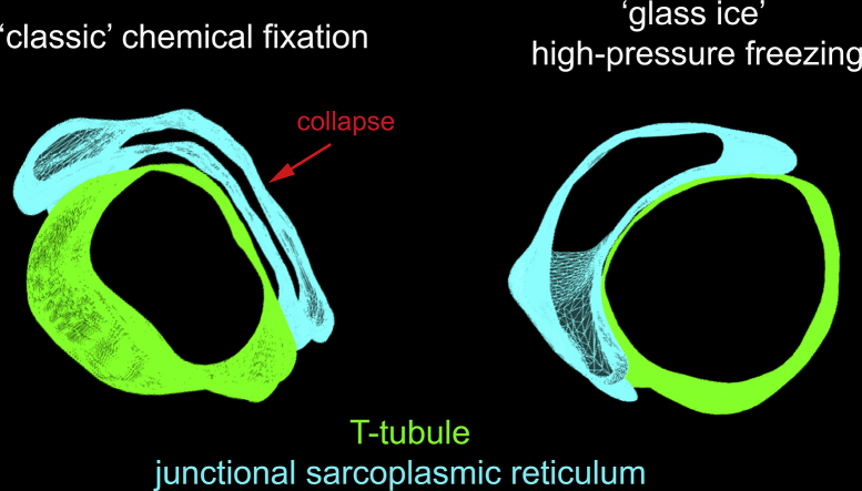
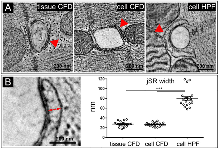
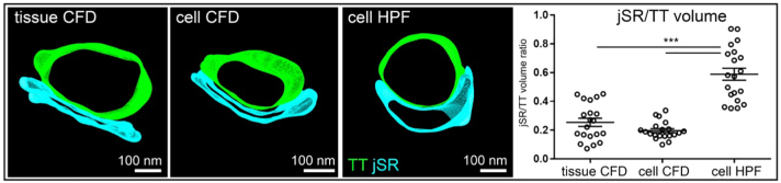
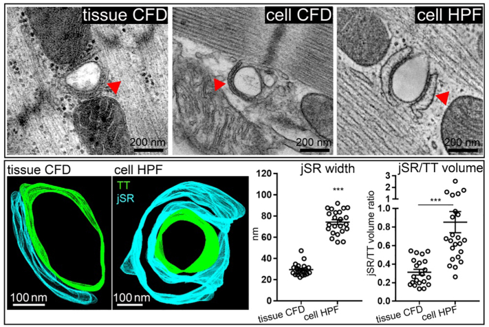
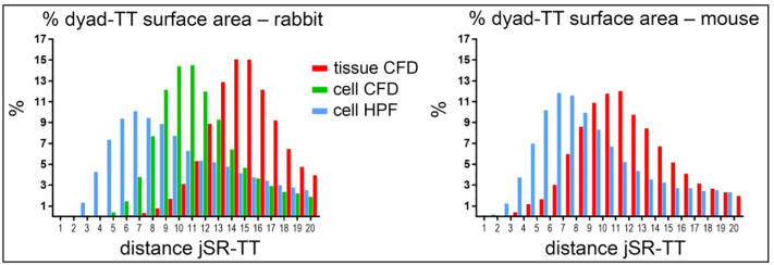
Detailed knowledge of the ultrastructure of intracellular compartments is a prerequisite for our understanding of how cells function. In cardiac muscle cells, close apposition of transverse (t)-tubule (TT) and sarcoplasmic reticulum (SR) membranes supports stable high-gain excitation-contraction coupling. Here, the fine structure of this key intracellular element is examined in rabbit and mouse ventricular cardiomyocytes, using ultra-rapid high-pressure freezing (HPF, omitting aldehyde fixation) and electron microscopy. 3D electron tomograms were used to quantify the dimensions of TT, terminal cisternae of the SR, and the space between SR and TT membranes (dyadic cleft). In comparison to conventional aldehyde-based chemical sample fixation, HPF-preserved samples of both species show considerably more voluminous SR terminal cisternae, both in absolute dimensions and in terms of junctional SR to TT volume ratio. In rabbit cardiomyocytes, the average dyadic cleft surface area of HPF and chemically fixed myocytes did not differ, but cleft volume was significantly smaller in HPF samples than in conventionally fixed tissue; in murine cardiomyocytes, the dyadic cleft surface area was higher in HPF samples with no difference in cleft volume. In both species, the apposition of the TT and SR membranes in the dyad was more likely to be closer than 10 nm in HPF samples compared to CFD, presumably resulting from avoidance of sample shrinkage associated with conventional fixation techniques. Overall, we provide a note of caution regarding quantitative interpretation of chemically-fixed ultrastructures, and offer novel insight into cardiac TT and SR ultrastructure with relevance for our understanding of cardiac physiology.

摘要

详细了解细胞内隔室的超微结构是理解细胞如何发挥功能的前提。在心肌细胞中,横管 (TT) 和肌浆网 (SR) 膜的紧密贴合支持稳定的高增益兴奋-收缩偶联。在这里,使用超快速高压冷冻 (HPF,省略醛固定) 和电子显微镜检查了兔和鼠心室心肌细胞中这一关键细胞内元件的精细结构。3D 电子断层图用于定量 TT、SR 的终末池和 SR 与 TT 膜之间的空间 (二联体裂) 的尺寸。与传统的基于醛的化学样品固定相比,两种物种的 HPF 保存的样品均显示出 SR 终末池体积明显更大,无论是在绝对尺寸还是在连接 SR 与 TT 体积比方面。在兔心肌细胞中,HPF 和化学固定心肌细胞的平均二联体裂表面面积没有差异,但裂体积在 HPF 样品中明显小于常规固定组织;在鼠心肌细胞中,HPF 样品中二联体裂表面面积较高,裂体积没有差异。在这两种物种中,与 CFD 相比,HPF 样品中二联体 TT 和 SR 膜的贴合更有可能小于 10nm,这可能是由于避免了与传统固定技术相关的样品收缩。总体而言,我们对化学固定超微结构的定量解释提出了警告,并为理解心脏 TT 和 SR 的超微结构提供了新的见解,这对我们理解心脏生理学具有重要意义。

https://cdn.ncbi.nlm.nih.gov/pmc/blobs/62f4/8035077/3297081c4b96/gr5.jpg
https://cdn.ncbi.nlm.nih.gov/pmc/blobs/62f4/8035077/4307c56062ea/ga1.jpg
https://cdn.ncbi.nlm.nih.gov/pmc/blobs/62f4/8035077/ffb1f62399eb/gr1.jpg
https://cdn.ncbi.nlm.nih.gov/pmc/blobs/62f4/8035077/405b24eab4c0/gr2.jpg
https://cdn.ncbi.nlm.nih.gov/pmc/blobs/62f4/8035077/07c4a5011873/gr3.jpg
https://cdn.ncbi.nlm.nih.gov/pmc/blobs/62f4/8035077/f2cb882f9a54/gr4.jpg
https://cdn.ncbi.nlm.nih.gov/pmc/blobs/62f4/8035077/3297081c4b96/gr5.jpg

相似文献

1
Nano-scale morphology of cardiomyocyte t-tubule/sarcoplasmic reticulum junctions revealed by ultra-rapid high-pressure freezing and electron tomography.超快速高压冷冻和电子断层扫描揭示的心肌细胞 T 小管/肌浆网连接的纳米级形态。
J Mol Cell Cardiol. 2021 Apr;153:86-92. doi: 10.1016/j.yjmcc.2020.12.006. Epub 2020 Dec 20.
2
Species differences in the morphology of transverse tubule openings in cardiomyocytes.心肌细胞横管开口形态的种间差异。
Europace. 2018 Nov 1;20(suppl_3):iii120-iii124. doi: 10.1093/europace/euy245.
3
Three-dimensional reconstruction of cardiac sarcoplasmic reticulum reveals a continuous network linking transverse-tubules: this organization is perturbed in heart failure.心肌肌浆网的三维重建显示其与横管相连形成连续网络:该结构在心力衰竭中发生紊乱。
Circ Res. 2013 Nov 8;113(11):1219-30. doi: 10.1161/CIRCRESAHA.113.301348. Epub 2013 Sep 17.
4
Sub-microscopic analysis of t-tubule geometry in living cardiac ventricular myocytes using a shape-based analysis method.使用基于形状的分析方法对活体心脏心室肌细胞中横管几何结构进行亚微观分析。
J Mol Cell Cardiol. 2017 Jul;108:1-7. doi: 10.1016/j.yjmcc.2017.05.003. Epub 2017 May 5.
5
Electron tomography of rabbit cardiomyocyte three-dimensional ultrastructure.兔心肌细胞三维超微结构的电子断层扫描
Prog Biophys Mol Biol. 2016 Jul;121(2):77-84. doi: 10.1016/j.pbiomolbio.2016.05.005. Epub 2016 May 19.
6
Different effects of cardiomyocyte contractile activity on transverse and axial tubular system luminal content dynamics.心肌细胞收缩活动对横向和轴向管状系统管腔内容物动力学的不同影响。
J Mol Cell Cardiol. 2024 Dec;197:125-135. doi: 10.1016/j.yjmcc.2024.10.014. Epub 2024 Nov 2.
7
A novel mechanism of tandem activation of ryanodine receptors by cytosolic and SR luminal Ca during excitation-contraction coupling in atrial myocytes.心房肌细胞兴奋-收缩偶联过程中,胞质和肌浆网腔Ca对兰尼碱受体串联激活的新机制。
J Physiol. 2017 Jun 15;595(12):3835-3845. doi: 10.1113/JP273611. Epub 2017 Feb 1.
8
Ultrastructure of the intercellular space in adult murine ventricle revealed by quantitative tomographic electron microscopy.定量断层扫描电子显微镜揭示成年小鼠心室细胞间空间的超微结构
Cardiovasc Res. 2015 Sep 1;107(4):442-52. doi: 10.1093/cvr/cvv182. Epub 2015 Jun 25.
9
Dynamic changes in sarcoplasmic reticulum structure in ventricular myocytes.心室肌细胞肌浆网结构的动态变化
J Biomed Biotechnol. 2011;2011:382586. doi: 10.1155/2011/382586. Epub 2011 Nov 1.
10
REEP5 (Receptor Accessory Protein 5) Acts as a Sarcoplasmic Reticulum Membrane Sculptor to Modulate Cardiac Function.REEP5(受体辅助蛋白 5)作为肌浆网膜雕刻师调节心脏功能。
J Am Heart Assoc. 2018 Feb 3;7(3):e007205. doi: 10.1161/JAHA.117.007205.

引用本文的文献

1
The Yin and Yang of Heartbeats: Magnesium-Calcium Antagonism Is Essential for Cardiac Excitation-Contraction Coupling.心跳的阴阳:镁钙拮抗作用对心脏兴奋-收缩偶联至关重要。
Cells. 2025 Aug 18;14(16):1280. doi: 10.3390/cells14161280.
2
Novel Ca2+ wave mechanisms in cardiac myocytes revealed by multiscale Ca2+ release model.多尺度钙离子释放模型揭示心肌细胞中的新型钙离子波机制
J Gen Physiol. 2025 May 5;157(3). doi: 10.1085/jgp.202413543. Epub 2025 Mar 6.
3
Modeling sarcoplasmic reticulum Ca in rat cardiomyocytes.大鼠心肌细胞肌浆网钙的建模

本文引用的文献

1
Beat-by-Beat Cardiomyocyte T-Tubule Deformation Drives Tubular Content Exchange.逐拍心肌细胞 T 小管变形驱动管状内容物交换。
Circ Res. 2021 Jan 22;128(2):203-215. doi: 10.1161/CIRCRESAHA.120.317266. Epub 2020 Nov 24.
2
Rabbit models of heart disease.心脏病的兔子模型。
Drug Discov Today Dis Models. 2008 Fall;5(3):185-193. doi: 10.1016/j.ddmod.2009.02.001. Epub 2009 Mar 17.
3
Dyadic Plasticity in Cardiomyocytes.心肌细胞中的二元可塑性。
Biophys Rep. 2024 Oct 31;10(5):328-335. doi: 10.52601/bpr.2024.240012.
4
Guidelines on antibody use in physiology research.抗体在生理学研究中的应用指南。
Am J Physiol Renal Physiol. 2024 Mar 1;326(3):F511-F533. doi: 10.1152/ajprenal.00347.2023. Epub 2024 Jan 18.
5
Timing mechanisms to control heart rhythm and initiate arrhythmias: roles for intracellular organelles, signalling pathways and subsarcolemmal Ca.控制心律和引发心律失常的时间机制:细胞内细胞器、信号通路和肌小节下 Ca 的作用。
Philos Trans R Soc Lond B Biol Sci. 2023 Jun 19;378(1879):20220170. doi: 10.1098/rstb.2022.0170. Epub 2023 May 1.
6
Evolution of the cardiac dyad.心脏偶联的演变。
Philos Trans R Soc Lond B Biol Sci. 2022 Nov 21;377(1864):20210329. doi: 10.1098/rstb.2021.0329. Epub 2022 Oct 3.
7
CardioVinci: building blocks for virtual cardiac cells using deep learning.CardioVinci:使用深度学习构建虚拟心脏细胞的积木。
Philos Trans R Soc Lond B Biol Sci. 2022 Nov 21;377(1864):20210469. doi: 10.1098/rstb.2021.0469. Epub 2022 Oct 3.
8
Mechanisms of spontaneous Ca release-mediated arrhythmia in a novel 3D human atrial myocyte model: I. Transverse-axial tubule variation.新型三维人心房肌细胞模型中自发性 Ca 释放介导的心律失常机制:I. 横-纵管变异性。
J Physiol. 2023 Jul;601(13):2655-2683. doi: 10.1113/JP283363. Epub 2022 Oct 27.
9
Fraction of the T-Tubular Membrane as an Important Parameter in Cardiac Cellular Electrophysiology: A New Way of Estimation.横管膜分数作为心脏细胞电生理学的一个重要参数:一种新的估计方法。
Front Physiol. 2022 May 10;13:837239. doi: 10.3389/fphys.2022.837239. eCollection 2022.
10
Electron microscopy of cardiac 3D nanodynamics: form, function, future.心脏 3D 纳米动力学的电子显微镜观察:形态、功能与未来。
Nat Rev Cardiol. 2022 Sep;19(9):607-619. doi: 10.1038/s41569-022-00677-x. Epub 2022 Apr 8.
Front Physiol. 2018 Dec 11;9:1773. doi: 10.3389/fphys.2018.01773. eCollection 2018.
4
Sample Preparation for Transmission Electron Microscopy.透射电子显微镜的样品制备
Methods Mol Biol. 2019;1897:417-424. doi: 10.1007/978-1-4939-8935-5_33.
5
Species differences in the morphology of transverse tubule openings in cardiomyocytes.心肌细胞横管开口形态的种间差异。
Europace. 2018 Nov 1;20(suppl_3):iii120-iii124. doi: 10.1093/europace/euy245.
6
Shining New Light on the Structural Determinants of Cardiac Couplon Function: Insights From Ten Years of Nanoscale Microscopy.为心脏偶联子功能的结构决定因素带来新曙光:十年纳米级显微镜研究的见解
Front Physiol. 2018 Oct 22;9:1472. doi: 10.3389/fphys.2018.01472. eCollection 2018.
7
Size Matters: Ryanodine Receptor Cluster Size Affects Arrhythmogenic Sarcoplasmic Reticulum Calcium Release.大小很重要:兰尼碱受体簇大小影响心律失常性肌浆网钙释放。
J Am Heart Assoc. 2018 Jun 21;7(13):e008724. doi: 10.1161/JAHA.118.008724.
8
True Molecular Scale Visualization of Variable Clustering Properties of Ryanodine Receptors.真分子水平可视化兰尼碱受体的变构簇性质。
Cell Rep. 2018 Jan 9;22(2):557-567. doi: 10.1016/j.celrep.2017.12.045.
9
Caveolae in Rabbit Ventricular Myocytes: Distribution and Dynamic Diminution after Cell Isolation.兔心室肌细胞中的小窝:细胞分离后的分布及动态减少
Biophys J. 2017 Sep 5;113(5):1047-1059. doi: 10.1016/j.bpj.2017.07.026.
10
A computational model of spatio-temporal cardiac intracellular calcium handling with realistic structure and spatial flux distribution from sarcoplasmic reticulum and t-tubule reconstructions.一个具有逼真结构以及来自肌浆网和横管重建的空间通量分布的时空心脏细胞内钙处理计算模型。
PLoS Comput Biol. 2017 Aug 31;13(8):e1005714. doi: 10.1371/journal.pcbi.1005714. eCollection 2017 Aug.