Department of Medicine, Division of Gastroenterology, Vanderbilt University Medical Center, Nashville, Tennessee; Program in Cancer Biology, Vanderbilt University, Nashville, Tennessee; Center for Mucosal Inflammation and Cancer, Vanderbilt University Medical Center, Nashville, Tennessee.
Department of Medicine, Division of Gastroenterology, Vanderbilt University Medical Center, Nashville, Tennessee; Program in Cancer Biology, Vanderbilt University, Nashville, Tennessee.
Gastroenterology. 2021 Apr;160(5):1694-1708.e3. doi: 10.1053/j.gastro.2020.12.059. Epub 2021 Jan 1.
BACKGROUND & AIMS: Patients with inflammatory bowel disease (IBD) demonstrate nutritional selenium deficiencies and are at greater risk of developing colon cancer. Previously, we determined that global reduction of the secreted antioxidant selenium-containing protein, selenoprotein P (SELENOP), substantially increased tumor development in an experimental colitis-associated cancer (CAC) model. We next sought to delineate tissue-specific contributions of SELENOP to intestinal inflammatory carcinogenesis and define clinical context.
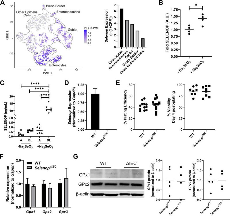
Selenop floxed mice crossed with Cre driver lines to delete Selenop from the liver, myeloid lineages, or intestinal epithelium were placed on an azoxymethane/dextran sodium sulfate experimental CAC protocol. SELENOP loss was assessed in human ulcerative colitis (UC) organoids, and expression was queried in human and adult UC samples.
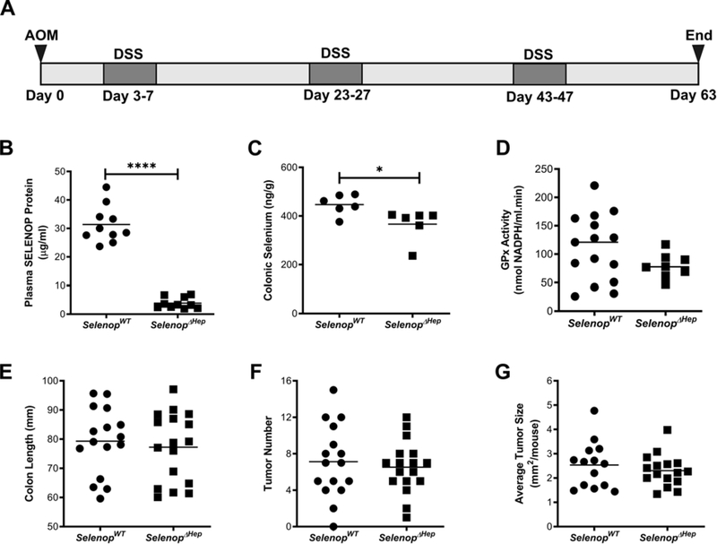
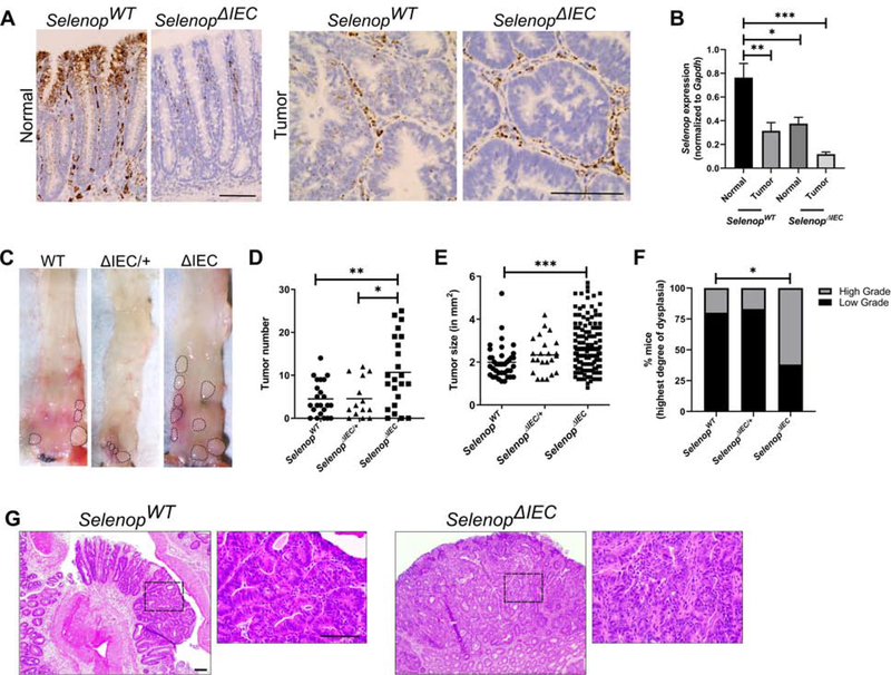
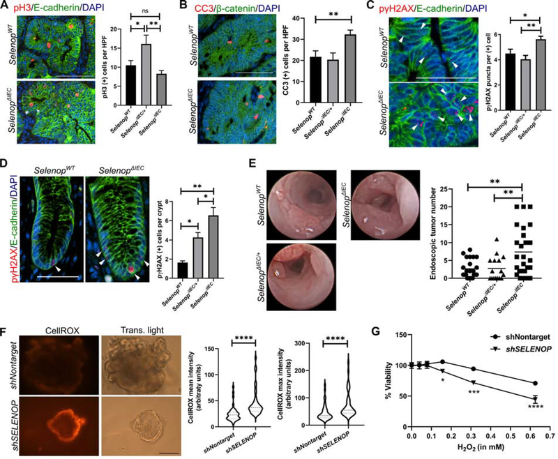
Although large sources of SELENOP, both liver- and myeloid-specific Selenop deletion failed to modify azoxymethane/dextran sodium sulfate-mediated tumorigenesis. Instead, epithelial-specific deletion increased CAC tumorigenesis, likely due to elevated oxidative stress with a resulting increase in genomic instability and augmented tumor initiation. SELENOP was down-regulated in UC colon biopsies and levels were inversely correlated with endoscopic disease severity and tissue S100A8 (calprotectin) gene expression.
Although global selenium status is typically assessed by measuring liver-derived plasma SELENOP levels, our results indicate that the peripheral SELENOP pool is dispensable for CAC. Colonic epithelial SELENOP is the main contributor to local antioxidant capabilities. Thus, colonic SELENOP is the most informative means to assess selenium levels and activity in IBD patients and may serve as a novel biomarker for UC disease severity and identify patients most predisposed to CAC development.
炎症性肠病(IBD)患者表现出营养性硒缺乏,并且患结肠癌的风险更高。此前,我们发现,分泌型抗氧化硒蛋白硒蛋白 P(SELENOP)的整体减少会显著增加实验性结肠炎相关癌症(CAC)模型中的肿瘤发展。我们接下来旨在描绘 SELENOP 对肠道炎症性致癌作用的组织特异性贡献,并确定临床背景。
将 Selenop 基因敲入小鼠与 Cre 驱动系杂交,使 Selenop 在肝脏、髓系谱系或肠道上皮细胞中缺失,然后将其置于氧化偶氮甲烷/葡聚糖硫酸钠实验性 CAC 方案中。在人类溃疡性结肠炎(UC)类器官中评估 SELENOP 的缺失,并在人类和成人 UC 样本中查询表达情况。
尽管 SELENOP 是一个大型来源,但肝脏和髓系特异性 Selenop 缺失均未能改变氧化偶氮甲烷/葡聚糖硫酸钠介导的肿瘤发生。相反,上皮细胞特异性缺失增加了 CAC 肿瘤的发生,这可能是由于氧化应激升高导致基因组不稳定性增加和肿瘤起始增加。在 UC 结肠活检中,SELENOP 下调,并且其水平与内镜疾病严重程度和组织 S100A8(钙卫蛋白)基因表达呈负相关。
虽然通常通过测量肝脏衍生的血浆 SELENOP 水平来评估整体硒状态,但我们的结果表明,外周 SELENOP 池对于 CAC 是可有可无的。结肠上皮细胞 SELENOP 是局部抗氧化能力的主要贡献者。因此,结肠 SELENOP 是评估 IBD 患者硒水平和活性的最具信息性的手段,并且可以作为 UC 疾病严重程度的新型生物标志物,并确定最易发生 CAC 发展的患者。