• 文献检索
  • 文档翻译
  • 深度研究
  • 学术资讯
  • Suppr Zotero 插件Zotero 插件
  • 邀请有礼
  • 套餐&价格
  • 历史记录
应用&插件
Suppr Zotero 插件Zotero 插件浏览器插件Mac 客户端Windows 客户端微信小程序
定价
高级版会员购买积分包购买API积分包
服务
文献检索文档翻译深度研究API 文档MCP 服务
关于我们
关于 Suppr公司介绍联系我们用户协议隐私条款
关注我们

Suppr 超能文献

核心技术专利:CN118964589B侵权必究
粤ICP备2023148730 号-1Suppr @ 2026

文献检索

告别复杂PubMed语法,用中文像聊天一样搜索,搜遍4000万医学文献。AI智能推荐,让科研检索更轻松。

立即免费搜索

文件翻译

保留排版,准确专业,支持PDF/Word/PPT等文件格式,支持 12+语言互译。

免费翻译文档

深度研究

AI帮你快速写综述,25分钟生成高质量综述,智能提取关键信息,辅助科研写作。

立即免费体验

开发并测试一种用于从三维超声图像中自动分割胎儿面部的算法。

Developing and testing an algorithm for automatic segmentation of the fetal face from three-dimensional ultrasound images.

作者信息

Clark A E, Biffi B, Sivera R, Dall'Asta A, Fessey L, Wong T-L, Paramasivam G, Dunaway D, Schievano S, Lees C C

机构信息

Queen Charlotte's and Chelsea Hospital, Imperial Healthcare NHS Trust, London, UK.

Imperial College London, London, UK.

出版信息

R Soc Open Sci. 2020 Nov 25;7(11):201342. doi: 10.1098/rsos.201342. eCollection 2020 Nov.

DOI:10.1098/rsos.201342
PMID:33391808
原文链接:https://pmc.ncbi.nlm.nih.gov/articles/PMC7735327/
Abstract

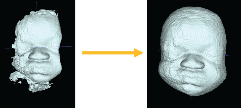
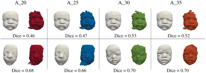
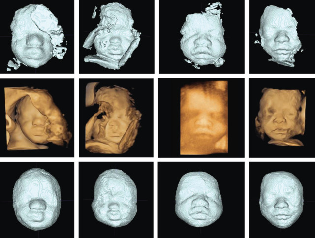
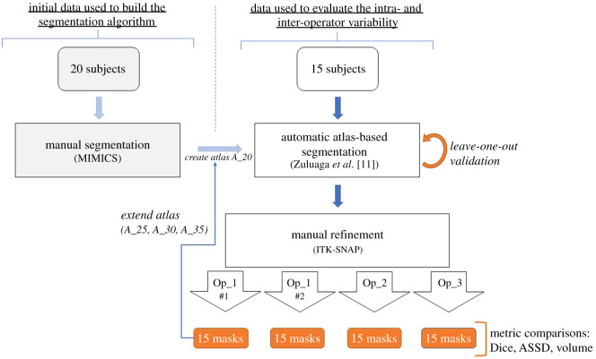
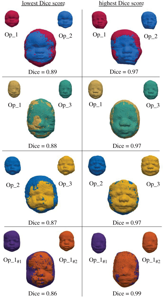
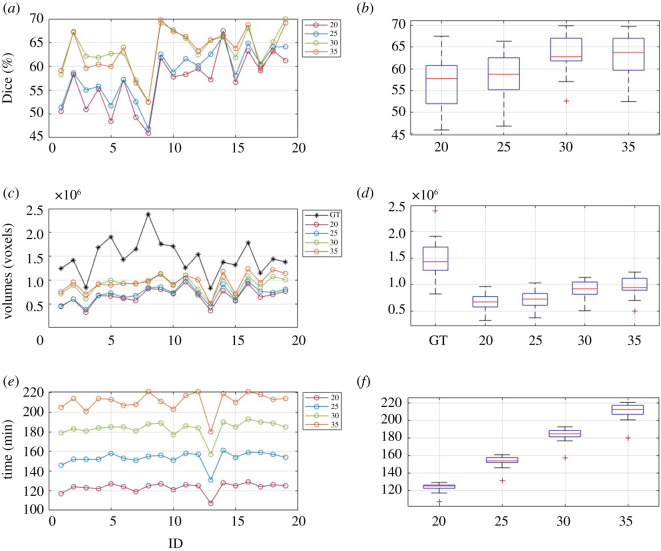
Fetal craniofacial abnormalities are challenging to detect and diagnose on prenatal ultrasound (US). Image segmentation and computer analysis of three-dimensional US volumes of the fetal face may provide an objective measure to quantify fetal facial features and identify abnormalities. We have developed and tested an atlas-based partially automated facial segmentation algorithm; however, the volumes require additional manual segmentation (MS), which is time and labour intensive and may preclude this method from clinical adoption. These manually refined segmentations can then be used as a reference (atlas) by the partially automated segmentation algorithm to improve algorithmic performance with the aim of eliminating the need for manual refinement and developing a fully automated system. This study assesses the inter- and intra-operator variability of MS and tests an optimized version of our automatic segmentation (AS) algorithm. The manual refinements of 15 fetal faces performed by three operators and repeated by one operator were assessed by Dice score, average symmetrical surface distance and volume difference. The performance of the partially automatic algorithm with difference size atlases was evaluated by Dice score and computational time. Assessment of the manual refinements showed low inter- and intra-operator variability demonstrating its suitability for optimizing the AS algorithm. The algorithm showed improved performance following an increase in the atlas size in turn reducing the need for manual refinement.

摘要

胎儿颅面部异常在产前超声检查中难以检测和诊断。对胎儿面部的三维超声容积进行图像分割和计算机分析,可能提供一种客观的方法来量化胎儿面部特征并识别异常。我们已经开发并测试了一种基于图谱的部分自动化面部分割算法;然而,这些容积需要额外的手动分割,这既耗时又费力,可能会阻碍该方法在临床上的应用。然后,这些经过手动优化的分割可以被部分自动化分割算法用作参考(图谱),以提高算法性能,目标是消除对手动优化的需求并开发出一个完全自动化的系统。本研究评估了手动分割的操作者间和操作者内变异性,并测试了我们自动分割(AS)算法的优化版本。通过Dice系数、平均对称表面距离和容积差异,评估了由三名操作者进行的15例胎儿面部手动优化以及一名操作者重复操作的情况。通过Dice系数和计算时间评估了使用不同大小图谱的部分自动算法的性能。对手动优化的评估显示操作者间和操作者内变异性较低,表明其适用于优化AS算法。随着图谱大小的增加,该算法性能有所改善,进而减少了对手动优化的需求。

https://cdn.ncbi.nlm.nih.gov/pmc/blobs/3ab5/7735327/e32d05d203df/rsos201342-g6.jpg
https://cdn.ncbi.nlm.nih.gov/pmc/blobs/3ab5/7735327/d738ce6a5a67/rsos201342-g1.jpg
https://cdn.ncbi.nlm.nih.gov/pmc/blobs/3ab5/7735327/5ec9cbc43f7c/rsos201342-g2.jpg
https://cdn.ncbi.nlm.nih.gov/pmc/blobs/3ab5/7735327/e4867e7635c5/rsos201342-g3.jpg
https://cdn.ncbi.nlm.nih.gov/pmc/blobs/3ab5/7735327/d204d1b29611/rsos201342-g4.jpg
https://cdn.ncbi.nlm.nih.gov/pmc/blobs/3ab5/7735327/133e6e01c7c9/rsos201342-g5.jpg
https://cdn.ncbi.nlm.nih.gov/pmc/blobs/3ab5/7735327/e32d05d203df/rsos201342-g6.jpg

相似文献

1
Developing and testing an algorithm for automatic segmentation of the fetal face from three-dimensional ultrasound images.开发并测试一种用于从三维超声图像中自动分割胎儿面部的算法。
R Soc Open Sci. 2020 Nov 25;7(11):201342. doi: 10.1098/rsos.201342. eCollection 2020 Nov.
2
Automated segmentation and measurement of the female pelvic floor from the mid-sagittal plane of 3D ultrasound volumes.自动分割和测量三维超声容积中矢状面的女性盆底。
Med Phys. 2023 Oct;50(10):6215-6227. doi: 10.1002/mp.16389. Epub 2023 Apr 6.
3
Multi-atlas segmentation of the whole hippocampus and subfields using multiple automatically generated templates.使用多个自动生成的模板对整个海马体及其子区进行多图谱分割。
Neuroimage. 2014 Nov 1;101:494-512. doi: 10.1016/j.neuroimage.2014.04.054. Epub 2014 Apr 29.
4
Minimally interactive placenta segmentation from three-dimensional ultrasound images.从三维超声图像中进行最小交互的胎盘分割。
J Med Imaging (Bellingham). 2020 Jan;7(1):014004. doi: 10.1117/1.JMI.7.1.014004. Epub 2020 Feb 22.
5
A framework for quantification and visualization of segmentation accuracy and variability in 3D lateral ventricle ultrasound images of preterm neonates.早产儿三维侧脑室超声图像分割准确性和变异性的量化与可视化框架。
Med Phys. 2015 Nov;42(11):6387-405. doi: 10.1118/1.4932366.
6
Fully automated lesion segmentation and visualization in automated whole breast ultrasound (ABUS) images.全乳腺自动超声(ABUS)图像中的全自动病灶分割与可视化
Quant Imaging Med Surg. 2020 Mar;10(3):568-584. doi: 10.21037/qims.2020.01.12.
7
Implementing a deep learning model for automatic tongue tumour segmentation in ex-vivo 3-dimensional ultrasound volumes.在离体三维超声容积中实现用于自动舌肿瘤分割的深度学习模型。
Br J Oral Maxillofac Surg. 2024 Apr;62(3):284-289. doi: 10.1016/j.bjoms.2023.12.017. Epub 2024 Jan 3.
8
Craniofacial phenotyping with fetal MRI: a feasibility study of 3D visualisation, segmentation, surface-rendered and physical models.胎儿 MRI 的颅面表型分析:3D 可视化、分割、表面渲染和物理模型的可行性研究。
BMC Med Imaging. 2024 Mar 1;24(1):52. doi: 10.1186/s12880-024-01230-7.
9
Automatic MRI-based Three-dimensional Models of Hip Cartilage Provide Improved Morphologic and Biochemical Analysis.基于 MRI 的髋关节软骨自动三维模型可提高形态学和生物化学分析的效果。
Clin Orthop Relat Res. 2019 May;477(5):1036-1052. doi: 10.1097/CORR.0000000000000755.
10
Layered deep learning for automatic mandibular segmentation in cone-beam computed tomography.基于分层深度学习的锥形束计算机断层扫描下颌骨自动分割。
J Dent. 2021 Nov;114:103786. doi: 10.1016/j.jdent.2021.103786. Epub 2021 Aug 20.

引用本文的文献

1
Automated craniofacial biometry with 3D T2w fetal MRI.利用三维T2加权胎儿磁共振成像进行自动颅面部生物测量
PLOS Digit Health. 2024 Dec 30;3(12):e0000663. doi: 10.1371/journal.pdig.0000663. eCollection 2024 Dec.
2
Automated Craniofacial Biometry with 3D T2w Fetal MRI.利用三维T2加权胎儿磁共振成像的自动颅面生物测量
medRxiv. 2024 Aug 14:2024.08.13.24311408. doi: 10.1101/2024.08.13.24311408.
3
Craniofacial phenotyping with fetal MRI: a feasibility study of 3D visualisation, segmentation, surface-rendered and physical models.胎儿 MRI 的颅面表型分析:3D 可视化、分割、表面渲染和物理模型的可行性研究。

本文引用的文献

1
SciPy 1.0: fundamental algorithms for scientific computing in Python.SciPy 1.0:Python 中的科学计算基础算法。
Nat Methods. 2020 Mar;17(3):261-272. doi: 10.1038/s41592-019-0686-2. Epub 2020 Feb 3.
2
Prenatal exome sequencing analysis in fetal structural anomalies detected by ultrasonography (PAGE): a cohort study.超声检查发现胎儿结构畸形的产前外显子组测序分析(PAGE):一项队列研究。
Lancet. 2019 Feb 23;393(10173):747-757. doi: 10.1016/S0140-6736(18)31940-8. Epub 2019 Jan 31.
3
Rapid prenatal diagnosis using targeted exome sequencing: a cohort study to assess feasibility and potential impact on prenatal counseling and pregnancy management.
BMC Med Imaging. 2024 Mar 1;24(1):52. doi: 10.1186/s12880-024-01230-7.
4
Fetal face shape analysis from prenatal 3D ultrasound images.胎儿面部形状分析来自产前三维超声图像。
Sci Rep. 2024 Feb 22;14(1):4411. doi: 10.1038/s41598-023-50386-9.
利用靶向外显子组测序进行快速产前诊断:一项评估可行性和对产前咨询及妊娠管理潜在影响的队列研究。
Genet Med. 2018 Nov;20(11):1430-1437. doi: 10.1038/gim.2018.30. Epub 2018 Mar 29.
4
Additive Manufacturing Techniques for the Reconstruction of 3D Fetal Faces.用于三维胎儿面部重建的增材制造技术
Appl Bionics Biomech. 2017;2017:9701762. doi: 10.1155/2017/9701762. Epub 2017 Dec 19.
5
Indexed Pain Journals.索引疼痛期刊。
J Pain Palliat Care Pharmacother. 2008;22(1):45-46. doi: 10.1080/15360280801989377.
6
Promises, pitfalls and practicalities of prenatal whole exome sequencing.产前全外显子组测序的承诺、陷阱和实际问题。
Prenat Diagn. 2018 Jan;38(1):10-19. doi: 10.1002/pd.5102. Epub 2017 Jul 25.
7
Quantitative analysis of fetal facial morphology using 3D ultrasound and statistical shape modeling: a feasibility study.使用三维超声和统计形状建模对胎儿面部形态进行定量分析:一项可行性研究。
Am J Obstet Gynecol. 2017 Jul;217(1):76.e1-76.e8. doi: 10.1016/j.ajog.2017.02.007. Epub 2017 Feb 14.
8
Cornelia de Lange syndrome: specific features for prenatal diagnosis.科妮莉亚·德朗热综合征:产前诊断的特定特征
Ultrasound Obstet Gynecol. 2017 May;49(5):668-670. doi: 10.1002/uog.15788.
9
User-guided segmentation of preterm neonate ventricular system from 3-D ultrasound images using convex optimization.使用凸优化从三维超声图像中进行用户引导的早产儿脑室系统分割。
Ultrasound Med Biol. 2015 Feb;41(2):542-56. doi: 10.1016/j.ultrasmedbio.2014.09.019. Epub 2014 Dec 23.
10
Cornelia de Lange syndrome.科妮莉亚·德·朗格综合征
Clin Genet. 2015 Jul;88(1):1-12. doi: 10.1111/cge.12499. Epub 2014 Oct 28.