Cai Ruyun, Lu Qian, Wang Da
Department of Colorectal Surgery, Sir Run Run Shaw Hospital, College of Medicine, Zhejiang University, Hangzhou, 310000, Zhejiang, China.
Department of Medical Oncology, Sir Run Run Shaw Hospital, College of Medicine, Zhejiang University, Hangzhou, 310000, Zhejiang, China.
World J Surg Oncol. 2021 Jan 4;19(1):7. doi: 10.1186/s12957-020-02107-z.
Colorectal cancer (CRC) is one of the most common cancers in the world, and liver metastasis is the leading cause of colorectal cancer-related deaths. However, the mechanism of liver metastasis in CRC has not been clearly elucidated.
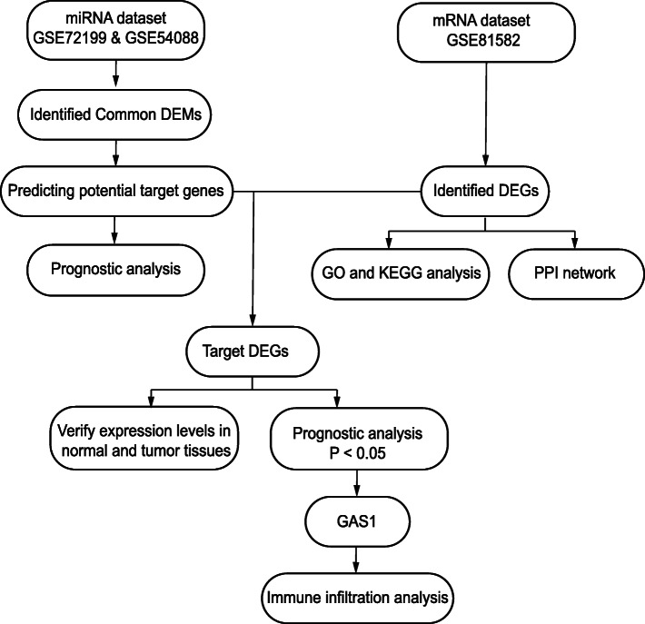
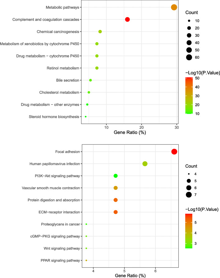
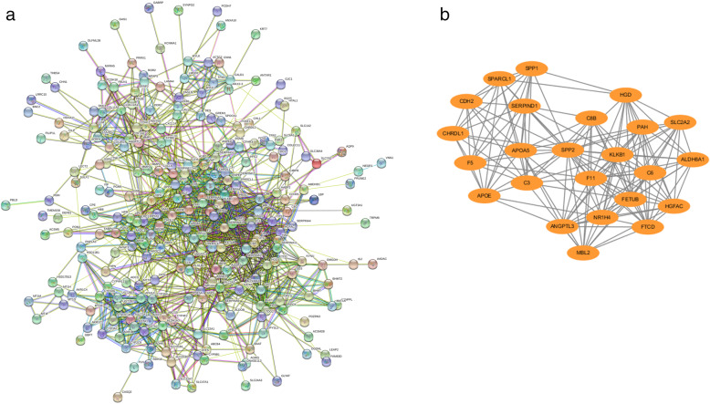
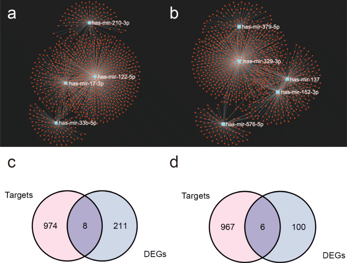
Three datasets from the Gene Expression Omnibus (GEO) database were analyzed to obtain differentially expressed genes (DEGs), which were subjected to functional enrichment analysis and protein-protein interaction analysis. Subsequently, mRNA-miRNA network was constructed, and the associated DEGs and DEMs were performed for prognostic analysis. Finally, we did infiltration analysis of growth arrest specific 1 (GAS1)-associated immune cells.
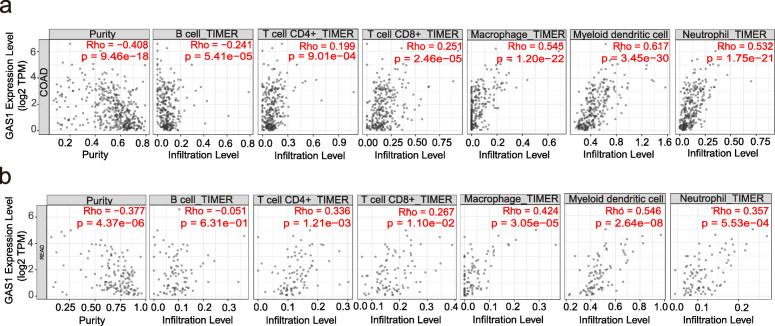
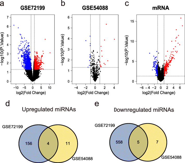
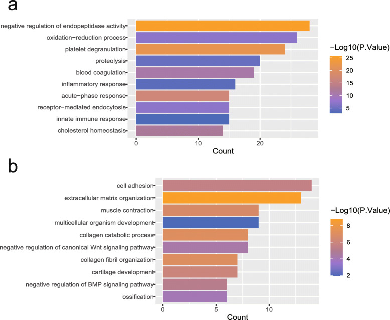
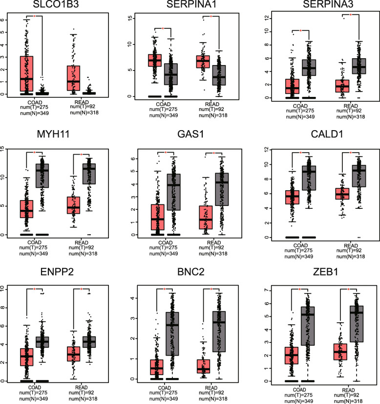
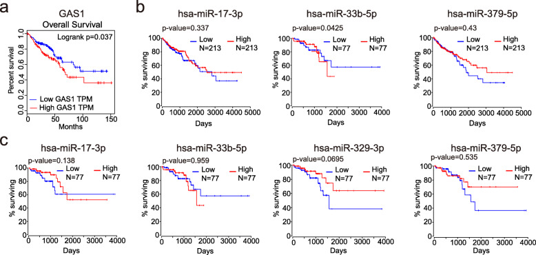
We obtained 325 DEGs and 9 differentially expressed miRNAs (DEMs) between primary CRC and liver metastases. Enrichment analysis and protein-protein interactions (PPI) further revealed the involvement of DEGs in the formation of the inflammatory microenvironment and epithelial-mesenchymal transition (EMT) during the liver metastases process in CRC. Survival analysis demonstrated that low-expressed GAS1 as well as low-expressed hsa-miR-33b-5p was a favorable prognostic indicator of overall survival. Further exploration of GAS1 revealed that its expression was interrelated with the infiltration of immune cells in tumor tissues.
In summary, DEGs, DEMs, and their interactions found in liver metastasis of CRC may provide a basis for further understanding of the mechanism of CRC metastasis.
结直肠癌(CRC)是世界上最常见的癌症之一,肝转移是结直肠癌相关死亡的主要原因。然而,CRC肝转移的机制尚未完全阐明。
分析来自基因表达综合数据库(GEO)的三个数据集,以获得差异表达基因(DEGs),并对其进行功能富集分析和蛋白质-蛋白质相互作用分析。随后,构建mRNA-miRNA网络,并对相关的DEGs和差异表达miRNA(DEMs)进行预后分析。最后,我们对生长停滞特异性蛋白1(GAS1)相关免疫细胞进行了浸润分析。
我们在原发性CRC和肝转移灶之间获得了325个DEGs和9个差异表达miRNA(DEMs)。富集分析和蛋白质-蛋白质相互作用(PPI)进一步揭示了DEGs在CRC肝转移过程中参与炎症微环境的形成和上皮-间质转化(EMT)。生存分析表明,低表达的GAS1以及低表达的hsa-miR-33b-5p是总生存的良好预后指标。对GAS1的进一步探索表明,其表达与肿瘤组织中免疫细胞的浸润相关。
总之,在CRC肝转移中发现的DEGs、DEMs及其相互作用可能为进一步了解CRC转移机制提供依据。