Department of Neurosurgery, Charité - Universitätsmedizin Berlin, Charitéplatz 1, 10117 Berlin, Germany.
Department of Neurosurgery, University of Turin, Turin, Italy.
Neuroimage Clin. 2021;29:102541. doi: 10.1016/j.nicl.2020.102541. Epub 2020 Dec 25.
Injury to major white matter pathways during language-area associated glioma surgery often leads to permanent loss of neurological function. The aim was to establish standardized tractography of language pathways as a predictor of language outcome in clinical neurosurgery.
We prospectively analyzed 50 surgical cases of patients with left perisylvian, diffuse gliomas. Standardized preoperative Diffusion-Tensor-Imaging (DTI)-based tractography of the 5 main language tracts (Arcuate Fasciculus [AF], Frontal Aslant Tract [FAT], Inferior Fronto-Occipital Fasciculus [IFOF], Inferior Longitudinal Fasciculus [ILF], Uncinate Fasciculus [UF]) and spatial analysis of tumor and tracts was performed. Postoperative imaging and the resulting resection map were analyzed for potential surgical injury of tracts. The language status was assessed preoperatively, postoperatively and after 3 months using the Aachen Aphasia Test and Berlin Aphasia Score. Correlation analyses, two-step cluster analysis and binary logistic regression were used to analyze associations of tractography results with language outcome after surgery.
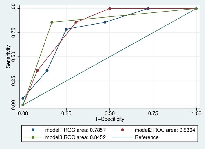
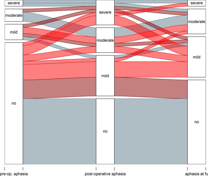
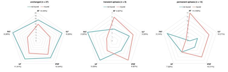
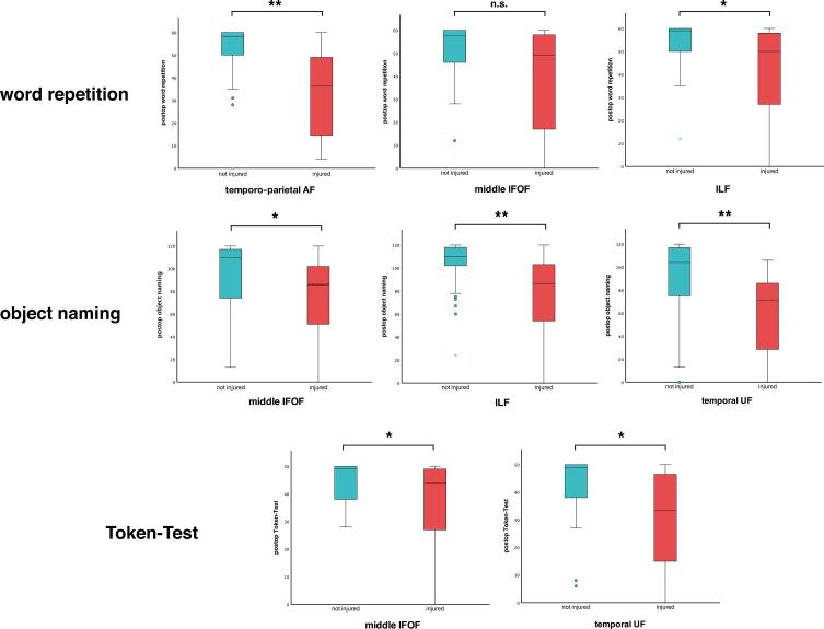
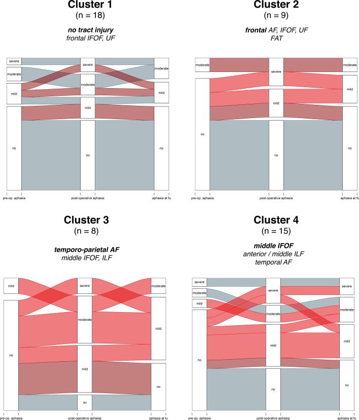
In 14 out of 50 patients (28%), new aphasic symptoms were detected 3 months after surgery. The preoperative infiltration of the AF was associated with functional worsening (cc = 0.314; p = 0.019). Cluster analysis of tract injury profiles revealed two areas particularly related to aphasia: the temporo-parieto-occipital junction (TPO; temporo-parietal AF, middle IFOF, middle ILF) and the temporal stem/peri-insular white matter (middle IFOF, anterior ILF, temporal UF, temporal AF). Injury to these areas (TPO: OR: 23.04; CI: 4.11 - 129.06; temporal stem: OR: 21.96; CI: 2.93 - 164.41) was associated with a higher-risk of persisting aphasia.
Tractography of language pathways can help to determine the individual aphasia risk profile pre-surgically. The TPO and temporal stem/peri-insular white matter were confirmed as functional nodes particularly sensitive to surgical injuries.
语言相关脑肿瘤术中损伤主要白质通路常导致永久性神经功能丧失。本研究旨在建立语言通路的标准化轨迹,作为临床神经外科中语言预后的预测指标。
我们前瞻性分析了 50 例左大脑外侧裂、弥漫性脑胶质瘤患者的手术病例。对 5 条主要语言束(弓状束[AF]、额斜束[FAT]、下额枕束[IFOF]、下额枕束[ILF]、钩束[UF])进行基于弥散张量成像(DTI)的标准化术前轨迹追踪,并对肿瘤和束进行空间分析。对术后影像学和由此产生的切除图进行分析,以确定束的潜在手术损伤。在术前、术后和 3 个月时使用 Aachen 失语症测试和柏林失语症评分评估语言状态。使用相关分析、两步聚类分析和二项逻辑回归分析来分析术前轨迹追踪结果与术后语言结果之间的相关性。
50 例患者中有 14 例(28%)在术后 3 个月时出现新的失语症状。术前 AF 浸润与功能恶化相关(cc=0.314,p=0.019)。束损伤图谱聚类分析显示,有两个区域与失语症特别相关:颞顶枕交界区(TPO;颞顶 AF、中 IFOF、中 ILF)和颞叶干/岛周白质(中 IFOF、前 ILF、颞叶 UF、颞叶 AF)。这些区域的损伤(TPO:OR:23.04;CI:4.11-129.06;颞叶干:OR:21.96;CI:2.93-164.41)与持续性失语的高风险相关。
语言通路的轨迹追踪有助于术前确定个体失语风险。TPO 和颞叶干/岛周白质被证实为对手术损伤特别敏感的功能节点。