Liu Rongtao, Zhang Shiyang, Zhao Chen, Yang Dong, Cui Tingting, Liu Yidong, Min Yonggang
School of Materials and Energy, Guangdong University of Technology (GDUT), Guangzhou, 510006, China.
Dongguan South China Design Innovation Institute, Dongguan, 523808, Guangdong, China.
Nanoscale Res Lett. 2021 Jan 6;16(1):4. doi: 10.1186/s11671-020-03457-z.
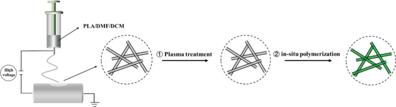
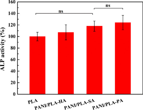
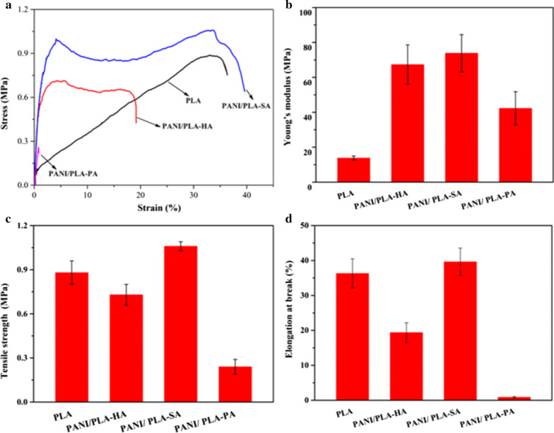
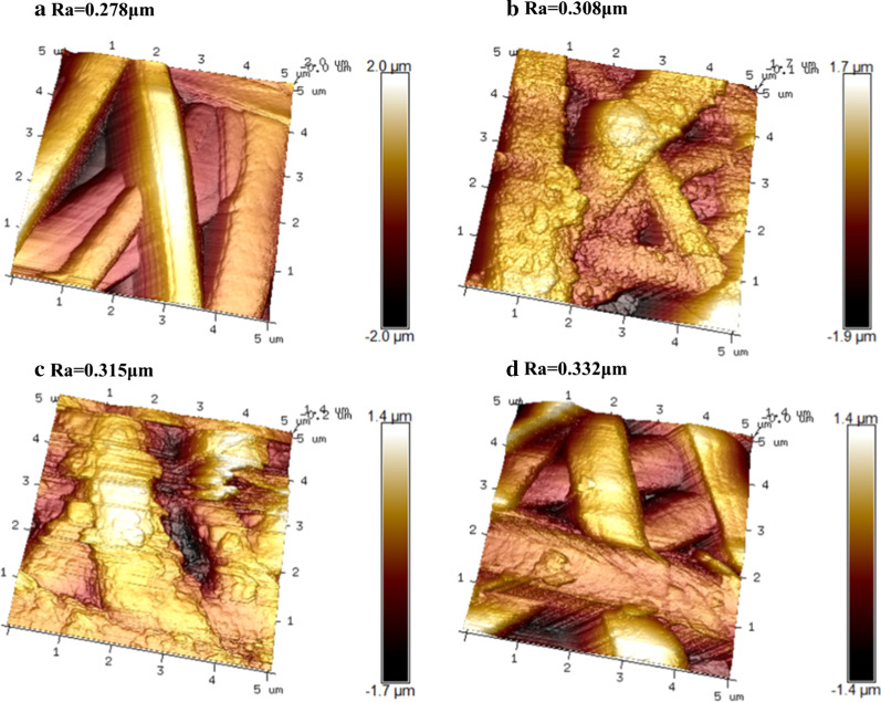
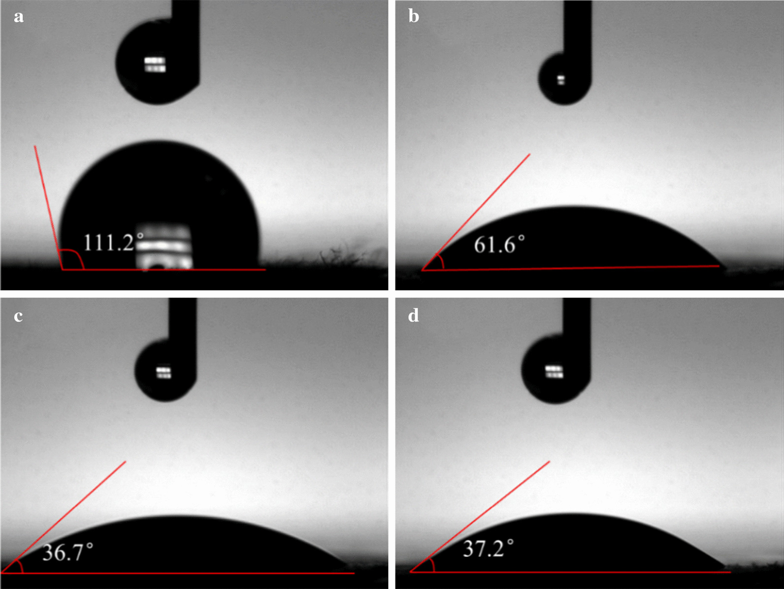
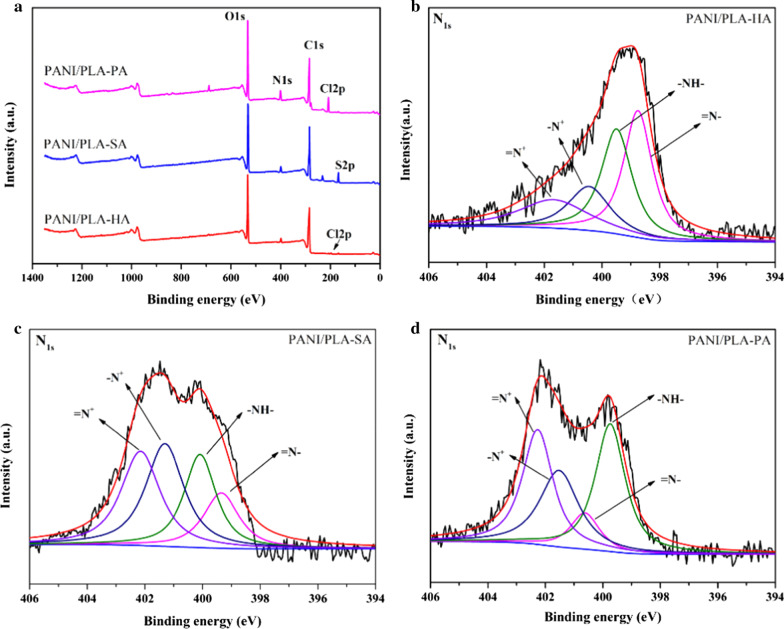
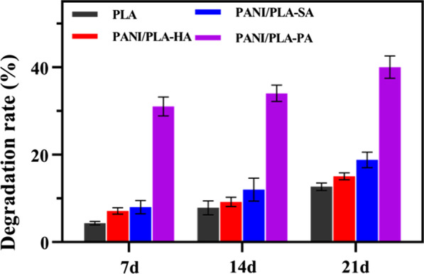
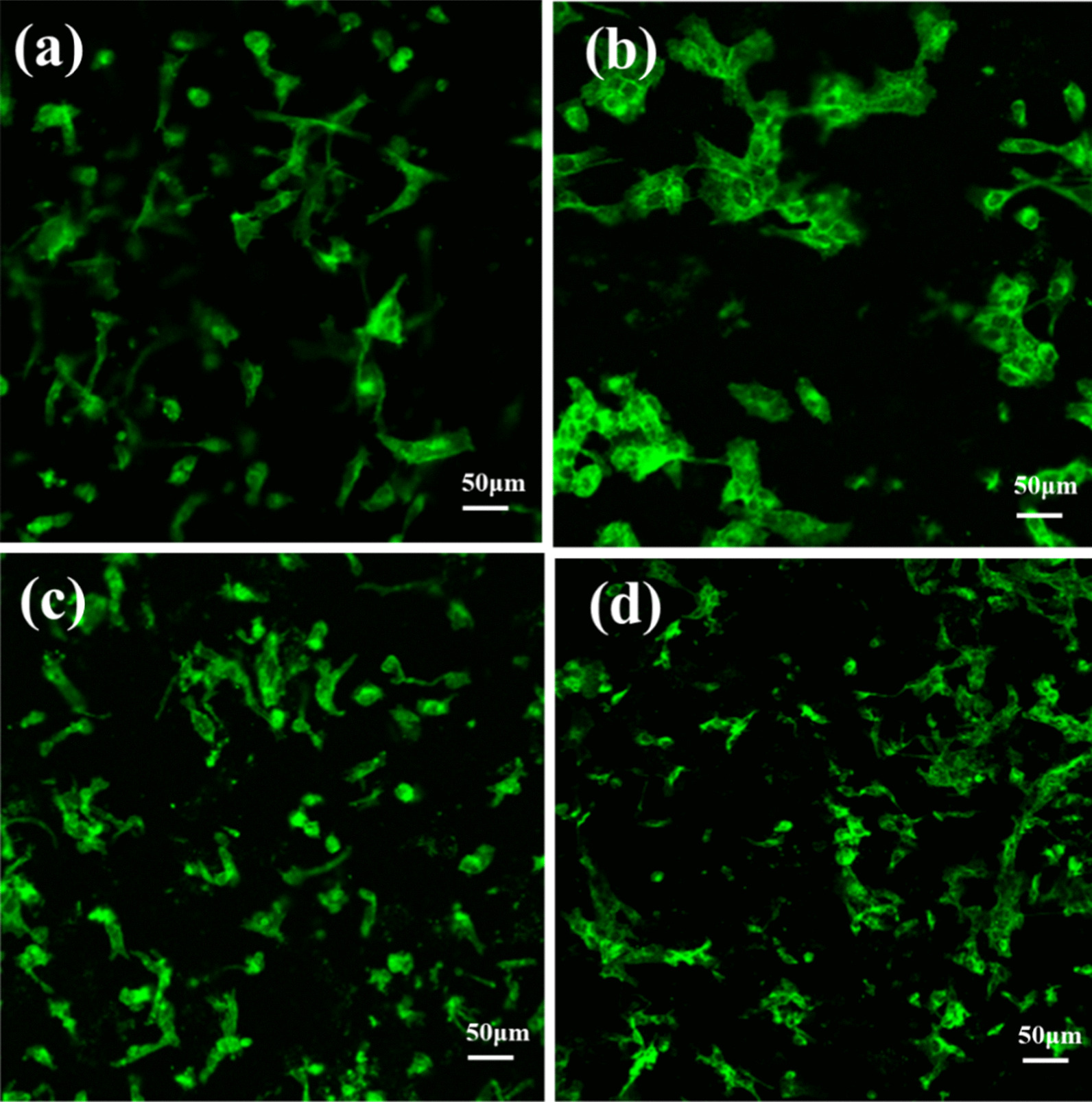
Conductive and degradable nanofibrous scaffolds have great potential in promoting cell growth, proliferation, and differentiation under an external electric field. Although the issue of inferior electrical conductivity in body fluids still exists, polyaniline (PANI)-based degradable nanofibers can promote cell adhesion, growth, and proliferation. To investigate whether the effect is caused by the PANI morphology, we selected three inorganic acids as dopants in the process of PANI in situ oxidative polymerization: hydrochloric acid, sulfuric acid, and perchloric acid. The obtained polyaniline/polylactic acid (PANI/PLA) composite nanofibers were characterized via SEM, FTIR, and XPS analysis, and we confirmed that the PLA nanofibers were successfully coated by PANI without any change to the porous structure of the PLA nanofibers. The in vitro mechanical properties and degradability indicated that the oxidation of acid dopants should be considered and that it was likely to have a higher oxidation degradation effect on PLA nanofibers. The contact angle test demonstrated that PANI/PLA composite nanofibers with different surface morphologies have good wettability, implying that they meet the requirements of bone tissue engineering scaffolds. The surface roughness and cell viability demonstrated that different PANI morphologies on the surface can promote cell proliferation. The higher the surface roughness of the PANI, the better the biocompatibility. Consequently, the regulated surface morphology of PANI/PLA composite nanofibers via different acids doping has positive effect on biocompatibility in tissue engineering.
导电且可降解的纳米纤维支架在外部电场作用下具有促进细胞生长、增殖和分化的巨大潜力。尽管体液中电导率较低的问题仍然存在,但基于聚苯胺(PANI)的可降解纳米纤维可促进细胞黏附、生长和增殖。为了研究这种效果是否由聚苯胺形态引起,我们在聚苯胺原位氧化聚合过程中选择了三种无机酸作为掺杂剂:盐酸、硫酸和高氯酸。通过扫描电子显微镜(SEM)、傅里叶变换红外光谱(FTIR)和X射线光电子能谱(XPS)分析对所得聚苯胺/聚乳酸(PANI/PLA)复合纳米纤维进行了表征,并且我们证实聚苯胺成功包覆了聚乳酸纳米纤维,而聚乳酸纳米纤维的多孔结构未发生任何变化。体外力学性能和降解性表明应考虑酸掺杂剂的氧化性,并且其可能对聚乳酸纳米纤维具有更高的氧化降解作用。接触角测试表明具有不同表面形态的聚苯胺/聚乳酸复合纳米纤维具有良好的润湿性,这意味着它们满足骨组织工程支架的要求。表面粗糙度和细胞活力表明表面不同的聚苯胺形态可促进细胞增殖。聚苯胺的表面粗糙度越高,生物相容性越好。因此,通过不同酸掺杂来调控聚苯胺/聚乳酸复合纳米纤维的表面形态对组织工程中的生物相容性具有积极作用。