O'Brien Christian, Souza Carolina A, Sheikh Adnan, Miguel Olivier, Wood Timothy
University of Ottawa Faculty of Medicine, University of Ottawa, 451 Smyth Road Ottawa, Ontario, K1H 8M5, Canada.
Department of Medical Imaging, Ottawa Hospital Research Institute (OHRI), The Ottawa Hospital, University of Ottawa, 501 Smyth Road, Ottawa, K1H 8L6, Canada.
3D Print Med. 2021 Jan 6;7(1):2. doi: 10.1186/s41205-020-00092-3.
This prospective study investigated whether the use of 3D-printed model facilitates novice learning of radiology anatomy on multiplanar computed tomography (CT) when compared to traditional 2D-based learning tools. Specifically, whether the use of a 3D printed model improved interpretation of multiplanar CT tracheobronchial anatomy.
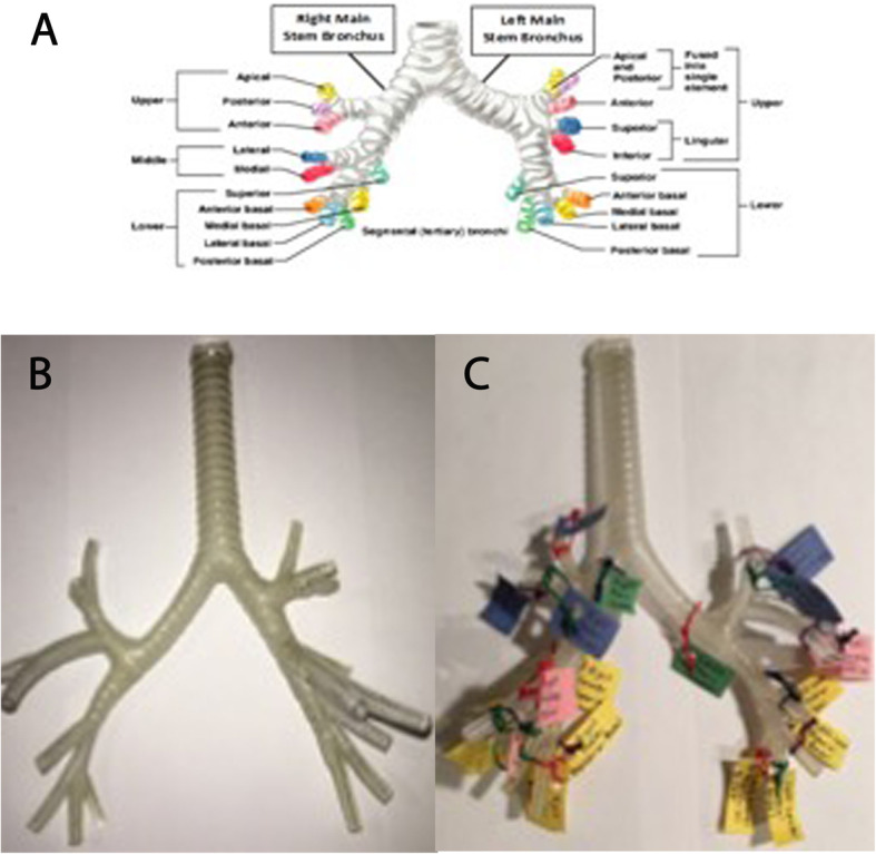
Thirty-one medical students (10F, 21 M) from years one to three were recruited, matched for gender and level of training and randomized to 2D or 3D group. Students underwent 20-min self-study session using 2D-printed image or 3D-printed model of the tracheobronchial tree. Immediately after, students answered 10 multiple-choice questions (Test 1) to identify tracheobronchial tree branches on multiplanar CT images. Two weeks later, identical test (Test 2) was used to assess retention of information. Mean scores of 2D and 3D groups were calculated. Student's t test was used to compare mean differences in tests scores and analysis of variance (ANOVA) was used to assess the interaction of gender, CT imaging plane and time on test scores between the two groups.
For test 1, 2D group had higher mean score than 3D group although not statistically significant (7.69 and 7.43, p = 0.39). Mean scores for Test 2 were significantly lower than for Test 1 (7 and 7.57, p = 0.03) with mean score decline for 2D group (Test 1 = 7.69, Test 2 = 6.63, p = 0.03), and similar score for 3D group (Test 1 and 2 = 7.43). There was no statistically significant interaction of gender and test score over time. Significant interaction between group and time of test was found for axial CT images but not for coronal images.
Use of a 3D-printed model of the tracheobronchial anatomy had no immediate advantage over traditional 2D-printed images for learning CT anatomy. However, use of a 3D model improved students' ability to retain learned information, irrespective of gender.
本前瞻性研究调查了与传统的基于二维的学习工具相比,使用3D打印模型是否有助于新手在多平面计算机断层扫描(CT)上学习放射解剖学。具体而言,使用3D打印模型是否能改善对多平面CT气管支气管解剖结构的解读。
招募了31名一至三年级的医学生(10名女性,21名男性),根据性别和培训水平进行匹配,并随机分为二维组或三维组。学生们使用气管支气管树的二维打印图像或三维打印模型进行了20分钟的自学。之后,学生们回答了10道多项选择题(测试1),以识别多平面CT图像上的气管支气管树分支。两周后,使用相同的测试(测试2)来评估信息保留情况。计算二维组和三维组的平均分数。使用学生t检验比较测试分数的平均差异,并使用方差分析(ANOVA)评估性别、CT成像平面和时间对两组测试分数的交互作用。
对于测试1,二维组的平均分数高于三维组,尽管无统计学意义(7.69和7.43,p = 0.39)。测试2的平均分数显著低于测试1(7和7.57,p = 0.03),二维组平均分数下降(测试1 = 7.69,测试2 = 6.63,p = 0.03),而三维组分数相似(测试1和2 = 7.43)。随着时间推移,性别与测试分数之间无统计学意义的交互作用。对于轴向CT图像,发现组与测试时间之间存在显著交互作用,但对于冠状图像则没有。
对于学习CT解剖学,使用气管支气管解剖结构的3D打印模型相对于传统的二维打印图像没有即时优势。然而,使用3D模型提高了学生保留所学信息的能力,与性别无关。