Satoh Yoko, Hirata Kenji, Tamada Daiki, Funayama Satoshi, Onishi Hiroshi
Yamanashi PET Imaging Clinic, Yamanashi, Japan.
Department of Radiology, University of Yamanashi, Yamanashi, Japan.
Front Med (Lausanne). 2020 Dec 23;7:603303. doi: 10.3389/fmed.2020.603303. eCollection 2020.
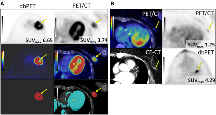
This retrospective study aimed to compare the ability to classify tumor characteristics of breast cancer (BC) of positron emission tomography (PET)-derived texture features between dedicated breast PET (dbPET) and whole-body PET/computed tomography (CT). Forty-four BCs scanned by both high-resolution ring-shaped dbPET and whole-body PET/CT were analyzed. The primary BC was extracted with a standardized uptake value (SUV) threshold segmentation method. On both dbPET and PET/CT images, 38 texture features were computed; their ability to classify tumor characteristics such as tumor (T)-category, lymph node (N)-category, molecular subtype, and Ki67 levels was compared. The texture features were evaluated using univariate and multivariate analyses following principal component analysis (PCA). AUC values were used to evaluate the diagnostic power of the computed texture features to classify BC characteristics. Some texture features of dbPET and PET/CT were different between Tis-1 and T2-4 and between Luminal A and other groups, respectively. No association with texture features was found in the N-category or Ki67 level. In contrast, receiver-operating characteristic analysis using texture features' principal components showed that the AUC for classification of any BC characteristics were equally good for both dbPET and whole-body PET/CT. PET-based texture analysis of dbPET and whole-body PET/CT may have equally good classification power for BC.
这项回顾性研究旨在比较专用乳腺正电子发射断层扫描(dbPET)和全身正电子发射断层扫描/计算机断层扫描(PET/CT)中基于正电子发射断层扫描(PET)的纹理特征对乳腺癌(BC)肿瘤特征的分类能力。分析了44例同时接受高分辨率环形dbPET和全身PET/CT扫描的BC病例。采用标准化摄取值(SUV)阈值分割方法提取原发性BC。在dbPET和PET/CT图像上,计算了38种纹理特征;比较了它们对肿瘤特征(如肿瘤(T)分期、淋巴结(N)分期、分子亚型和Ki67水平)的分类能力。在主成分分析(PCA)之后,使用单变量和多变量分析对纹理特征进行评估。AUC值用于评估计算出的纹理特征对BC特征的诊断能力。dbPET和PET/CT的一些纹理特征在Tis-1和T2-4之间以及管腔A型和其他组之间分别存在差异。在N分期或Ki67水平上未发现与纹理特征有关联。相比之下,使用纹理特征主成分的受试者操作特征分析表明,dbPET和全身PET/CT对任何BC特征分类的AUC同样良好。基于PET的dbPET和全身PET/CT纹理分析对BC可能具有同样良好的分类能力。