• 文献检索
  • 文档翻译
  • 深度研究
  • 学术资讯
  • Suppr Zotero 插件Zotero 插件
  • 邀请有礼
  • 套餐&价格
  • 历史记录
应用&插件
Suppr Zotero 插件Zotero 插件浏览器插件Mac 客户端Windows 客户端微信小程序
定价
高级版会员购买积分包购买API积分包
服务
文献检索文档翻译深度研究API 文档MCP 服务
关于我们
关于 Suppr公司介绍联系我们用户协议隐私条款
关注我们

Suppr 超能文献

核心技术专利:CN118964589B侵权必究
粤ICP备2023148730 号-1Suppr @ 2026

文献检索

告别复杂PubMed语法,用中文像聊天一样搜索,搜遍4000万医学文献。AI智能推荐,让科研检索更轻松。

立即免费搜索

文件翻译

保留排版,准确专业,支持PDF/Word/PPT等文件格式,支持 12+语言互译。

免费翻译文档

深度研究

AI帮你快速写综述,25分钟生成高质量综述,智能提取关键信息,辅助科研写作。

立即免费体验

主动脉加速度作为小鼠左心室收缩力的无创指数。

Aortic acceleration as a noninvasive index of left ventricular contractility in the mouse.

机构信息

Section of Cardiovascular Research, Department of Medicine, Baylor College of Medicine, One Baylor Plaza, MS:BCM285, Houston, TX, 77030, USA.

Texas A&M University, Houston, TX, USA.

出版信息

Sci Rep. 2021 Jan 12;11(1):536. doi: 10.1038/s41598-020-79866-y.

DOI:10.1038/s41598-020-79866-y
PMID:33436716
原文链接:https://pmc.ncbi.nlm.nih.gov/articles/PMC7804023/
Abstract

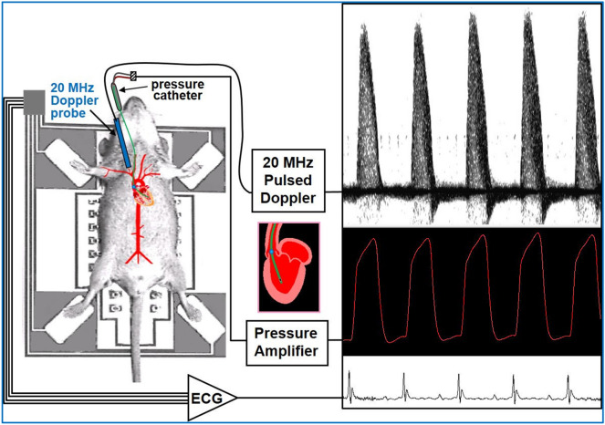
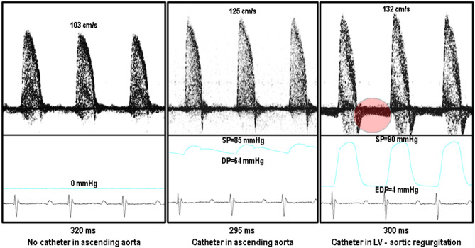
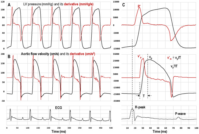
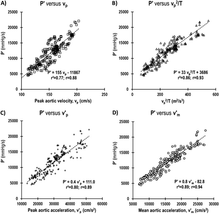
The maximum value of the first derivative of the invasively measured left ventricular (LV) pressure (+ dP/dt or P') is often used to quantify LV contractility, which in mice is limited to a single terminal study. Thus, determination of P' in mouse longitudinal/serial studies requires a group of mice at each desired time point resulting in "pseudo" serial measurements. Alternatively, a noninvasive surrogate for P' will allow for repeated measurements on the same group of mice, thereby minimizing physiological variability and requiring fewer animals. In this study we evaluated aortic acceleration and other parameters of aortic flow velocity as noninvasive indices of LV contractility in mice. We simultaneously measured LV pressure invasively with an intravascular pressure catheter and aortic flow velocity noninvasively with a pulsed Doppler probe in mice, at baseline and after the administration of the positive inotrope, dobutamine. Regression analysis of P' versus peak aortic velocity (v), peak velocity squared/rise time (v/T), peak (+ dv/dt or v') and mean (+ dv/dt or v') aortic acceleration showed a high degree of association (P' versus: v, r = 0.77; v/T, r = 0.86; v', r = 0.80; and v', r = 0.89). The results suggest that mean or peak aortic acceleration or the other parameters may be used as a noninvasive index of LV contractility.

摘要

左心室(LV)压力的一阶导数的最大值(+ dP/dt 或 P')常被用来量化 LV 收缩性,而在小鼠中,这种方法仅限于单次终末研究。因此,在小鼠纵向/系列研究中确定 P' 需要在每个所需的时间点对一组小鼠进行测量,从而导致“伪”系列测量。或者,P'的非侵入性替代物将允许对同一组小鼠进行重复测量,从而最大限度地减少生理变异性并减少所需的动物数量。在这项研究中,我们评估了主动脉加速度和其他主动脉血流速度参数作为小鼠 LV 收缩性的非侵入性指标。我们在小鼠中同时使用血管内压力导管进行 LV 压力侵入性测量,使用脉冲多普勒探头进行主动脉血流速度非侵入性测量,在基础状态和正性变力药多巴酚丁胺给药后进行测量。P'与峰值主动脉速度(v)、速度平方/上升时间(v/T)、峰值 (+ dV/dt 或 v')和平均 (+ dV/dt 或 v')主动脉加速度的回归分析显示出高度相关性(P'与:v,r=0.77;v/T,r=0.86;v',r=0.80;v',r=0.89)。结果表明,平均或峰值主动脉加速度或其他参数可用作 LV 收缩性的非侵入性指标。

https://cdn.ncbi.nlm.nih.gov/pmc/blobs/1c26/7804023/bfd1893c98a0/41598_2020_79866_Fig8_HTML.jpg
https://cdn.ncbi.nlm.nih.gov/pmc/blobs/1c26/7804023/699cd478929d/41598_2020_79866_Fig1_HTML.jpg
https://cdn.ncbi.nlm.nih.gov/pmc/blobs/1c26/7804023/c7f2b859c4e3/41598_2020_79866_Fig2_HTML.jpg
https://cdn.ncbi.nlm.nih.gov/pmc/blobs/1c26/7804023/82d11dceac37/41598_2020_79866_Fig3_HTML.jpg
https://cdn.ncbi.nlm.nih.gov/pmc/blobs/1c26/7804023/18e8f2519418/41598_2020_79866_Fig4_HTML.jpg
https://cdn.ncbi.nlm.nih.gov/pmc/blobs/1c26/7804023/7f1df2fad695/41598_2020_79866_Fig5_HTML.jpg
https://cdn.ncbi.nlm.nih.gov/pmc/blobs/1c26/7804023/cff0b0df8434/41598_2020_79866_Fig6_HTML.jpg
https://cdn.ncbi.nlm.nih.gov/pmc/blobs/1c26/7804023/943a99ffc9ea/41598_2020_79866_Fig7_HTML.jpg
https://cdn.ncbi.nlm.nih.gov/pmc/blobs/1c26/7804023/bfd1893c98a0/41598_2020_79866_Fig8_HTML.jpg

相似文献

1
Aortic acceleration as a noninvasive index of left ventricular contractility in the mouse.主动脉加速度作为小鼠左心室收缩力的无创指数。
Sci Rep. 2021 Jan 12;11(1):536. doi: 10.1038/s41598-020-79866-y.
2
Doppler echocardiographic evaluation of left ventricular function.左心室功能的多普勒超声心动图评估。
Echocardiography. 1990 Jan;7(1):21-25. doi: 10.1111/j.1540-8175.1990.tb00344.x.
3
Peak systolic velocity of mitral annular longitudinal movement measured by pulsed tissue Doppler imaging as an index of global left ventricular contractility.脉冲组织多普勒成像测量的二尖瓣环纵向运动的收缩期峰值速度可作为整体左心室收缩功能的指标。
Am J Physiol Heart Circ Physiol. 2010 May;298(5):H1608-15. doi: 10.1152/ajpheart.01231.2009. Epub 2010 Mar 5.
4
Noninvasive assessment of left ventricular isovolumic contraction and relaxation with continuous wave Doppler aortic regurgitant velocity signals: an in vivo validation study.利用连续波多普勒主动脉反流速度信号对左心室等容收缩和舒张进行无创评估:一项体内验证研究。
J Am Soc Echocardiogr. 2001 Jul;14(7):715-22. doi: 10.1067/mje.2001.111477.
5
Estimation of left ventricular performance through temporal pressure variations measured by MR velocity and acceleration mappings.通过磁共振速度和加速度映射测量的时间压力变化来估计左心室功能。
J Magn Reson Imaging. 2002 Sep;16(3):246-52. doi: 10.1002/jmri.10162.
6
[Noninvasive estimation of max (dP/dt) of the left ventricle based on maximum aortic flow acceleration as measured by pulsed Doppler echocardiography].基于脉冲多普勒超声心动图测量的最大主动脉血流加速度对左心室最大(dP/dt)进行无创估计
J Cardiol. 1990;20(4):977-89.
7
Noninvasive assessment of left ventricular relaxation using continuous-wave Doppler aortic regurgitant velocity curve. Its comparative value to the mitral regurgitation method.使用连续波多普勒主动脉反流速度曲线对左心室舒张功能进行无创评估。其与二尖瓣反流法的比较价值。
Circulation. 1995 Jan 1;91(1):192-200. doi: 10.1161/01.cir.91.1.192.
8
Relationship of maximum rate of pressure rise between aorta and left ventricle in pediatric patients. Implication for ventricular-vascular interaction with the potential for noninvasive determination of left ventricular contractility.儿科患者主动脉与左心室之间压力上升最大速率的关系。对心室-血管相互作用的影响以及无创测定左心室收缩力的可能性。
Circ J. 2009 Sep;73(9):1698-704. doi: 10.1253/circj.cj-08-0954. Epub 2009 Jul 13.
9
Assessment of left ventricular systolic wall motion velocity with pulsed tissue Doppler imaging: comparison with peak dP/dt of the left ventricular pressure curve.用脉冲组织多普勒成像评估左心室收缩期壁运动速度:与左心室压力曲线的峰值dP/dt比较。
J Am Soc Echocardiogr. 1998 May;11(5):442-9. doi: 10.1016/s0894-7317(98)70024-0.
10
Evaluation of a theoretical Doppler index to noninvasively estimate peak dP/dt using continuous wave Doppler ultrasound of ascending aortic flow in man.使用连续波多普勒超声对人体升主动脉血流进行理论多普勒指数评估,以无创估计峰值dP/dt。
Cathet Cardiovasc Diagn. 1991 Jul;23(3):219-22. doi: 10.1002/ccd.1810230317.

引用本文的文献

1
Haemodynamic Effects of Pimobendan during General Anaesthesia in Healthy Senior Dogs: A Prospective, Randomised, Triple-Blinded, Placebo-Controlled Clinical Study.匹莫苯丹在健康老年犬全身麻醉期间的血流动力学效应:一项前瞻性、随机、三盲、安慰剂对照的临床研究。
Animals (Basel). 2023 Jun 26;13(13):2110. doi: 10.3390/ani13132110.
2
Echocardiography protocol: A tool for infrequently used parameters in mice.超声心动图方案:一种用于小鼠中不常用参数的工具。
Front Cardiovasc Med. 2022 Dec 21;9:1038385. doi: 10.3389/fcvm.2022.1038385. eCollection 2022.
3
Adaptation of a mouse Doppler echocardiograph system for assessing cardiac function and thermal performance in a juvenile salmonid.

本文引用的文献

1
Ischemia and reperfusion injury following cardioplegic arrest is attenuated by age and testosterone deficiency in male but not female mice.心脏停搏后缺血再灌注损伤在雄性而非雌性小鼠中被年龄和睾酮缺乏所减轻。
Biol Sex Differ. 2019 Aug 23;10(1):42. doi: 10.1186/s13293-019-0256-4.
2
The Left and Right Ventricles Respond Differently to Variation of Pacing Delays in Cardiac Resynchronization Therapy: A Combined Experimental- Computational Approach.心脏再同步治疗中左右心室对起搏延迟变化的反应不同:一种实验与计算相结合的方法
Front Physiol. 2019 Feb 1;10:17. doi: 10.3389/fphys.2019.00017. eCollection 2019.
3
Performance comparison of ventricular and arterial dP/dt for assessing left ventricular systolic function during different experimental loading and contractile conditions.
一种用于评估幼年鲑科鱼类心脏功能和热性能的小鼠多普勒超声心动图系统的适应性调整。
Conserv Physiol. 2021 Sep 3;9(1):coab070. doi: 10.1093/conphys/coab070. eCollection 2021.
比较心室和动脉 dp/dt 在不同实验负荷和收缩条件下评估左心室收缩功能的性能。
Crit Care. 2018 Nov 29;22(1):325. doi: 10.1186/s13054-018-2260-1.
4
Minimally invasive transtracheal cardiac plexus block for sympathetic neuromodulation.经气管微创心丛神经阻滞用于交感神经调节。
Heart Rhythm. 2019 Jan;16(1):117-124. doi: 10.1016/j.hrthm.2018.07.037. Epub 2018 Jul 31.
5
Invasive Hemodynamics of Myocardial Disease: Systolic and Diastolic Dysfunction (and Hypertrophic Obstructive Cardiomyopathy).心肌病的有创血流动力学:收缩和舒张功能障碍(以及肥厚型梗阻性心肌病)。
Interv Cardiol Clin. 2017 Jul;6(3):297-307. doi: 10.1016/j.iccl.2017.03.001. Epub 2017 Apr 21.
6
AICAR-dependent AMPK activation improves scar formation in the aged heart in a murine model of reperfused myocardial infarction.AICAR 依赖性 AMPK 激活可改善再灌注心肌梗死小鼠模型中衰老心脏的瘢痕形成。
J Mol Cell Cardiol. 2013 Oct;63:26-36. doi: 10.1016/j.yjmcc.2013.07.005. Epub 2013 Jul 19.
7
Assessment of spectral Doppler in preclinical ultrasound using a small-size rotating phantom.使用小型旋转体模评估临床前超声中的谱多普勒。
Ultrasound Med Biol. 2013 Aug;39(8):1491-9. doi: 10.1016/j.ultrasmedbio.2013.03.013. Epub 2013 May 24.
8
Young little mice express a premature cardiovascular aging phenotype.幼龄小鼠表现出过早的心血管衰老表型。
J Gerontol A Biol Sci Med Sci. 2014 Feb;69(2):152-9. doi: 10.1093/gerona/glt055. Epub 2013 May 16.
9
SRC-2 coactivator deficiency decreases functional reserve in response to pressure overload of mouse heart.SRC-2 共激活剂缺乏降低了小鼠心脏对压力超负荷的功能储备。
PLoS One. 2012;7(12):e53395. doi: 10.1371/journal.pone.0053395. Epub 2012 Dec 31.
10
Assessment of strain and strain rate by two-dimensional speckle tracking in mice: comparison with tissue Doppler echocardiography and conductance catheter measurements.二维斑点追踪技术评估小鼠应变和应变率:与组织多普勒超声心动图和导丝压力测量的比较。
Eur Heart J Cardiovasc Imaging. 2013 Aug;14(8):765-73. doi: 10.1093/ehjci/jes274. Epub 2012 Dec 2.