Arranz-Romera Alicia, Hernandez Maria, Checa-Casalengua Patricia, Garcia-Layana Alfredo, Molina-Martinez Irene T, Recalde Sergio, Young Michael J, Tucker Budd A, Herrero-Vanrell Rocío, Fernandez-Robredo Patricia, Bravo-Osuna Irene
Pharmaceutical Innovation in Ophthalmology (InnOftal), Research Group (UCM 920415), Department of Pharmaceutics and Food Technology, Faculty of Pharmacy, Complutense University, Plaza de Ramón y Cajal, s/n, 28040 Madrid, Spain.
Retinal Pathologies and New Therapies Group, Experimental Ophthalmology Laboratory, Department of Ophthalmology, Clínica Universidad de Navarra, 31008 Pamplona, Spain.
Pharmaceuticals (Basel). 2021 Jan 11;14(1):50. doi: 10.3390/ph14010050.
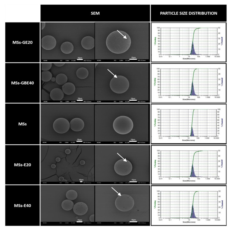
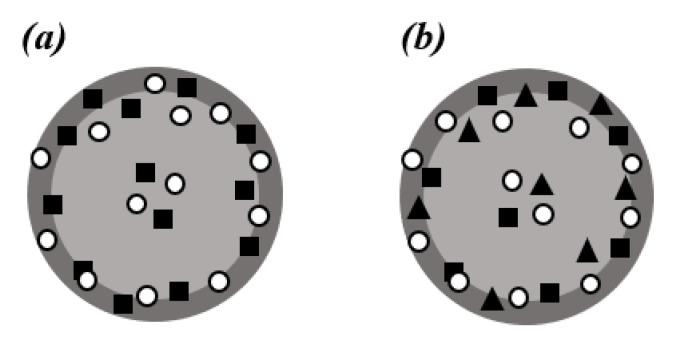
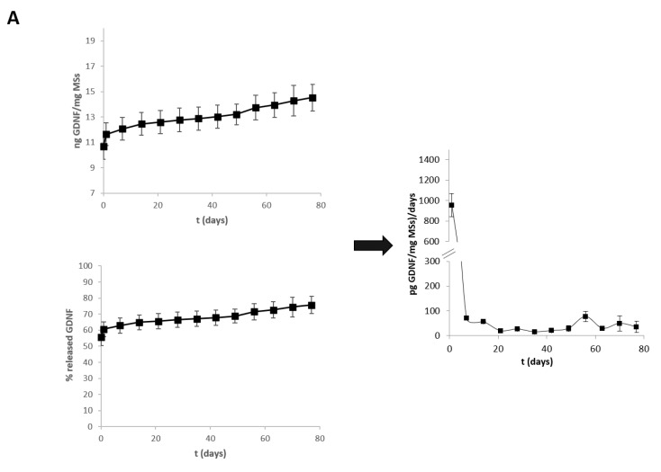
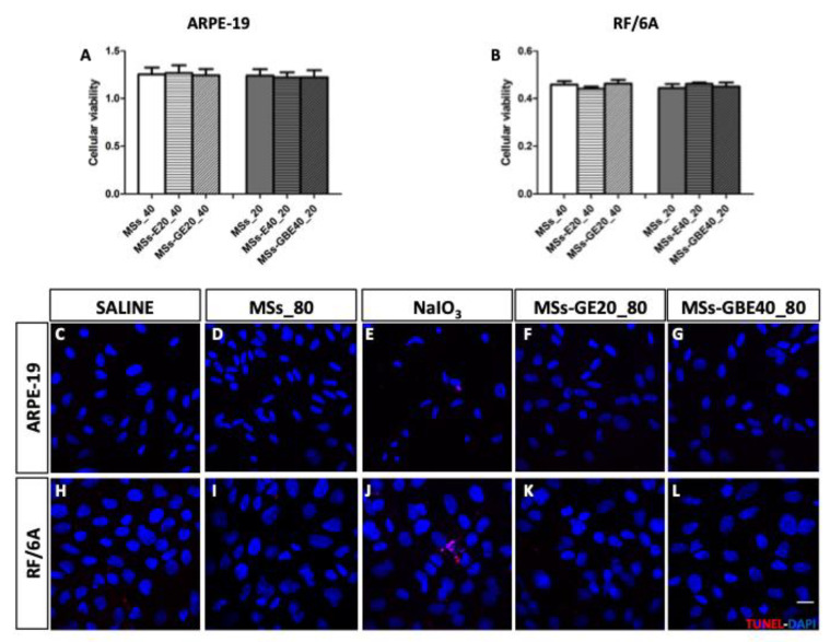
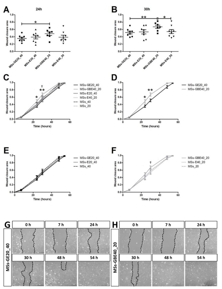
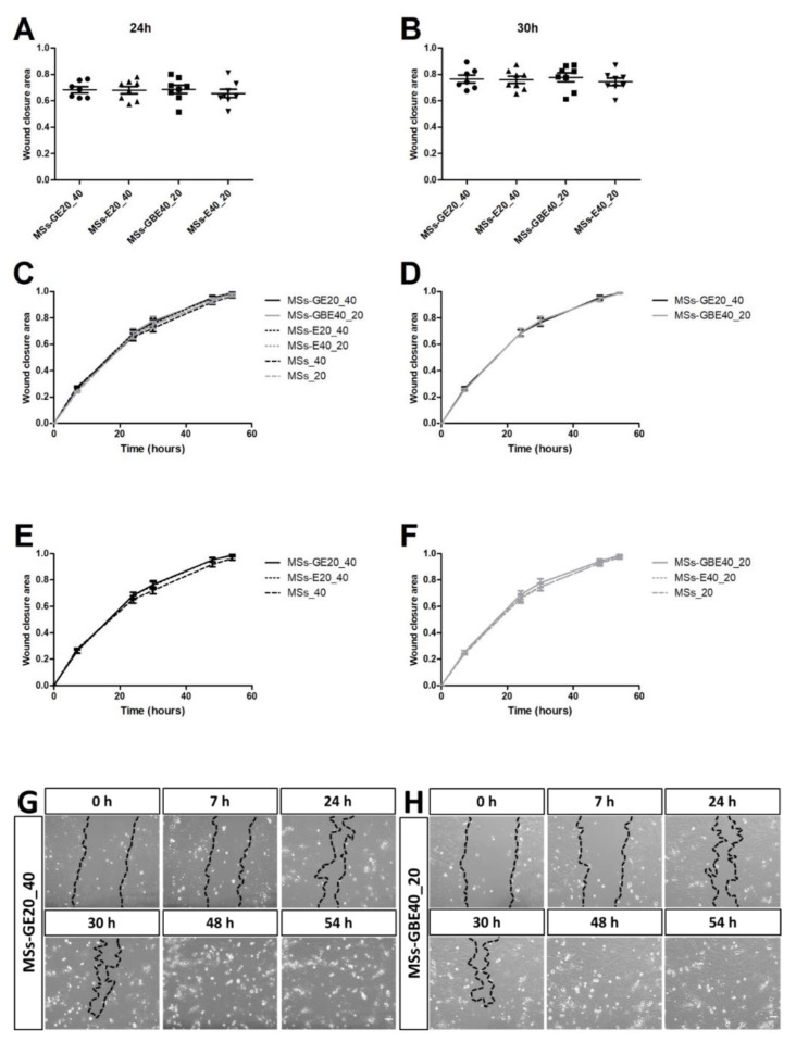
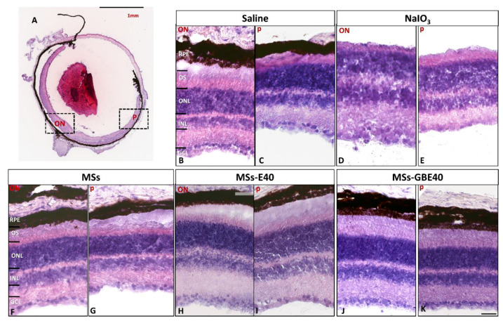
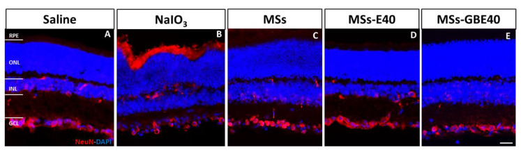
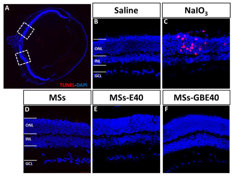
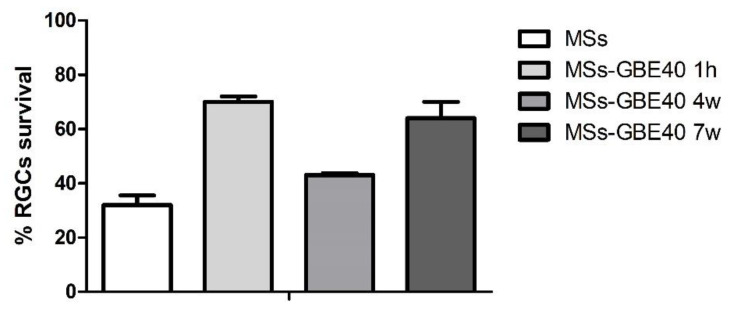
We assessed the sustained delivery effect of poly (lactic-co-glycolic) acid (PLGA)/vitamin E (VitE) microspheres (MSs) loaded with glial cell-derived neurotrophic factor (GDNF) alone (GDNF-MSs) or combined with brain-derived neurotrophic factor (BDNF; GDNF/BDNF-MSs) on migration of the human adult retinal pigment epithelial cell-line-19 (ARPE-19) cells, primate choroidal endothelial (RF/6A) cells, and the survival of isolated mouse retinal ganglion cells (RGCs). The morphology of the MSs, particle size, and encapsulation efficiencies of the active substances were evaluated. In vitro release, 3-(4,5-dimethylthiazol-2-yl)-2,5-diphenyltetrazolium bromide (MTT) cell viability, terminal deoxynucleotidyl transferase (TdT) deoxyuridine dUTP nick-end labelling (TUNEL) apoptosis, functional wound healing migration (ARPE-19; migration), and (RF/6A; angiogenesis) assays were conducted. The safety of MS intravitreal injection was assessed using hematoxylin and eosin, neuronal nuclei (NeuN) immunolabeling, and TUNEL assays, and RGC in vitro survival was analyzed. MSs delivered GDNF and co-delivered GDNF/BDNF in a sustained manner over 77 days. The BDNF/GDNF combination increased RPE cell migration, whereas no effect was observed on RF/6A. MSs did not alter cell viability, apoptosis was absent in vitro, and RGCs survived in vitro for seven weeks. In mice, retinal toxicity and apoptosis was absent in histologic sections. This delivery strategy could be useful as a potential co-therapy in retinal degenerations and glaucoma, in line with future personalized long-term intravitreal treatment as different amounts (doses) of microparticles can be administered according to patients' needs.
我们评估了单独负载胶质细胞源性神经营养因子(GDNF)的聚乳酸-羟基乙酸共聚物(PLGA)/维生素E(VitE)微球(MSs)(GDNF-MSs)或与脑源性神经营养因子(BDNF;GDNF/BDNF-MSs)联合使用对人成年视网膜色素上皮细胞系-19(ARPE-19)细胞、灵长类脉络膜内皮(RF/6A)细胞迁移以及分离的小鼠视网膜神经节细胞(RGCs)存活的持续递送效果。评估了MSs的形态、粒径以及活性物质的包封效率。进行了体外释放、3-(4,5-二甲基噻唑-2-基)-2,5-二苯基四氮唑溴盐(MTT)细胞活力、末端脱氧核苷酸转移酶(TdT)介导的脱氧尿苷三磷酸缺口末端标记(TUNEL)凋亡、功能性伤口愈合迁移(ARPE-19;迁移)以及(RF/6A;血管生成)测定。使用苏木精和伊红、神经元细胞核(NeuN)免疫标记以及TUNEL测定评估了MS玻璃体内注射的安全性,并分析了RGC的体外存活情况。MSs在77天内持续递送GDNF并共同递送GDNF/BDNF。BDNF/GDNF组合增加了RPE细胞迁移,而对RF/6A未观察到影响。MSs未改变细胞活力,体外无凋亡发生,RGCs在体外存活了7周。在小鼠中,组织学切片中无视网膜毒性和凋亡。这种递送策略作为视网膜变性和青光眼的潜在联合治疗可能有用,这与未来根据患者需求给予不同量(剂量)微粒的个性化长期玻璃体内治疗一致。