Department of Respiratory Medicine, Saitama Medical University, 38 Morohongo, Moroyama-machi, Iruma-gun, Saitama, 350-0495, Japan. t.shirahata+
Department of Respiratory Medicine, Saitama Medical University, 38 Morohongo, Moroyama-machi, Iruma-gun, Saitama, 350-0495, Japan.
Respir Res. 2021 Jan 15;22(1):18. doi: 10.1186/s12931-021-01621-2.
Physical inactivity due to cachexia and muscle wasting is well recognized as a sign of poor prognosis in chronic obstructive pulmonary disease (COPD). However, there have been no reports on the relationship between trunk muscle measurements and energy expenditure parameters, such as the total energy expenditure (TEE) and physical activity level (PAL), in COPD. In this study, we investigated the associations of computed tomography (CT)-derived muscle area and density measurements with clinical parameters, including TEE and PAL, in patients with or at risk for COPD, and examined whether these muscle measurements serve as an indicator of TEE and PAL.
The study population consisted of 36 male patients with (n = 28, stage 1-4) and at risk for (n = 8) COPD aged over 50 years. TEE was measured by the doubly labeled water method, and PAL was calculated as the TEE/basal metabolic rate estimated by the indirect method. The cross-sectional areas and densities of the pectoralis muscles, rectus abdominis muscles, and erector spinae muscles were measured. We evaluated the relationship between these muscle measurements and clinical outcomes, including body composition, lung function, muscle strength, TEE, and PAL.
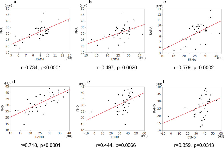
All the muscle areas were significantly associated with TEE, severity of emphysema, and body composition indices such as body mass index, fat-free mass, and trunk muscle mass. All trunk muscle densities were correlated with PAL. The product of the rectus abdominis muscle area and density showed the highest association with TEE (r = 0.732) and PAL (r = 0.578). Several trunk muscle measurements showed significant correlations with maximal inspiratory and expiratory pressures, indicating their roles in respiration.
CT-derived measurements for trunk muscles are helpful in evaluating physical status and function in patients with or at risk for COPD. Particularly, trunk muscle evaluation may be a useful marker reflecting TEE and PAL.
由于恶病质和肌肉消耗导致的身体活动减少,被认为是慢性阻塞性肺疾病(COPD)预后不良的一个标志。然而,目前还没有关于 CT 测量的躯干肌肉面积和密度与能量消耗参数(如总能量消耗(TEE)和体力活动水平(PAL))之间关系的报告。在这项研究中,我们调查了 CT 衍生的肌肉面积和密度测量值与临床参数(包括 TEE 和 PAL)在 COPD 患者和高危患者中的相关性,并检验了这些肌肉测量值是否可以作为 TEE 和 PAL 的指标。
研究人群由 36 名年龄超过 50 岁的男性组成,其中 28 名为 COPD 患者(1-4 期),8 名为 COPD 高危患者。TEE 通过双标记水法测量,PAL 通过间接法估计的基础代谢率计算。测量了胸肌、腹直肌和竖脊肌的横截面积和密度。我们评估了这些肌肉测量值与临床结局之间的关系,包括身体成分、肺功能、肌肉力量、TEE 和 PAL。
所有肌肉面积均与 TEE、肺气肿严重程度以及身体成分指数(如 BMI、去脂体重和躯干肌肉质量)显著相关。所有躯干肌肉密度均与 PAL 相关。腹直肌面积和密度的乘积与 TEE(r=0.732)和 PAL(r=0.578)的相关性最高。几个躯干肌肉测量值与最大吸气和呼气压力显著相关,表明它们在呼吸中起作用。
CT 衍生的躯干肌肉测量值有助于评估 COPD 患者和高危患者的身体状况和功能。特别是,躯干肌肉评估可能是反映 TEE 和 PAL 的有用标志物。