Department of Radiology, Sun Yat-sen Memorial Hospital, Sun Yat-sen University, Guangzhou, Guangdong 510120, China.
Guangdong Provincial Key Laboratory of Malignant Tumor Epigenetics and Gene Regulation, Medical Research Center, Sun Yat-sen Memorial Hospital, Sun Yat-sen University, Guangzhou, Guangdong 510120, China.
Theranostics. 2021 Jan 1;11(6):2917-2931. doi: 10.7150/thno.50825. eCollection 2021.
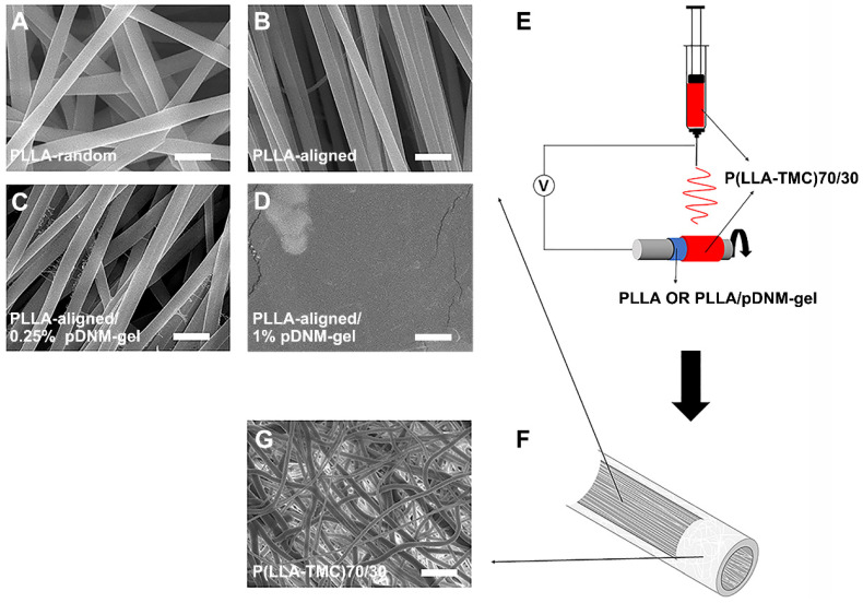
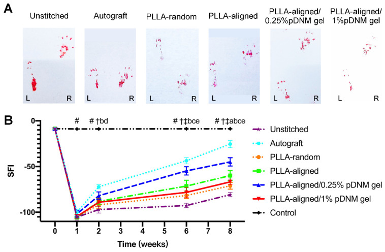
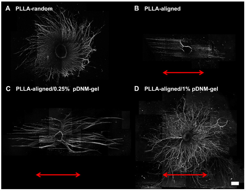
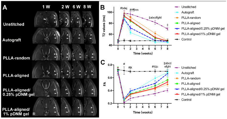
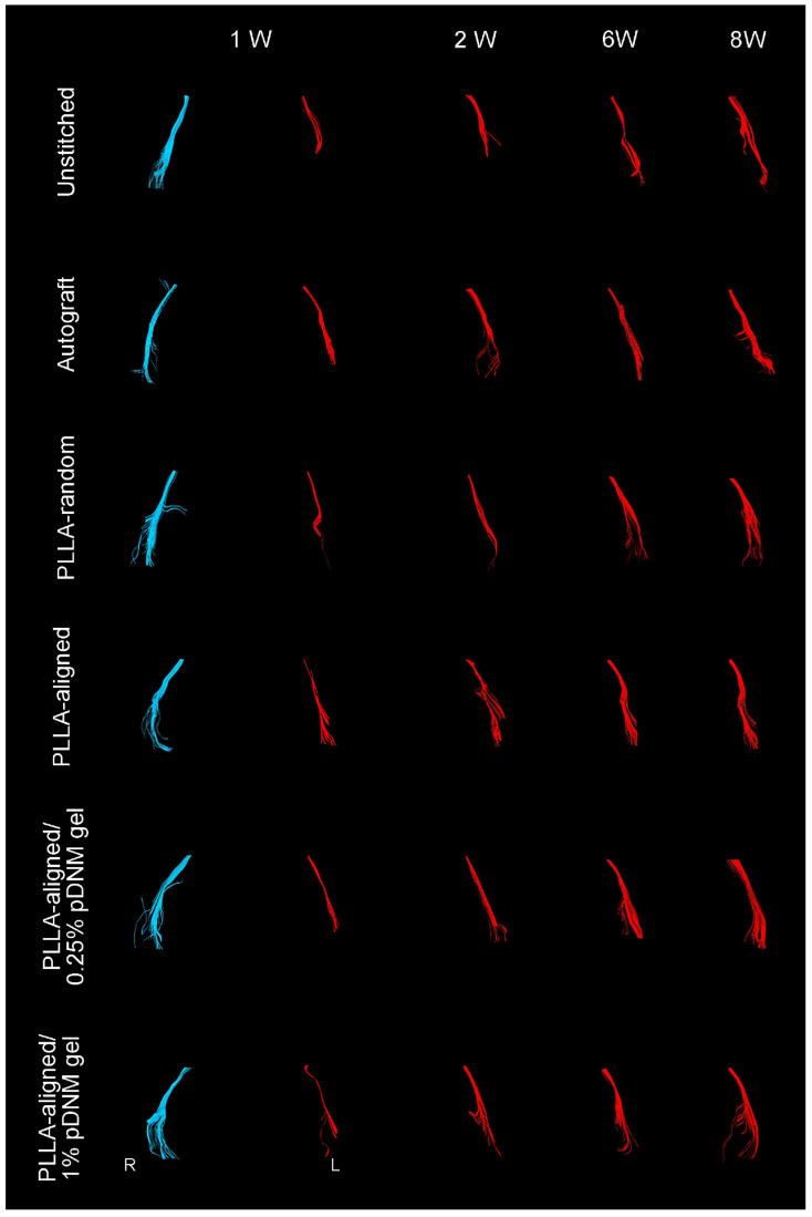
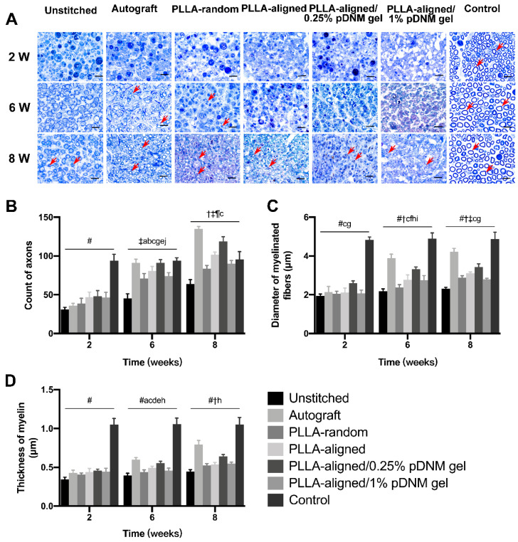
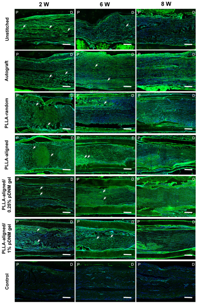
Peripheral nerve injury (PNI) is a great challenge for regenerative medicine. Nerve autograft is the gold standard for clinical PNI repair. Due to its significant drawbacks, artificial nerve guidance conduits (NGCs) have drawn much attention as replacement therapies. We developed a combinatorial NGC consisting of longitudinally aligned electrospun nanofibers and porcine decellularized nerve matrix hydrogel (pDNM gel). The in vivo capacity for facilitating nerve tissue regeneration and functional recovery was evaluated in a rat sciatic nerve defect model. Poly (-lactic acid) (PLLA) was electrospun into randomly oriented (PLLA-random) and longitudinally aligned (PLLA-aligned) nanofibers. PLLA-aligned were further coated with pDNM gel at concentrations of 0.25% (PLLA-aligned/0.25% pDNM gel) and 1% (PLLA-aligned/1% pDNM gel). Axonal extension and Schwann cells migration were evaluated by immunofluorescence staining of dorsal root ganglia cultured on the scaffolds. To fabricate implantable NGCs, the nanofibrous scaffolds were rolled and covered with an electrospun protection tube. The fabricated NGCs were then implanted into a 5 mm sciatic nerve defect model in adult male Sprague-Dawley rats. Nerves treated with NGCs were compared to contralateral uninjured nerves (control group), injured but untreated nerves (unstitched group), and autografted nerves. Nerve regeneration was monitored by an established set of assays, including T2 values and diffusion tensor imaging (DTI) derived from multiparametric magnetic resonance imaging (MRI), histological assessments, and immunostaining. Nerve functional recovery was evaluated by walking track analysis. PLLA-aligned/0.25% pDNM gel scaffold exhibited the best performance in facilitating directed axonal extension and Schwann cells migration in vitro due to the combined effects of the topological cues provided by the aligned nanofibers and the biochemical cues retained in the pDNM gel. Consistent results were obtained in animal experiments with the fabricated NGCs. Both the T2 and fractional anisotropy values of the PLLA-aligned/0.25% pDNM gel group were the closest to those of the autografted group, and returned to normal much faster than those of the other NGCs groups. Histological assessment indicated that the implanted PLLA-aligned/0.25% pDNM gel NGC resulted in the largest number of axons and the most extensive myelination among all fabricated NGCs. Further, the PLLA-aligned/0.25% pDNM gel group exhibited the highest sciatic nerve function index, which was comparable to that of the autografted group, at 8 weeks post-surgery. NGCs composed of aligned PLLA nanofibers decorated with 0.25% pDNM gel provided both topological and biochemical guidance for directing and promoting axonal extension, nerve fiber myelination, and functional recovery. Moreover, T2-mapping and DTI metrics were found to be useful non-invasive monitoring techniques for PNI treatment.
周围神经损伤 (PNI) 是再生医学的一大挑战。神经自体移植是临床 PNI 修复的金标准。由于其明显的缺点,人工神经引导导管 (NGC) 作为替代疗法引起了广泛关注。我们开发了一种组合 NGC,由纵向排列的静电纺纳米纤维和猪去细胞神经基质水凝胶 (pDNM 凝胶) 组成。在大鼠坐骨神经缺损模型中评估了促进神经组织再生和功能恢复的体内能力。聚 (乳酸) (PLLA) 被静电纺成随机取向 (PLLA-random) 和纵向排列 (PLLA-aligned) 的纳米纤维。PLLA-aligned 进一步用浓度为 0.25% (PLLA-aligned/0.25% pDNM 凝胶) 和 1% (PLLA-aligned/1% pDNM 凝胶) 的 pDNM 凝胶包被。通过对培养在支架上的背根神经节进行免疫荧光染色来评估轴突延伸和施万细胞迁移。为了制造可植入的 NGC,将纳米纤维支架卷起并用静电纺丝保护管覆盖。然后将制备好的 NGC 植入成年雄性 Sprague-Dawley 大鼠的 5mm 坐骨神经缺损模型中。用 NGC 处理的神经与对侧未受伤的神经 (对照组)、受伤但未处理的神经 (未缝合组) 和自体移植的神经进行比较。通过一组已建立的检测方法监测神经再生,包括来自多参数磁共振成像 (MRI) 的 T2 值和扩散张量成像 (DTI)、组织学评估和免疫染色。通过步态分析评估神经功能恢复。由于纵向纳米纤维提供的拓扑线索和 pDNM 凝胶保留的生化线索的综合作用,PLLA-aligned/0.25% pDNM 凝胶支架在体外促进定向轴突延伸和施万细胞迁移方面表现出最佳性能。在动物实验中,用制备的 NGC 获得了一致的结果。PLLA-aligned/0.25% pDNM 凝胶组的 T2 值和各向异性分数值最接近自体移植组,并且比其他 NGC 组更快地恢复正常。组织学评估表明,植入的 PLLA-aligned/0.25% pDNM 凝胶 NGC 导致所有制备的 NGC 中轴突数量最多,髓鞘化最广泛。此外,PLLA-aligned/0.25% pDNM 凝胶组在术后 8 周时表现出最高的坐骨神经功能指数,与自体移植组相当。由排列整齐的 PLLA 纳米纤维组成的 NGC,用 0.25% pDNM 凝胶装饰,为定向和促进轴突延伸、神经纤维髓鞘化和功能恢复提供了拓扑和生化指导。此外,T2 映射和 DTI 指标被发现是用于 PNI 治疗的有用的非侵入性监测技术。