Abdelqader Altaweel Alaa, Aziz Baiomy Abdullah Baiomy Abdel, Abdel-Hameed Elsayed Shadia
Faculty of Dental Medicine, Al-Azhar University for Boys, 11727 Cairo, Egypt.
Alfarabi Private College for Dentistry and Nursing, Jeddah, Saudi Arabia.
Saudi Dent J. 2021 Jan;33(1):27-33. doi: 10.1016/j.sdentj.2019.11.008. Epub 2019 Dec 2.
This study aimed to assess the clinical and radiographic findings obtained by using amniotic membrane (AM) to cover nano-hydroxyapatite (nHA) bone grafts coated with platelet-rich fibrin (PRF) and thereby evaluate the osseointegration of posterior mandibular implants inserted simultaneously during alveolar piezoelectric ridge splitting technique (RST).
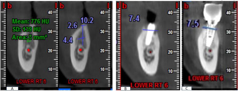
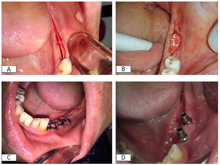
A prospective cohort study was implemented with thirty patients who had a narrow posterior mandibular alveolar ridge and required implant restoration. Patients were distributed randomly into three groups (group I treated by piezoelectric RST and immediate implant insertion, augmented by the nHA bone graft only; group II treated by piezoelectric RST augmented by nHA bone graft and covered by AM; while group III was treated by piezoelectric RST augmented with PRF and nHA graft and covered by AM). Patients were evaluated clinically to assess the implant stability quotient (ISQ) and radiographically to assess horizontal ridge dimension, crestal bone level (CBL), and bone densitometric (BD) parameters.
ISQ results showed a non-significant clinical difference between groups while CBL values showed a high statistically significant difference over the 12-month interval when comparing groups III and II with group I. BD outcomes showed statistically significant differences at all intervals in comparisons of group III with groups I and II.
The results of this study suggest that concomitant use of PRF with nHA graft covered with AM for augmentation around the dental implant in a narrow posterior mandible after piezoelectric alveolar ridge splitting accelerate osseointegration and significantly increase bone density around the osseointegrated implant and decrease bone resorption in comparison to that achieved with the graft alone.
本研究旨在评估使用羊膜(AM)覆盖富含血小板纤维蛋白(PRF)涂层的纳米羟基磷灰石(nHA)骨移植材料所获得的临床和影像学结果,从而评估在牙槽骨压电嵴劈开技术(RST)期间同时植入的下颌后牙种植体的骨整合情况。
对30例下颌后牙牙槽嵴狭窄且需要种植修复的患者进行了一项前瞻性队列研究。患者被随机分为三组(第一组采用压电RST并立即植入种植体,仅用nHA骨移植材料增强;第二组采用压电RST并用nHA骨移植材料增强,并用AM覆盖;第三组采用压电RST并用PRF和nHA移植材料增强,并用AM覆盖)。对患者进行临床评估以评估种植体稳定性商数(ISQ),并进行影像学评估以评估水平嵴尺寸、嵴顶骨水平(CBL)和骨密度(BD)参数。
ISQ结果显示各组之间临床差异不显著,而在比较第三组和第二组与第一组时,CBL值在12个月期间显示出高度统计学显著差异。BD结果显示,在所有时间间隔内,第三组与第一组和第二组比较均有统计学显著差异。
本研究结果表明,在压电牙槽嵴劈开术后的狭窄下颌后牙区,将PRF与覆盖有AM的nHA移植材料同时用于种植体周围增强,与单独使用移植材料相比,可加速骨整合,显著增加骨整合种植体周围的骨密度,并减少骨吸收。