Wang Yao-Kuang, Syu Hao-Yi, Chen Yi-Hsun, Chung Chen-Shuan, Tseng Yu Sheng, Ho Shinn-Ying, Huang Chien-Wei, Wu I-Chen, Wang Hsiang-Chen
Division of Gastroenterology, Department of Internal Medicine, Kaohsiung Medical University Hospital, Kaohsiung Medical University, No.100, Tzyou 1st Rd., Sanmin Dist., Kaohsiung City 80756, Taiwan.
Graduate Institute of Clinical Medicine, College of Medicine, Kaohsiung Medical University, No.100, Tzyou 1st Rd., Sanmin Dist., Kaohsiung City 80756, Taiwan.
Cancers (Basel). 2021 Jan 17;13(2):321. doi: 10.3390/cancers13020321.
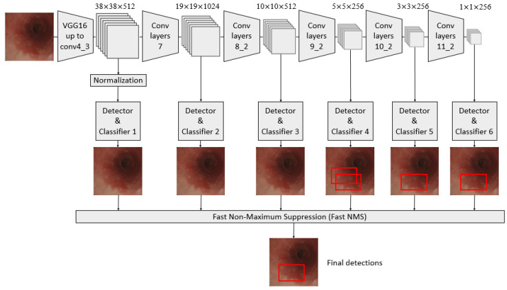
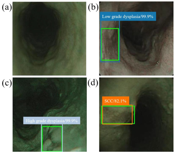
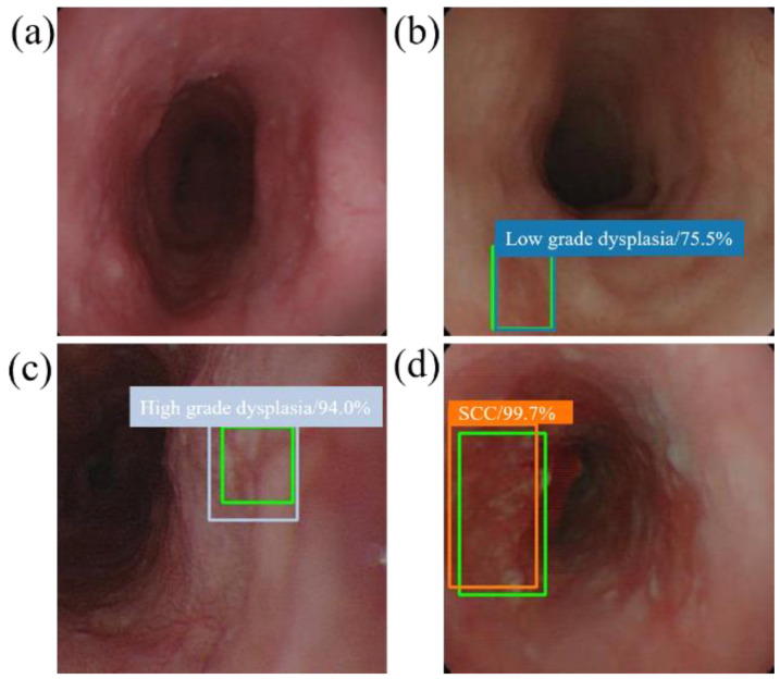
Diagnosis of early esophageal neoplasia, including dysplasia and superficial cancer, is a great challenge for endoscopists. Recently, the application of artificial intelligence (AI) using deep learning in the endoscopic field has made significant advancements in diagnosing gastrointestinal cancers. In the present study, we constructed a single-shot multibox detector using a convolutional neural network for diagnosing different histological grades of esophageal neoplasms and evaluated the diagnostic accuracy of this computer-aided system. A total of 936 endoscopic images were used as training images, and these images included 498 white-light imaging (WLI) and 438 narrow-band imaging (NBI) images. The esophageal neoplasms were divided into three classifications: squamous low-grade dysplasia, squamous high-grade dysplasia, and squamous cell carcinoma, based on pathological diagnosis. This AI system analyzed 264 test images in 10 s, and the sensitivity, specificity, and diagnostic accuracy of this system in detecting esophageal neoplasms were 96.2%, 70.4%, and 90.9%, respectively. The accuracy of this AI system in differentiating the histological grade of esophageal neoplasms was 92%. Our system showed better accuracy in diagnosing NBI (95%) than WLI (89%) images. Our results showed the great potential of AI systems in identifying esophageal neoplasms as well as differentiating histological grades.
早期食管肿瘤(包括发育异常和浅表癌)的诊断对内窥镜医师来说是一项巨大挑战。近年来,深度学习人工智能(AI)在内窥镜领域的应用在胃肠道癌诊断方面取得了显著进展。在本研究中,我们构建了一种基于卷积神经网络的单发多框检测器,用于诊断不同组织学分级的食管肿瘤,并评估了该计算机辅助系统的诊断准确性。总共936张内镜图像用作训练图像,这些图像包括498张白光成像(WLI)图像和438张窄带成像(NBI)图像。根据病理诊断,食管肿瘤分为三类:鳞状低级别发育异常、鳞状高级别发育异常和鳞状细胞癌。该AI系统在10秒内分析了264张测试图像,该系统检测食管肿瘤的灵敏度、特异度和诊断准确性分别为96.2%、70.4%和90.9%。该AI系统区分食管肿瘤组织学分级的准确性为92%。我们的系统在诊断NBI图像(95%)时比WLI图像(89%)显示出更高的准确性。我们的结果表明,AI系统在识别食管肿瘤以及区分组织学分级方面具有巨大潜力。