Eye Hospital, School of Ophthalmology and Optometry, Wenzhou Medical University, 270 Xueyuan Road, Wenzhou, 325027, Zhejiang, China.
R&D Vision Sciences AMERA, Essilor International, Singapore, Singapore.
Sci Rep. 2021 Jan 21;11(1):2015. doi: 10.1038/s41598-021-81770-y.
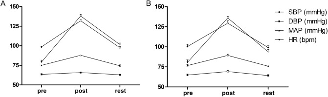
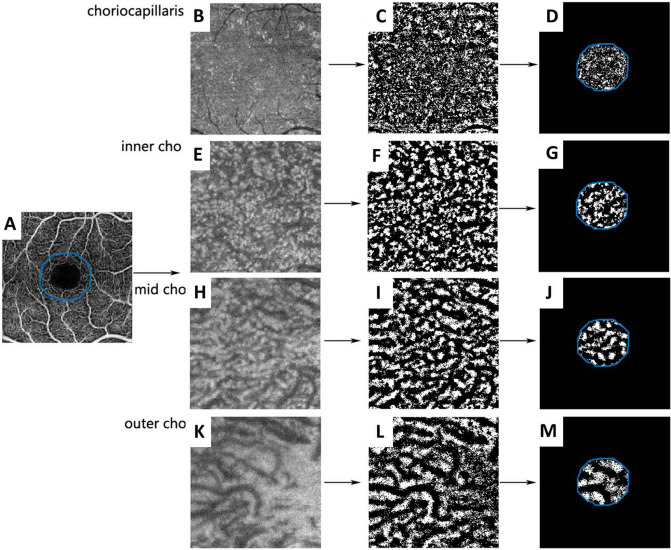
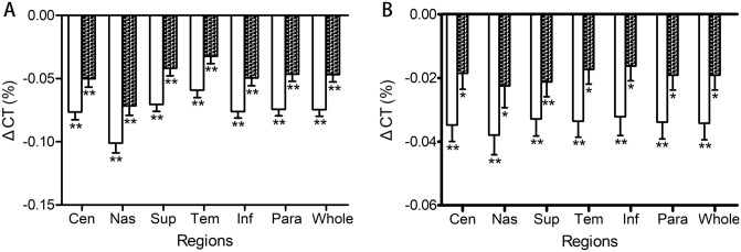
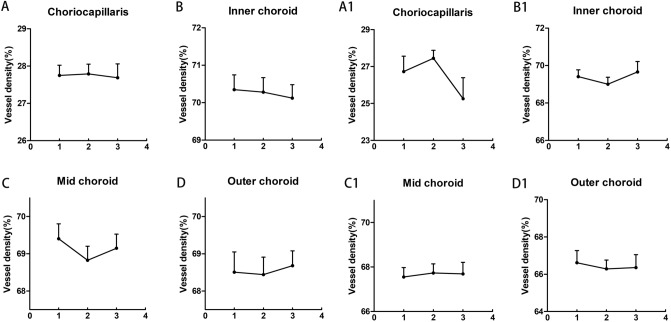
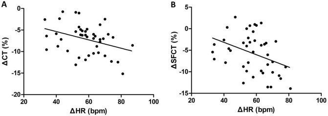
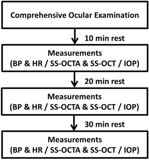
We used swept-source (SS) optical coherence tomography (OCT) and OCT angiography (OCTA) to investigate the effects of moderate physical exercise on retinal and choroidal vessel densities (VDs) and thicknesses in children. One eye in each of 40 myopic children (mean age, 11.70 years) and 18 emmetropic children (mean age, 11.06 years) were included. SS-OCT 6 × 6-mm radial scans and SS-OCTA 3 × 3-mm images were centered on the macula. Heart rate (HR), systolic and diastolic blood pressure, and intraocular pressure (IOP) were recorded before and immediately after a 20-min stationary cycling exercise and after a 30-min rest. The subfoveal choroidal thickness (SFCT), choroidal thickness (CT), and VD at the superficial and deep retinal layers, choriocapillaris, and deeper choroidal vessels were determined. SFCT and CT were significantly lower at all locations immediately after exercise (p < 0.001) and did not fully recover after rest (p < 0.05). VD was lower in the deep retinal layer after exercise (p = 0.02) and higher in the superficial layer after rest (p = 0.03) in myopic eyes while it was higher in the superficial (p < 0.01) and deep layer (p < 0.01) after rest in emmetropic eyes. No significant exercise-related changes in the superficial retinal VD, choroidal VD, or IOP were observed. ΔCT% and ΔSFCT% were significantly correlated with increases in HR in myopic group (p = 0.04 and p = 0.03, respectively). Exercise increased retinal VD after rest in emmetropic eyes, and caused significant CT thinning that lasted for at least 30 min in both emmetropic and myopic eyes.
我们使用扫频源(SS)光学相干断层扫描(OCT)和 OCT 血管造影(OCTA)来研究适度的体育锻炼对儿童视网膜和脉络膜血管密度(VD)和厚度的影响。每只眼纳入 40 名近视儿童(平均年龄 11.70 岁)和 18 名正视儿童(平均年龄 11.06 岁)各一只眼。SS-OCT 6×6mm 径向扫描和 SS-OCTA 3×3mm 图像以黄斑为中心。在 20 分钟的固定自行车运动前和运动后立即以及 30 分钟的休息后,记录心率(HR)、收缩压和舒张压以及眼内压(IOP)。确定了中心凹下脉络膜厚度(SFCT)、脉络膜厚度(CT)以及浅层和深层视网膜、脉络膜毛细血管和更深脉络膜血管的 VD。运动后所有部位的 SFCT 和 CT 均显著降低(p<0.001),休息后并未完全恢复(p<0.05)。运动后近视眼的深层视网膜层 VD 降低(p=0.02),休息后浅层视网膜层 VD 升高(p=0.03),而正视眼的浅层(p<0.01)和深层(p<0.01)视网膜层 VD 升高。未观察到与运动相关的浅层视网膜 VD、脉络膜 VD 或 IOP 显著变化。近视组的 ΔCT%和 ΔSFCT%与 HR 增加显著相关(p=0.04 和 p=0.03)。休息后,正视眼的视网膜 VD 增加,而在正视眼和近视眼中,CT 明显变薄,持续至少 30 分钟。