Instituto de Ciências Biomédicas, Universidade Federal do Rio de Janeiro, Rio de Janeiro, Brazil.
PLoS One. 2021 Jan 22;16(1):e0245795. doi: 10.1371/journal.pone.0245795. eCollection 2021.
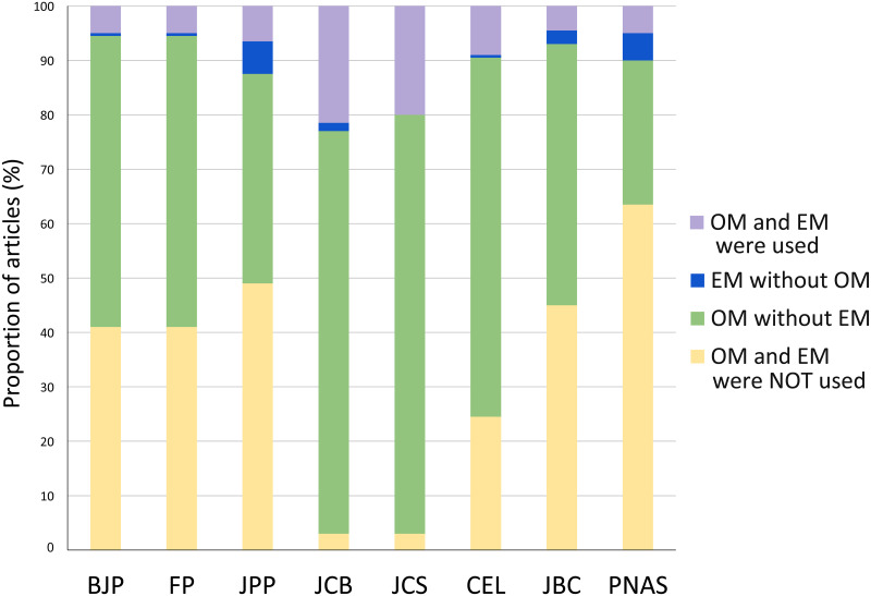
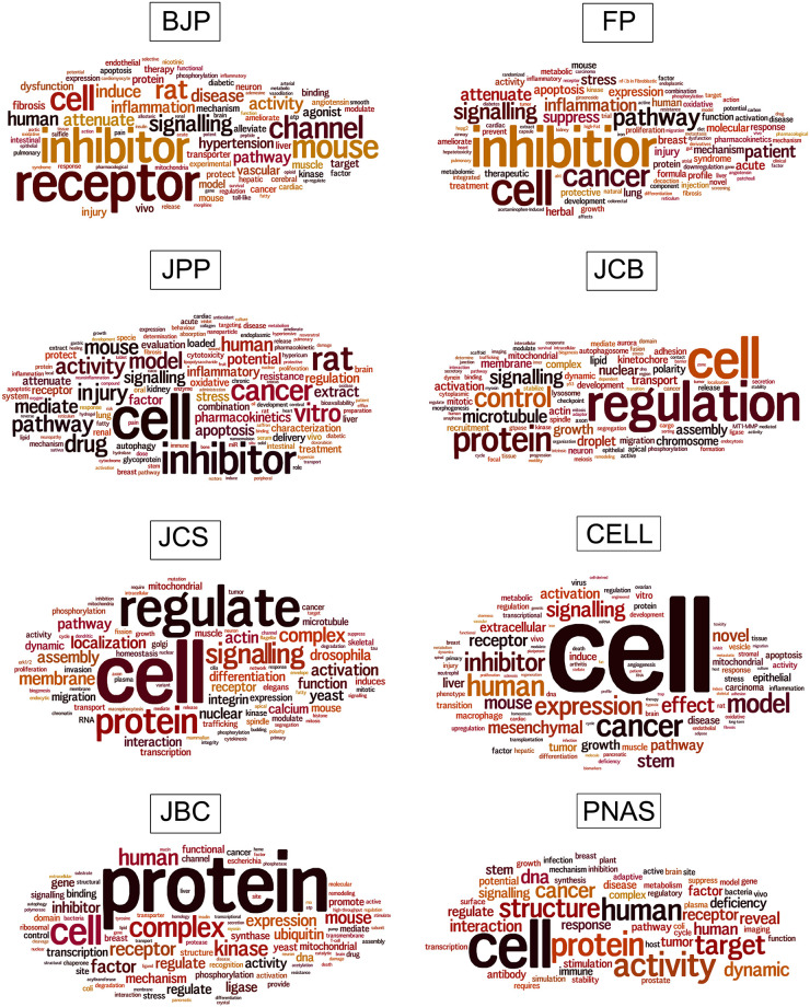
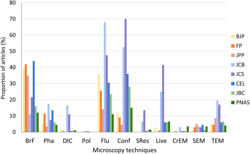
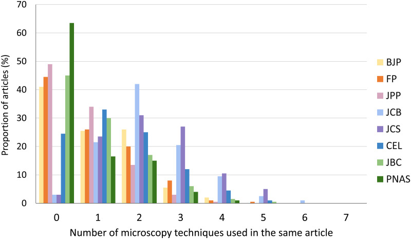
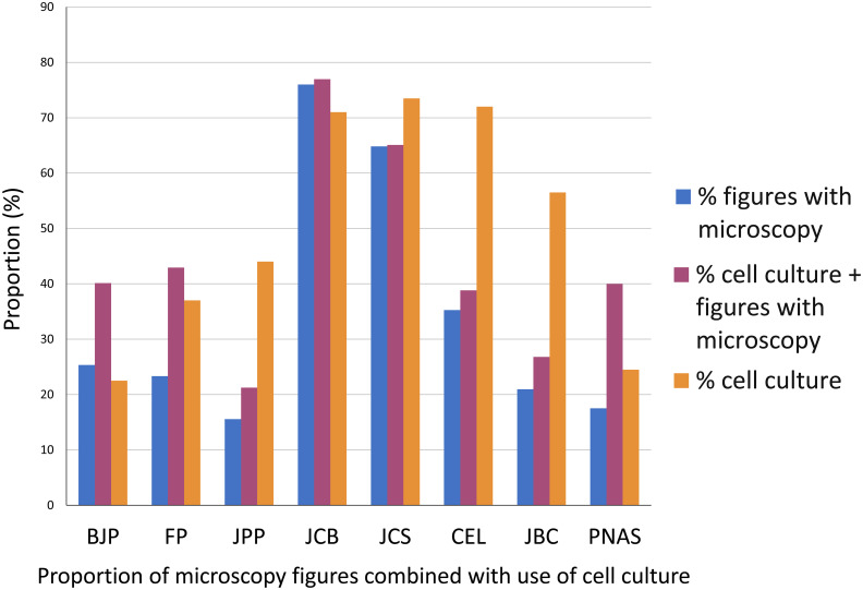
Microscopy is the main technique to visualize and study the structure and function of cells. The impact of optical and electron microscopy techniques is enormous in all fields of biomedical research. It is possible that different research areas rely on microscopy in diverse ways. Here, we analyzed comparatively the use of microscopy in pharmacology and cell biology, among other biomedical sciences fields. We collected data from articles published in several major journals in these fields. We analyzed the frequency of use of different optical and electron microscopy techniques: bright field, phase contrast, differential interference contrast, polarization, conventional fluorescence, confocal, live cell imaging, super resolution, transmission and scanning electron microscopy, and cryoelectron microscopy. Our analysis showed that the use of microscopy has a distinctive pattern in each research area, and that nearly half of the articles from pharmacology journals did not use any microscopy method, compared to the use of microscopy in almost all the articles from cell biology journals. The most frequent microscopy methods in all the journals in all areas were bright field and fluorescence (conventional and confocal). Again, the pattern of use was different: while the most used microscopy methods in pharmacology were bright field and conventional fluorescence, in cell biology the most used methods were conventional and confocal fluorescence, and live cell imaging. We observed that the combination of different microscopy techniques was more frequent in cell biology, with up to 6 methods in the same article. To correlate the use of microscopy with the research theme of each article, we analyzed the proportion of microscopy figures with the use of cell culture. We analyzed comparatively the vocabulary of each biomedical sciences field, by the identification of the most frequent words in the articles. The collection of data described here shows a vast difference in the use of microscopy among different fields of biomedical sciences. The data presented here could be valuable in other scientific and educational contexts.
显微镜是可视化和研究细胞结构和功能的主要技术。光学和电子显微镜技术在生物医学研究的各个领域都具有巨大的影响。不同的研究领域可能以不同的方式依赖显微镜。在这里,我们比较分析了药理学和细胞生物学等生物医学科学领域中显微镜的使用情况。我们从这些领域的几个主要期刊上发表的文章中收集了数据。我们分析了不同光学和电子显微镜技术的使用频率:明场、相差、微分干涉对比、偏振、常规荧光、共聚焦、活细胞成像、超分辨率、透射和扫描电子显微镜以及冷冻电子显微镜。我们的分析表明,显微镜的使用在每个研究领域都有独特的模式,与细胞生物学期刊几乎所有文章都使用显微镜方法相比,药理学期刊的文章中有近一半没有使用任何显微镜方法。在所有期刊的所有领域中,最常用的显微镜方法都是明场和荧光(常规和共聚焦)。同样,使用模式也不同:在药理学中最常用的显微镜方法是明场和常规荧光,而在细胞生物学中最常用的方法是常规和共聚焦荧光以及活细胞成像。我们观察到,在细胞生物学中,不同显微镜技术的组合更为频繁,同一篇文章中最多可以使用 6 种方法。为了将显微镜的使用与每篇文章的研究主题相关联,我们分析了带有细胞培养的显微镜图片的比例。我们通过识别文章中最常用的词来比较分析每个生物医学科学领域的词汇。这里描述的数据显示了生物医学科学不同领域之间在显微镜使用方面存在巨大差异。这里呈现的数据在其他科学和教育背景下可能具有价值。