• 文献检索
  • 文档翻译
  • 深度研究
  • 学术资讯
  • Suppr Zotero 插件Zotero 插件
  • 邀请有礼
  • 套餐&价格
  • 历史记录
应用&插件
Suppr Zotero 插件Zotero 插件浏览器插件Mac 客户端Windows 客户端微信小程序
定价
高级版会员购买积分包购买API积分包
服务
文献检索文档翻译深度研究API 文档MCP 服务
关于我们
关于 Suppr公司介绍联系我们用户协议隐私条款
关注我们

Suppr 超能文献

核心技术专利:CN118964589B侵权必究
粤ICP备2023148730 号-1Suppr @ 2026

文献检索

告别复杂PubMed语法,用中文像聊天一样搜索,搜遍4000万医学文献。AI智能推荐,让科研检索更轻松。

立即免费搜索

文件翻译

保留排版,准确专业,支持PDF/Word/PPT等文件格式,支持 12+语言互译。

免费翻译文档

深度研究

AI帮你快速写综述,25分钟生成高质量综述,智能提取关键信息,辅助科研写作。

立即免费体验

原发性甲状旁腺功能亢进症合并股骨病理性骨折患者的多发棕色瘤

Multiple Brown tumors in a Case of Primary Hyperparathyroidism with Pathological Fracture in Femur.

作者信息

Dhaniwala Nareshkumar Satyanarayan, Dhaniwala Mukund Naresh

机构信息

Department of Orthopedics, Jawaharlal Nehru Medical College and Acharya Vinoba Bhave Rural Hospital, Datta Meghe Institute of Medical Sciences, Wardha, Maharashtra, India.

Department of Orthopedics, Mahatma Gandhi Institute of Medical Sciences, Wardha, Maharashtra, India.

出版信息

J Orthop Case Rep. 2020 Sep;10(6):49-53. doi: 10.13107/jocr.2020.v10.i06.1872.

DOI:10.13107/jocr.2020.v10.i06.1872
PMID:33489969
原文链接:https://pmc.ncbi.nlm.nih.gov/articles/PMC7815671/
Abstract

INTRODUCTION

Pathological fractures in long bones are commonly caused by simple bone cyst or Osteogenesis imperfecta in children and by metastatic tumors from primary carcinoma, multiple myeloma, osteoporosis, and bone tumors in adults. Hyperparathyroidism causing pathological fractures, though a well-known entity, is seen infrequently in clinical practice. The fractures occur in the expansile fibro-cystic bone lesions called as "Brown tumor." The reported case describes a patient with classical features of primary hyperparathyroidism (PHPT) having multiple lytic lesions in pelvis and bilateral femur with pathological fracture. It is being reported due to its rarity.

CASE REPORT

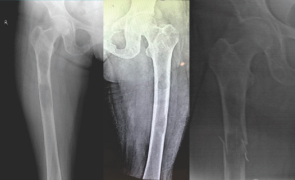
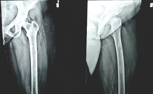
A 28-year-old young married lady presented with chronic dull aching pain in both thighs and difficulty in walking for 2 years. Examination revealed tenderness in both thighs, with antalgic gait. X-ray pelvis with both thighs showed multiple lytic lesions of variable size in both femora and pelvis. Blood investigations showed raised levels of serum calcium, with highly raised levels of serum parathyroid hormone (PTH). Contrast-enhanced computerized tomography (CT) scan of neck demonstrated parathyroid adenoma. The patient admitted for prophylactic nailing for right femur, developed a fracture while indoor and was managed by right proximal femoral nailing, followed by parathyroid adenoma excision. Follow-up showed dramatic clinical and radiological improvement with good healing of fracture. Lytic lesions healed gradually and blood parameters returned to normal. The patient remains asymptomatic at 2 years follow-up.

CONCLUSION

Advanced case of symptomatic PHPT affecting bones is rare and it should be considered as a differential diagnosis in cases of a solitary and or multiple osteolytic lesions. Serum calcium and PTH level estimation at an early stage prevents missing the diagnosis and progression of disease. Early diagnosis and appropriate treatment help in rapid clinical improvement with almost total reversal of bony changes, thus avoiding devastating complications.

摘要

引言

儿童长骨病理性骨折通常由单纯性骨囊肿或成骨不全引起,而成人则由原发性癌转移瘤、多发性骨髓瘤、骨质疏松症和骨肿瘤引起。甲状旁腺功能亢进导致病理性骨折,虽然是一个众所周知的病症,但在临床实践中很少见。骨折发生在被称为“棕色瘤”的膨胀性纤维囊性骨病变中。本文报道的病例描述了一名具有原发性甲状旁腺功能亢进(PHPT)典型特征的患者,其骨盆和双侧股骨有多个溶骨性病变并伴有病理性骨折。因其罕见性而予以报道。

病例报告

一名28岁已婚年轻女性,双大腿慢性钝痛伴行走困难2年。检查发现双大腿压痛,有跛行步态。骨盆及双大腿X线检查显示双侧股骨和骨盆有多个大小不一的溶骨性病变。血液检查显示血清钙水平升高,血清甲状旁腺激素(PTH)水平显著升高。颈部增强计算机断层扫描(CT)显示甲状旁腺腺瘤。患者因右股骨预防性钉固定入院,在住院期间发生骨折,随后行右股骨近端钉固定,接着切除甲状旁腺腺瘤。随访显示临床和影像学有显著改善,骨折愈合良好。溶骨性病变逐渐愈合,血液参数恢复正常。在2年随访时患者仍无症状。

结论

有症状的晚期PHPT累及骨骼的病例罕见,在出现孤立性和/或多发性溶骨性病变的病例中应将其作为鉴别诊断。早期测定血清钙和PTH水平可防止漏诊和疾病进展。早期诊断和适当治疗有助于临床快速改善,几乎完全逆转骨改变,从而避免严重并发症。

https://cdn.ncbi.nlm.nih.gov/pmc/blobs/a187/7815671/06d446a921e0/JOCR-10-49-g007.jpg
https://cdn.ncbi.nlm.nih.gov/pmc/blobs/a187/7815671/a1bb94b822d8/JOCR-10-49-g001.jpg
https://cdn.ncbi.nlm.nih.gov/pmc/blobs/a187/7815671/0223593e0c44/JOCR-10-49-g002.jpg
https://cdn.ncbi.nlm.nih.gov/pmc/blobs/a187/7815671/5cec6aeeafe3/JOCR-10-49-g003.jpg
https://cdn.ncbi.nlm.nih.gov/pmc/blobs/a187/7815671/66c999d2f89e/JOCR-10-49-g005.jpg
https://cdn.ncbi.nlm.nih.gov/pmc/blobs/a187/7815671/c4d0bbbcbb29/JOCR-10-49-g006.jpg
https://cdn.ncbi.nlm.nih.gov/pmc/blobs/a187/7815671/06d446a921e0/JOCR-10-49-g007.jpg

相似文献

1
Multiple Brown tumors in a Case of Primary Hyperparathyroidism with Pathological Fracture in Femur.原发性甲状旁腺功能亢进症合并股骨病理性骨折患者的多发棕色瘤
J Orthop Case Rep. 2020 Sep;10(6):49-53. doi: 10.13107/jocr.2020.v10.i06.1872.
2
Brown tumor due to primary hyperparathyroidism in a familial case: a case report.家族性原发性甲状旁腺功能亢进致棕色瘤 1 例报告。
BMC Endocr Disord. 2023 Oct 8;23(1):214. doi: 10.1186/s12902-023-01475-3.
3
Multiple Brown Tumors in Primary Hyperparathyroidism Causing Pathological Fracture: A Case Report of a 21-Year-Old Adult Male.原发性甲状旁腺功能亢进症中导致病理性骨折的多发性棕色瘤:一名21岁成年男性的病例报告
Cureus. 2023 Mar 10;15(3):e35979. doi: 10.7759/cureus.35979. eCollection 2023 Mar.
4
Osteitis Fibrosa Cystica and pathological fractures-the classic but neglected skeletal manifestation of primary hyperparathyroidism: a case report.纤维囊性骨炎和病理性骨折——原发性甲状旁腺功能亢进症的经典但被忽视的骨骼表现:一例报告。
BMC Musculoskelet Disord. 2021 May 14;22(1):443. doi: 10.1186/s12891-021-04326-1.
5
Osteitis fibrosa cystica-a forgotten radiological feature of primary hyperparathyroidism.囊性纤维性骨炎——原发性甲状旁腺功能亢进症被遗忘的放射学特征。
Endocrine. 2017 Nov;58(2):380-385. doi: 10.1007/s12020-017-1414-2. Epub 2017 Sep 12.
6
Unusual body weight loss due to primary hyperparathyroidism: A case study with literature review.原发性甲状旁腺功能亢进导致的异常体重减轻:一例病例报告并文献复习
Heliyon. 2024 Mar 16;10(6):e28333. doi: 10.1016/j.heliyon.2024.e28333. eCollection 2024 Mar 30.
7
Osteolytic lesions (brown tumors) of primary hyperparathyroidism misdiagnosed as multifocal giant cell tumor of the distal ulna and radius: a case report.原发性甲状旁腺功能亢进症的溶骨性病变(棕色瘤)被误诊为尺骨和桡骨远端多灶性巨细胞瘤:一例报告
J Med Case Rep. 2018 Jun 25;12(1):176. doi: 10.1186/s13256-018-1723-y.
8
Primary Hyperparathyroidism Mimicking Skeletal Metastasis - A Diagnostic Dilemma.酷似骨转移的原发性甲状旁腺功能亢进——诊断难题
J Orthop Case Rep. 2024 Jan;14(1):103-108. doi: 10.13107/jocr.2024.v14.i01.4158.
9
Osteitis fibrosa cystica mistaken for malignant disease.纤维囊性骨炎被误诊为恶性疾病。
Clin Exp Otorhinolaryngol. 2013 Jun;6(2):110-3. doi: 10.3342/ceo.2013.6.2.110. Epub 2011 Sep 15.
10
Giant parathyroid adenoma diagnosed by brown tumour, a clinical manifestation of primary hyperparathyroidism: A case report.棕色瘤诊断的巨大甲状旁腺腺瘤,原发性甲状旁腺功能亢进症的一种临床表现:病例报告。
J Pak Med Assoc. 2021 Apr;71(4):1266-1269. doi: 10.47391/JPMA.393.

引用本文的文献

1
Primary hyperparathyroidism due to a giant parathyroid adenoma presenting with pathological fractures and multiple brown tumors.因巨大甲状旁腺腺瘤导致的原发性甲状旁腺功能亢进,表现为病理性骨折和多发棕色瘤。
Endocrinol Diabetes Metab Case Rep. 2024 Dec 19;2024(4). doi: 10.1530/EDM-24-0054. Print 2024 Oct 1.
2
[Hypercalcium crisis and postoperative hungry bone syndrome caused by primary hyperparathyroidism: a case report].[原发性甲状旁腺功能亢进症所致高钙血症危象及术后饥饿骨综合征:一例报告]
Lin Chuang Er Bi Yan Hou Tou Jing Wai Ke Za Zhi. 2023 May;37(5):389-392. doi: 10.13201/j.issn.2096-7993.2023.05.014.
3
Brown tumor of the knee as the first presentation of primary hyperparathyroidism caused by parathyroid adenoma: A case report.

本文引用的文献

1
Osseous Manifestations of Primary Hyperparathyroidism: Imaging Findings.原发性甲状旁腺功能亢进的骨表现:影像学发现
Int J Endocrinol. 2020 Feb 21;2020:3146535. doi: 10.1155/2020/3146535. eCollection 2020.
2
Osteitis fibrosa cystica masquerading as bone neoplasm.伪装成骨肿瘤的纤维囊性骨炎
BMJ Case Rep. 2018 May 7;2018:bcr-2018-224546. doi: 10.1136/bcr-2018-224546.
3
Osteitis fibrosa cystica-a forgotten radiological feature of primary hyperparathyroidism.囊性纤维性骨炎——原发性甲状旁腺功能亢进症被遗忘的放射学特征。
膝关节棕色瘤作为甲状旁腺腺瘤所致原发性甲状旁腺功能亢进的首发表现:一例报告
Radiol Case Rep. 2023 Mar 6;18(5):1852-1855. doi: 10.1016/j.radcr.2023.01.080. eCollection 2023 May.
4
[Case of clinically "aggressive" course of primary hyperparathyroidism, algorithm of differential diagnosis].[原发性甲状旁腺功能亢进症临床“侵袭性”病程病例,鉴别诊断算法]
Probl Endokrinol (Mosk). 2022 Sep 19;68(6):59-66. doi: 10.14341/probl13159.
5
Diagnosis and Treatment of Brown Tumor in the Femur Induced by Parathyroid Carcinoma: A Case Report.甲状旁腺癌所致股骨棕色瘤的诊断与治疗:1例报告
Case Rep Oncol. 2022 Nov 28;15(3):1074-1080. doi: 10.1159/000527637. eCollection 2022 Sep-Dec.
Endocrine. 2017 Nov;58(2):380-385. doi: 10.1007/s12020-017-1414-2. Epub 2017 Sep 12.
4
A case of adolescent giant parathyroid adenoma presenting multiple osteolytic fractures and postoperative hungry bone syndrome.一例青少年巨大甲状旁腺腺瘤伴多发溶骨性骨折及术后饥饿骨综合征
Clin Case Rep. 2015 Oct;3(10):835-40. doi: 10.1002/ccr3.360. Epub 2015 Sep 2.
5
Bilateral femoral fractures in a 21-year-old man following a simple fall.一名21岁男性在一次简单摔倒后发生双侧股骨骨折。
BMJ Case Rep. 2013 Sep 7;2013:bcr2013009753. doi: 10.1136/bcr-2013-009753.
6
Bone quality, as measured by trabecular bone score, in patients with primary hyperparathyroidism.原发性甲状旁腺功能亢进症患者的骨质量(以骨小梁评分衡量)。
Eur J Endocrinol. 2013 Jun 29;169(2):155-62. doi: 10.1530/EJE-13-0305. Print 2013 Aug.
7
Emphasis on the MR imaging findings of brown tumor: a report of five cases.强调棕色瘤的磁共振成像表现:五例报告。
Skeletal Radiol. 2011 Feb;40(2):205-13. doi: 10.1007/s00256-010-0979-0. Epub 2010 Jun 13.
8
Brown tumor of the maxillary sinus in a patient with primary hyperparathyroidism: a case report.原发性甲状旁腺功能亢进患者上颌窦棕色瘤:病例报告
J Med Case Rep. 2009 Jul 6;3:7495. doi: 10.4076/1752-1947-3-7495.
9
Sellar-parasellar brown tumor: case report and review of literature.鞍区-鞍旁棕色瘤:病例报告及文献复习
Skull Base. 2004 Aug;14(3):163-8; discussion 168. doi: 10.1055/s-2004-832261.
10
Unique clinical characteristics of primary hyperparathyroidism in India.印度原发性甲状旁腺功能亢进症的独特临床特征。
Br J Surg. 2001 May;88(5):708-14. doi: 10.1046/j.0007-1323.2001.01775.x.