• 文献检索
  • 文档翻译
  • 深度研究
  • 学术资讯
  • Suppr Zotero 插件Zotero 插件
  • 邀请有礼
  • 套餐&价格
  • 历史记录
应用&插件
Suppr Zotero 插件Zotero 插件浏览器插件Mac 客户端Windows 客户端微信小程序
定价
高级版会员购买积分包购买API积分包
服务
文献检索文档翻译深度研究API 文档MCP 服务
关于我们
关于 Suppr公司介绍联系我们用户协议隐私条款
关注我们

Suppr 超能文献

核心技术专利:CN118964589B侵权必究
粤ICP备2023148730 号-1Suppr @ 2026

文献检索

告别复杂PubMed语法,用中文像聊天一样搜索,搜遍4000万医学文献。AI智能推荐,让科研检索更轻松。

立即免费搜索

文件翻译

保留排版,准确专业,支持PDF/Word/PPT等文件格式,支持 12+语言互译。

免费翻译文档

深度研究

AI帮你快速写综述,25分钟生成高质量综述,智能提取关键信息,辅助科研写作。

立即免费体验

意大利的2019冠状病毒病(COVID-19):胸部CT检查的双重解读

Coronavirus Disease 2019 (COVID-19) in Italy: Double Reading of Chest CT Examination.

作者信息

Reginelli Alfonso, Grassi Roberta, Feragalli Beatrice, Belfiore Maria Paola, Montanelli Alessandro, Patelli Gianluigi, La Porta Michelearcangelo, Urraro Fabrizio, Fusco Roberta, Granata Vincenza, Petrillo Antonella, Giacobbe Giuliana, Russo Gaetano Maria, Sacco Palmino, Grassi Roberto, Cappabianca Salvatore

机构信息

Department of Precision Medicine, Università degli Studi della Campania Luigi Vanvitelli, 80121 Naples, Italy.

Oral and Biotechnological Sciences-Radiology Unit "G. D'Annunzio", Department of Medical, University of Chieti-Pescara, 66100 Chieti, Italy.

出版信息

Biology (Basel). 2021 Jan 25;10(2):89. doi: 10.3390/biology10020089.

DOI:10.3390/biology10020089
PMID:33504028
原文链接:https://pmc.ncbi.nlm.nih.gov/articles/PMC7911408/
Abstract

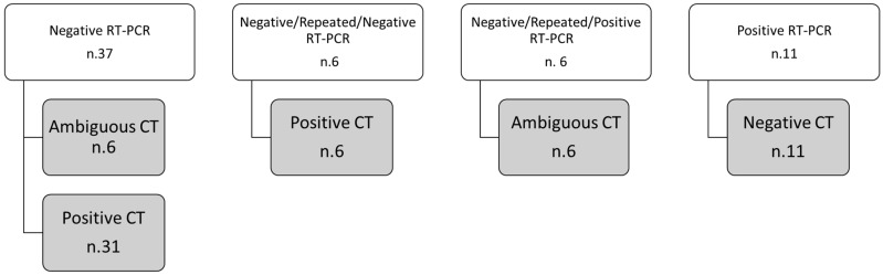
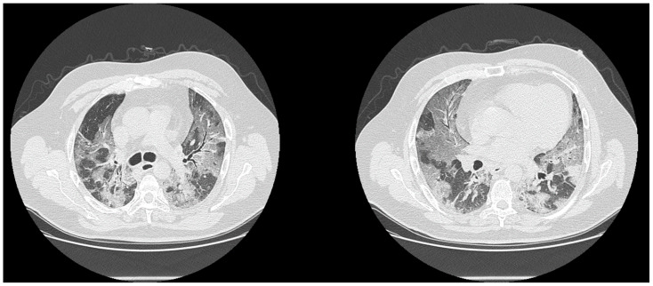
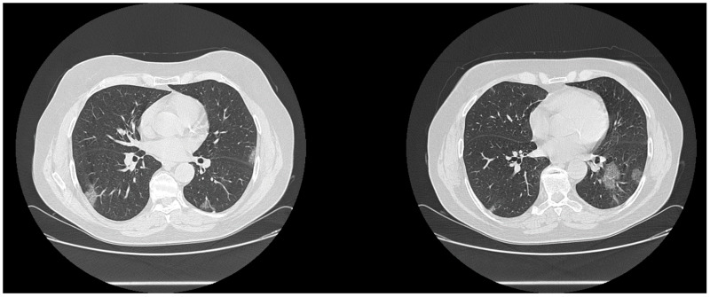
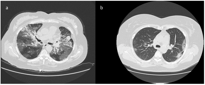
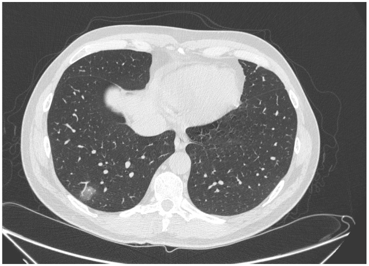
To assess the performance of the second reading of chest compute tomography (CT) examinations by expert radiologists in patients with discordance between the reverse transcription real-time fluorescence polymerase chain reaction (RT-PCR) test for COVID-19 viral pneumonia and the CT report. Three hundred and seventy-eight patients were included in this retrospective study (121 women and 257 men; 71 years median age, with a range of 29-93 years) and subjected to RT-PCR tests for suspicious COVID-19 infection. All patients were subjected to CT examination in order to evaluate the pulmonary disease involvement by COVID-19. CT images were reviewed first by two radiologists who identified COVID-19 typical CT patterns and then reanalyzed by another two radiologists using a CT structured report for COVID-19 diagnosis. Weighted к values were used to evaluate the inter-reader agreement. The median temporal window between RT-PCRs execution and CT scan was zero days with a range of (-9,11) days. The RT-PCR test was positive in 328/378 (86.8%). Discordance between RT-PCR and CT findings for viral pneumonia was revealed in 60 cases. The second reading changed the CT diagnosis in 16/60 (26.7%) cases contributing to an increase the concordance with the RT-PCR. Among these 60 cases, eight were false negative with positive RT-PCR, and 36 were false positive with negative RT-PCR. Sensitivity, specificity, positive predictive value and negative predictive value of CT were respectively of 97.3%, 53.8%, 89.0%, and 88.4%. Double reading of CT scans and expert second readers could increase the diagnostic confidence of radiological interpretation in COVID-19 patients.

摘要

评估在新型冠状病毒肺炎逆转录实时荧光聚合酶链反应(RT-PCR)检测结果与胸部计算机断层扫描(CT)报告不一致的患者中,专家放射科医生对胸部CT检查进行二次解读的表现。本回顾性研究纳入了378例患者(121例女性和257例男性;中位年龄71岁,范围29-93岁),对其进行了可疑新型冠状病毒感染的RT-PCR检测。所有患者均接受CT检查以评估新型冠状病毒肺炎累及的肺部疾病。CT图像首先由两名识别出新型冠状病毒肺炎典型CT表现的放射科医生进行阅片,然后由另外两名放射科医生使用针对新型冠状病毒肺炎诊断的CT结构化报告进行重新分析。采用加权к值评估阅片者间的一致性。RT-PCR检测与CT扫描之间的中位时间间隔为0天,范围为(-9,11)天。RT-PCR检测结果为阳性的有328/378(86.8%)。60例患者的RT-PCR与病毒性肺炎的CT表现存在不一致。二次解读改变了16/60(26.7%)例患者的CT诊断结果,使与RT-PCR的一致性有所提高。在这60例患者中,8例RT-PCR阳性但为假阴性,36例RT-PCR阴性但为假阳性。CT的敏感性、特异性、阳性预测值和阴性预测值分别为97.3%、53.8%、89.0%和88.4%。对CT扫描进行双重阅片以及专家二次阅片可提高对新型冠状病毒肺炎患者放射学解读的诊断信心。

https://cdn.ncbi.nlm.nih.gov/pmc/blobs/27ea/7911408/668c10092da2/biology-10-00089-g005.jpg
https://cdn.ncbi.nlm.nih.gov/pmc/blobs/27ea/7911408/5b098bfd2bac/biology-10-00089-g001.jpg
https://cdn.ncbi.nlm.nih.gov/pmc/blobs/27ea/7911408/cb32a690f2c4/biology-10-00089-g002.jpg
https://cdn.ncbi.nlm.nih.gov/pmc/blobs/27ea/7911408/b49e71f2fc05/biology-10-00089-g003.jpg
https://cdn.ncbi.nlm.nih.gov/pmc/blobs/27ea/7911408/b2ca97a76f31/biology-10-00089-g004.jpg
https://cdn.ncbi.nlm.nih.gov/pmc/blobs/27ea/7911408/668c10092da2/biology-10-00089-g005.jpg

相似文献

1
Coronavirus Disease 2019 (COVID-19) in Italy: Double Reading of Chest CT Examination.意大利的2019冠状病毒病(COVID-19):胸部CT检查的双重解读
Biology (Basel). 2021 Jan 25;10(2):89. doi: 10.3390/biology10020089.
2
Diagnostic accuracy and false-positive rate of chest CT as compared to RT-PCR in coronavirus disease 2019 (COVID-19) pneumonia: A prospective cohort of 612 cases from India and review of literature.2019冠状病毒病(COVID-19)肺炎中胸部CT与逆转录聚合酶链反应(RT-PCR)相比的诊断准确性和假阳性率:来自印度的612例前瞻性队列研究及文献综述
Indian J Radiol Imaging. 2021 Jan;31(Suppl 1):S161-S169. doi: 10.4103/ijri.IJRI_377_20. Epub 2021 Jan 23.
3
Positive Chest CT Features in Patients With COVID-19 Pneumonia and Negative Real-Time Polymerase Chain Reaction Test.新型冠状病毒肺炎患者胸部CT呈阳性特征但实时聚合酶链反应检测呈阴性
Cureus. 2020 Aug 22;12(8):e9942. doi: 10.7759/cureus.9942.
4
Thoracic imaging tests for the diagnosis of COVID-19.用于诊断新型冠状病毒肺炎的胸部影像学检查
Cochrane Database Syst Rev. 2020 Sep 30;9:CD013639. doi: 10.1002/14651858.CD013639.pub2.
5
Chest CT in the Emergency Department for Diagnosis of COVID-19 Pneumonia: Dutch Experience.急诊科胸部 CT 诊断 COVID-19 肺炎:荷兰经验。
Radiology. 2021 Feb;298(2):E98-E106. doi: 10.1148/radiol.2020203465. Epub 2020 Nov 17.
6
Can Chest CT Features Distinguish Patients With Negative From Those With Positive Initial RT-PCR Results for Coronavirus Disease (COVID-19)?胸部 CT 特征能否区分新冠病毒(COVID-19)初始 RT-PCR 结果阴性与阳性患者?
AJR Am J Roentgenol. 2021 Jan;216(1):66-70. doi: 10.2214/AJR.20.23012. Epub 2020 May 5.
7
Diagnostic Efficacy of Chest Computed Tomography for Coronavirus Disease 2019.胸部计算机断层扫描对2019冠状病毒病的诊断效能
J Med Signals Sens. 2023 May 29;13(2):129-135. doi: 10.4103/jmss.jmss_118_21. eCollection 2023 Apr-Jun.
8
Radiological Society of North America Chest CT Classification System for Reporting COVID-19 Pneumonia: Interobserver Variability and Correlation with Reverse-Transcription Polymerase Chain Reaction.北美放射学会用于报告新型冠状病毒肺炎的胸部CT分类系统:观察者间差异及与逆转录聚合酶链反应的相关性
Radiol Cardiothorac Imaging. 2020 Jun 11;2(3):e200213. doi: 10.1148/ryct.2020200213. eCollection 2020 Jun.
9
Initial Results of the Use of a Standardized Diagnostic Criteria for Chest Computed Tomography Findings in Coronavirus Disease 2019.2019年冠状病毒病胸部计算机断层扫描结果标准化诊断标准的初步应用结果
J Comput Assist Tomogr. 2020 Sep/Oct;44(5):647-651. doi: 10.1097/RCT.0000000000001054.
10
RSNA-STR-ACR Consensus Statement for COVID-19 CT Patterns: Interreader Agreement in 240 Consecutive Patients and Association With RT-PCR Status.RSNA-STR-ACR 关于 COVID-19 CT 模式的共识声明:240 例连续患者的读者间一致性及与 RT-PCR 状态的关联。
J Comput Assist Tomogr. 2021;45(3):485-489. doi: 10.1097/RCT.0000000000001162.

引用本文的文献

1
Diagnostic Efficacy of Chest Computed Tomography with a Dual-Reviewer Approach in Patients Diagnosed with Pneumonia Secondary to Severe Acute Respiratory Syndrome Coronavirus 2.双评阅者胸部计算机断层扫描对诊断严重急性呼吸综合征冠状病毒 2 引起肺炎的患者的诊断效能。
Tomography. 2023 Aug 24;9(5):1617-1628. doi: 10.3390/tomography9050129.
2
Qualitative and semi-quantitative ultrasound assessment in delta and Omicron Covid-19 patients: data from high volume reference center.德尔塔和奥密克戎新冠患者的定性和半定量超声评估:来自大容量参考中心的数据。
Infect Agent Cancer. 2023 May 27;18(1):34. doi: 10.1186/s13027-023-00515-w.
3

本文引用的文献

1
False-negative results of initial RT-PCR assays for COVID-19: A systematic review.COVID-19 初始 RT-PCR 检测的假阴性结果:系统评价。
PLoS One. 2020 Dec 10;15(12):e0242958. doi: 10.1371/journal.pone.0242958. eCollection 2020.
2
Can quantitative RT-PCR for SARS-CoV-2 help in better management of patients and control of coronavirus disease 2019 pandemic.针对严重急性呼吸综合征冠状病毒2的定量逆转录聚合酶链反应能否有助于更好地管理患者并控制2019冠状病毒病大流行?
Indian J Med Microbiol. 2020 Jul-Dec;38(3 & 4):284-287. doi: 10.4103/ijmm.IJMM_20_380.
3
Coronavirus disease 2019 (COVID-19) in Italy: features on chest computed tomography using a structured report system.
CARdioimaging in Lung Cancer PatiEnts Undergoing Radical RadioTherapy: CARE-RT Trial.
接受根治性放疗的肺癌患者的心脏成像:CARE-RT试验
Diagnostics (Basel). 2023 May 12;13(10):1717. doi: 10.3390/diagnostics13101717.
4
Radiation Recall Pneumonitis: The Open Challenge in Differential Diagnosis of Pneumonia Induced by Oncological Treatments.放射性回忆性肺炎:肿瘤治疗所致肺炎鉴别诊断中的公开挑战
J Clin Med. 2023 Feb 10;12(4):1442. doi: 10.3390/jcm12041442.
5
Robustness of Radiomics in Pre-Surgical Computer Tomography of Non-Small-Cell Lung Cancer.非小细胞肺癌术前计算机断层扫描中影像组学的稳健性
J Pers Med. 2022 Dec 29;13(1):83. doi: 10.3390/jpm13010083.
6
Coronavirus Disease 2019 (COVID-19).2019冠状病毒病(COVID-19)
Biology (Basel). 2022 Aug 22;11(8):1250. doi: 10.3390/biology11081250.
7
Imaging Severity COVID-19 Assessment in Vaccinated and Unvaccinated Patients: Comparison of the Different Variants in a High Volume Italian Reference Center.接种疫苗和未接种疫苗患者的COVID-19影像严重程度评估:意大利一家大型参考中心不同变体的比较
J Pers Med. 2022 Jun 10;12(6):955. doi: 10.3390/jpm12060955.
8
Thoracic imaging tests for the diagnosis of COVID-19.用于 COVID-19 诊断的胸部影像学检查。
Cochrane Database Syst Rev. 2022 May 16;5(5):CD013639. doi: 10.1002/14651858.CD013639.pub5.
9
Pulmonary Lymphangitis Poses a Major Challenge for Radiologists in an Oncological Setting during the COVID-19 Pandemic.在新冠疫情期间,肺部淋巴管炎给肿瘤学领域的放射科医生带来了重大挑战。
J Pers Med. 2022 Apr 12;12(4):624. doi: 10.3390/jpm12040624.
10
Not only lymphadenopathy: case of chest lymphangitis assessed with MRI after COVID 19 vaccine.不仅仅是淋巴结病:19 岁后接种新冠疫苗后经 MRI 评估的胸部淋巴管炎病例 。 你提供的原文中“COVID 19 vaccine”表述有误,应该是“COVID-19 vaccine”,正确译文如下: 不仅仅是淋巴结病:接种新冠疫苗后经 MRI 评估的胸部淋巴管炎病例 。
Infect Agent Cancer. 2022 Mar 17;17(1):8. doi: 10.1186/s13027-022-00419-1.
意大利 2019 年冠状病毒病(COVID-19):使用结构化报告系统的胸部计算机断层扫描的特征。
Sci Rep. 2020 Oct 14;10(1):17236. doi: 10.1038/s41598-020-73788-5.
4
Chest CT Computerized Aided Quantification of PNEUMONIA Lesions in COVID-19 Infection: A Comparison among Three Commercial Software.胸部 CT 计算机辅助 COVID-19 感染性肺炎病变定量分析:三种商业软件比较
Int J Environ Res Public Health. 2020 Sep 22;17(18):6914. doi: 10.3390/ijerph17186914.
5
Routine blood analysis greatly reduces the false-negative rate of RT-PCR testing for Covid-19.常规血液分析大大降低了新冠病毒RT-PCR检测的假阴性率。
Acta Biomed. 2020 Sep 7;91(3):e2020003. doi: 10.23750/abm.v91i3.9843.
6
Variation in False-Negative Rate of Reverse Transcriptase Polymerase Chain Reaction-Based SARS-CoV-2 Tests by Time Since Exposure.基于时间的暴露后,逆转录聚合酶链反应(RT-PCR)检测 SARS-CoV-2 的假阴性率的变化。
Ann Intern Med. 2020 Aug 18;173(4):262-267. doi: 10.7326/M20-1495. Epub 2020 May 13.
7
Imaging features and evolution on CT in 100 COVID-19 pneumonia patients in Wuhan, China.中国武汉100例新冠肺炎患者的CT影像特征及演变
Eur Radiol. 2020 Oct;30(10):5446-5454. doi: 10.1007/s00330-020-06879-6. Epub 2020 May 4.
8
Diagnostic value and key features of computed tomography in Coronavirus Disease 2019.计算机断层扫描在 2019 年冠状病毒病中的诊断价值和关键特征。
Emerg Microbes Infect. 2020 Dec;9(1):787-793. doi: 10.1080/22221751.2020.1750307.
9
Proposal of a low-dose, long-pitch, dual-source chest CT protocol on third-generation dual-source CT using a tin filter for spectral shaping at 100 kVp for CoronaVirus Disease 2019 (COVID-19) patients: a feasibility study.提出一种使用锡滤器在 100kVp 下对第三代双源 CT 进行能谱塑形的低剂量、长螺距、双源胸部 CT 方案,用于 2019 年冠状病毒病(COVID-19)患者:一项可行性研究。
Radiol Med. 2020 Apr;125(4):365-373. doi: 10.1007/s11547-020-01179-x. Epub 2020 Apr 1.
10
Facing the COVID-19 emergency: we can and we do.面对新冠疫情紧急情况:我们能做到,而且我们正在做。
Radiol Med. 2020 Apr;125(4):337-338. doi: 10.1007/s11547-020-01178-y.