Pharmacology and Experimental Oncology Unit, National Cancer Institute, Cairo University, Cairo, 11796, Egypt.
Department of Biochemistry, Faculty of Sciences, King Abdulaziz University, Experimental Biochemistry Unit, King Fahad Medical Research Centre, Jeddah, Saudi Arabia.
BMC Pharmacol Toxicol. 2021 Jan 28;22(1):8. doi: 10.1186/s40360-021-00473-2.
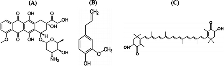
Hormonal receptor positive (HR+) breast cancer is the most commonly diagnosed molecular subtype of breast cancer; which showed good response to doxorubicin (DOX)-based chemotherapy. Eugenol (EUG) and astaxanthin (AST) are natural compounds with proved epigenetic and immunomodulatory effects in several cancer cell lines. This study has been initiated to investigate the molecular mechanism (s) whereby EUG and AST could enhance DOX cytotoxicity in MCF7 cells.
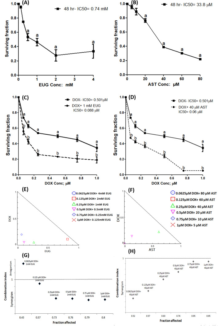
Cytotoxic activity of DOX alone and combined with either 1 mM EUG or 40 μM AST was performed using sulphorhodamine-B assay in MCF7 cells. Global histones acetylation and some immunological markers were investigated using ELISA, western blotting and quantitative RT-PCR techniques. Functional assay of multidrug resistance was performed using rhodamine 123 and Hoechst 3342 dyes. Flow cytometry with annexin V and propidium iodide were used to assess the change in cell cycle and apoptosis along with the expression of some differentiation, apoptosis and autophagy proteins.
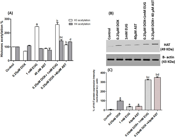
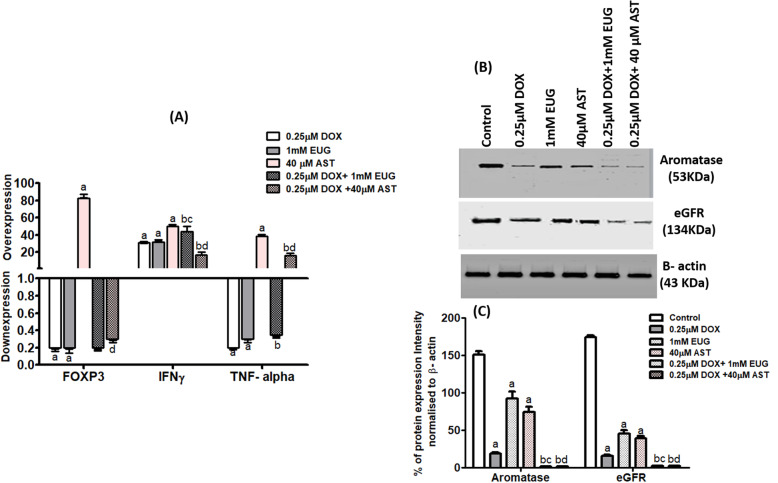
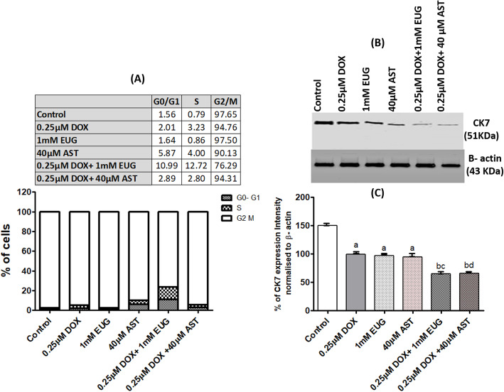
DOX alone resulted in concentration-dependent cytotoxicity with IC of 0.5 μM. Both EUG and AST significantly increased DOX cytotoxicity which is manifested as a significant decrease in DOX IC from 0.5 μM to 0.088 μM with EUG and to 0.06 μM with AST. Combinations of DOX with 1 mM EUG or 40 μM AST significantly increased the level of histones acetylation and histone acetyl transferase expression, while reduced the expression of aromatase and epidermal growth factor receptor (EGFR) when compared with 0.25 μM DOX alone. Also both combinations showed higher uptake of rhodamine but lower of Hoechst stains, along with increased the percentage of caspase 3, and decreased the expression of CK7 and LC3BI/II ratio. EUG combination induced IFγ but reduced TNFα causing shifting of cells from G2/M to S and G0/ G1 phases. Combination of DOX with EUG induced apoptosis through the higher BAX/ BCl2 ratio, while with AST was through the increase in caspase 8 expressions.
EUG and AST potentiated the anticancer activity of DOX through epigenetic histones acetylation along with the immunonomodulation of different apoptotic approaches in MCF7 cells.
激素受体阳性(HR+)乳腺癌是最常见的乳腺癌分子亚型;它对多柔比星(DOX)为基础的化疗有很好的反应。丁香酚(EUG)和虾青素(AST)是两种天然化合物,在几种癌细胞系中具有已证实的表观遗传和免疫调节作用。本研究旨在探讨 EUG 和 AST 增强 MCF7 细胞中 DOX 细胞毒性的分子机制(s)。
采用磺酰罗丹明 B 法在 MCF7 细胞中检测 DOX 单独及与 1mM EUG 或 40μM AST 联合的细胞毒性。采用 ELISA、Western blot 和定量 RT-PCR 技术检测整体组蛋白乙酰化和一些免疫标志物。采用罗丹明 123 和 Hoechst 3342 染料进行多药耐药功能测定。采用 Annexin V 和碘化丙啶进行流式细胞术,评估细胞周期和凋亡的变化以及一些分化、凋亡和自噬蛋白的表达。
DOX 单独作用时呈浓度依赖性细胞毒性,IC 为 0.5μM。EUG 和 AST 均显著增加 DOX 的细胞毒性,表现为 EUG 时 DOX 的 IC 从 0.5μM 降至 0.088μM,AST 时降至 0.06μM。DOX 与 1mM EUG 或 40μM AST 联合应用可显著增加组蛋白乙酰化水平和组蛋白乙酰转移酶表达,同时降低芳香酶和表皮生长因子受体(EGFR)的表达,与单独使用 0.25μM DOX 相比。两种组合均表现出更高的罗丹明摄取率,但 Hoechst 染色率较低,同时 caspase 3 百分比增加,CK7 表达减少,LC3BI/II 比值降低。EUG 联合诱导 IFγ,但减少 TNFα,导致细胞从 G2/M 期转移到 S 和 G0/G1 期。DOX 与 EUG 联合诱导细胞凋亡通过更高的 BAX/BCL2 比值,而与 AST 联合通过增加 caspase 8 的表达。
EUG 和 AST 通过表观遗传组蛋白乙酰化增强 DOX 的抗癌活性,同时通过 MCF7 细胞中不同的凋亡途径的免疫调节作用增强 DOX 的抗癌活性。