Ahmed M K, Zayed M A, El-Dek S I, Hady Mayssa Abdel, El Sherbiny Doaa H, Uskoković Vuk
Faculty of Nanotechnology for Postgraduate studies, Cairo University, El‑Sheikh Zayed 12588, Egypt.
Department of Physics, Faculty of Science, Suez University, Suez 43518, Egypt.
Bioact Mater. 2021 Jan 9;6(7):2070-2088. doi: 10.1016/j.bioactmat.2020.12.026. eCollection 2021 Jul.
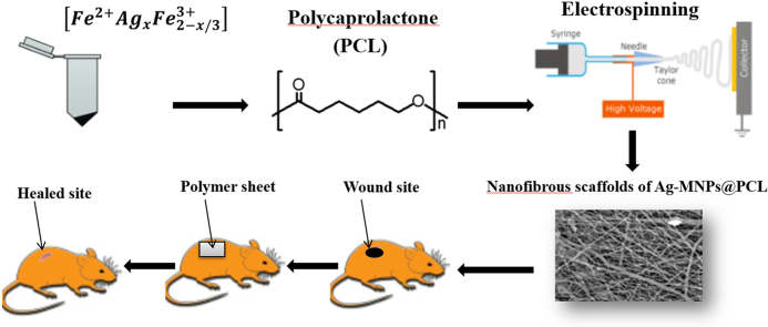
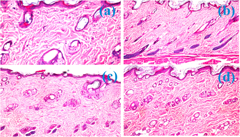
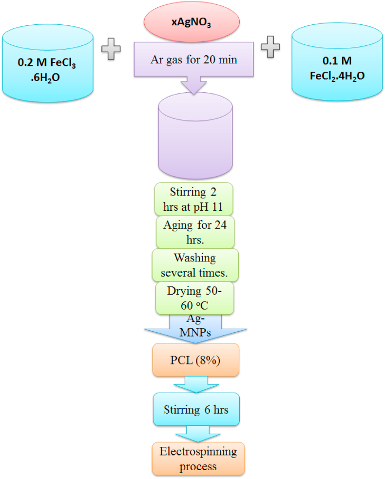
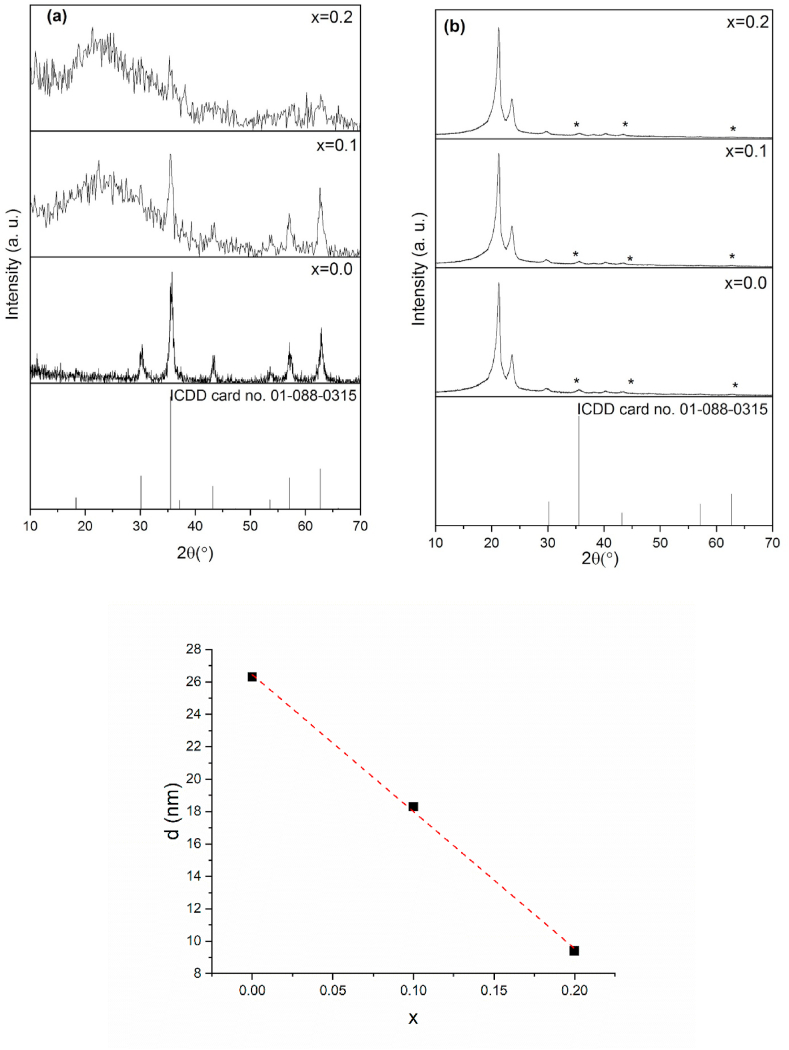
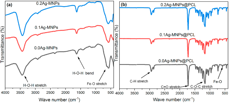
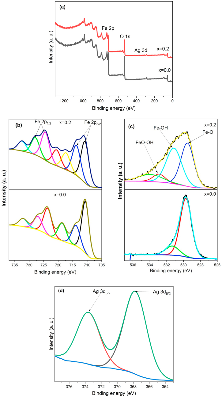
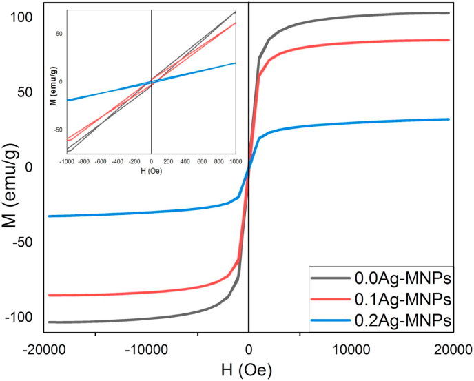
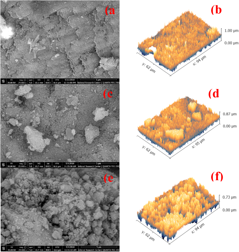
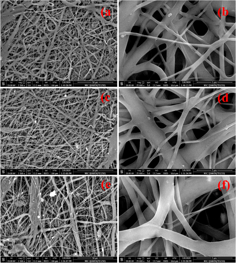
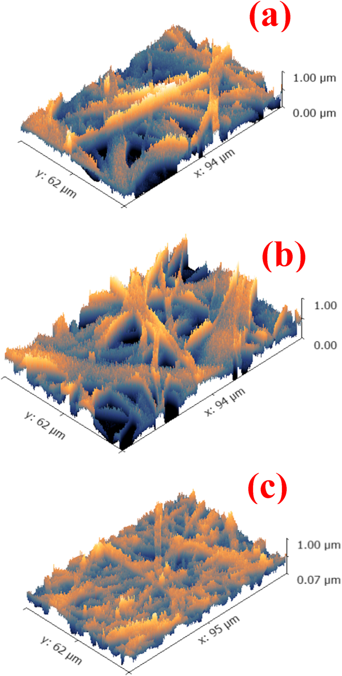
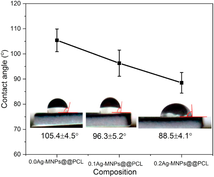
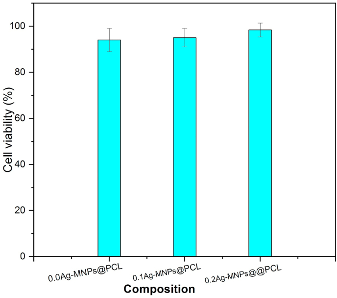
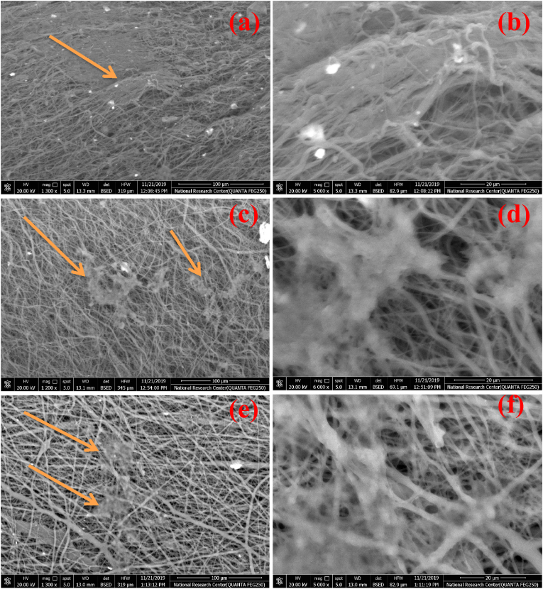
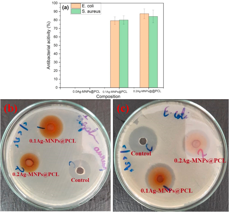
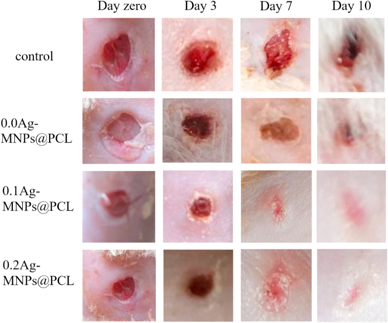
Skin wounds can lead to numerous complications with dangerous health consequences. In this work, magnetite nanoparticles were doped with different concentrations of antimicrobial silver (Ag) ions and incorporated into the electrospun nanofibrous ε-polycaprolactone (PCL) scaffolds. Nanoparticles and scaffolds with various Ag contents were characterized using a range of physicochemical techniques. Ag entered magnetite as cations and preferentially positioned at tetrahedral sites, introducing lattice distortions and topographic irregularities. Amorphization of the structure due to accommodation of Ag expanded the lattice in the bulk and contracted it on the surface, where broadened distribution of Fe-O coordinations was detected. Promoting spin canting and diminishing the double exchange interaction through altered distribution of ferric and ferrous ions, Ag softened the magnetism of magnetite. By making the nanoparticle structure more defective, Ag modified the interface with the polymer and promoted the protrusion of the nanoparticles from the surface of the polymeric nanofibers, thus increasing their roughness and hydrophilicity, with positive repercussions on cell adhesion and growth. Both the viability of human melanocytes and the antibacterial activity against and increased with the concentration of Ag in the magnetite phase of the scaffolds. Skin wound healing rate in rats also increased in direct proportion with the concentration of Ag in the magnetite phase, and no abnormalities in the dermal and epidermal tissues were visible on day 10 in the treatment group. These results imply an excellent potential of these composite nanofibrous scaffolds for use as wound dressings and in other reconstructive skin therapies.
皮肤伤口会引发多种并发症,对健康造成危险后果。在这项工作中,将磁铁矿纳米颗粒掺杂不同浓度的抗菌银(Ag)离子,并将其掺入电纺纳米纤维ε-聚己内酯(PCL)支架中。使用一系列物理化学技术对具有不同银含量的纳米颗粒和支架进行了表征。银以阳离子形式进入磁铁矿,并优先占据四面体位置,引入晶格畸变和形貌不规则性。由于银的掺入导致结构非晶化,使整体晶格膨胀而表面晶格收缩,在表面检测到铁-氧配位分布变宽。通过改变三价铁离子和二价铁离子的分布促进自旋倾斜并减少双交换相互作用,银使磁铁矿的磁性减弱。通过使纳米颗粒结构更具缺陷,银修饰了与聚合物的界面,并促进纳米颗粒从聚合物纳米纤维表面突出,从而增加其粗糙度和亲水性,对细胞粘附和生长产生积极影响。支架磁铁矿相中银的浓度增加,人黑素细胞的活力以及对金黄色葡萄球菌和大肠杆菌的抗菌活性均随之提高。大鼠皮肤伤口愈合率也与支架磁铁矿相中银的浓度成正比,在治疗组第10天时,真皮和表皮组织未见异常。这些结果表明这些复合纳米纤维支架在用作伤口敷料和其他皮肤重建治疗方面具有巨大潜力。