• 文献检索
  • 文档翻译
  • 深度研究
  • 学术资讯
  • Suppr Zotero 插件Zotero 插件
  • 邀请有礼
  • 套餐&价格
  • 历史记录
应用&插件
Suppr Zotero 插件Zotero 插件浏览器插件Mac 客户端Windows 客户端微信小程序
定价
高级版会员购买积分包购买API积分包
服务
文献检索文档翻译深度研究API 文档MCP 服务
关于我们
关于 Suppr公司介绍联系我们用户协议隐私条款
关注我们

Suppr 超能文献

核心技术专利:CN118964589B侵权必究
粤ICP备2023148730 号-1Suppr @ 2026

文献检索

告别复杂PubMed语法,用中文像聊天一样搜索,搜遍4000万医学文献。AI智能推荐,让科研检索更轻松。

立即免费搜索

文件翻译

保留排版,准确专业,支持PDF/Word/PPT等文件格式,支持 12+语言互译。

免费翻译文档

深度研究

AI帮你快速写综述,25分钟生成高质量综述,智能提取关键信息,辅助科研写作。

立即免费体验

COVID-19 肺炎的超声、X 线和 CT 表现:全面的影像学图片综述。

COVID-19 pneumonia-ultrasound, radiographic, and computed tomography findings: a comprehensive pictorial essay.

机构信息

Department of Radiology, ASST Fatebenefratelli Sacco, P.zza Principessa Clotilde, 3, 20121, Milan, Italy.

Department of Radiology, San Raffaele Hospital, via Olgettina 60, 20123, Milan, Italy.

出版信息

Emerg Radiol. 2021 Jun;28(3):519-526. doi: 10.1007/s10140-021-01905-6. Epub 2021 Jan 30.

DOI:10.1007/s10140-021-01905-6
PMID:33517546
原文链接:https://pmc.ncbi.nlm.nih.gov/articles/PMC7847301/
Abstract

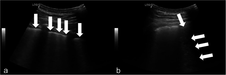
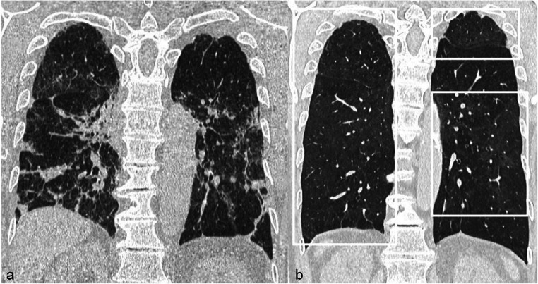
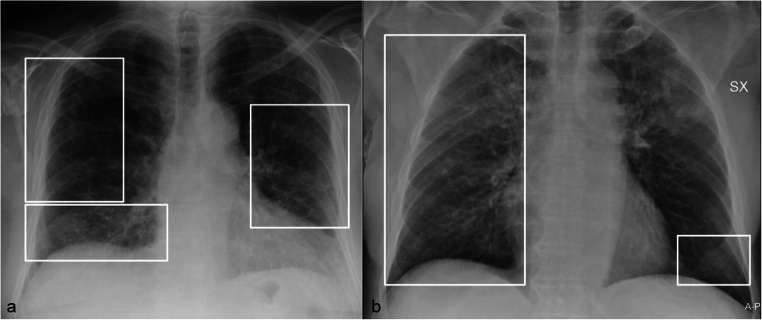
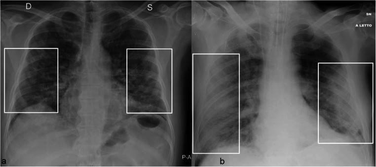
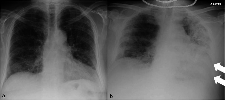
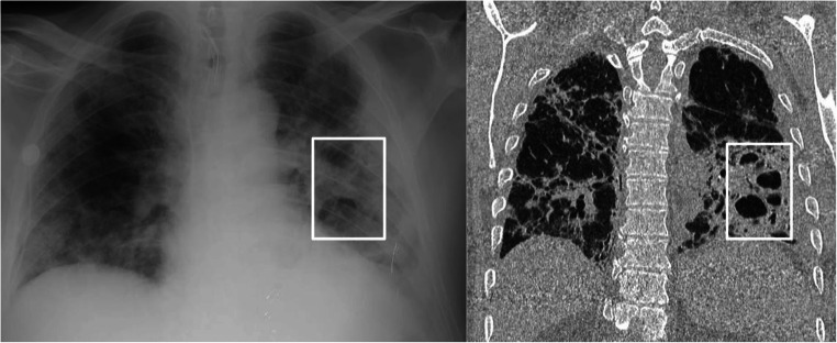
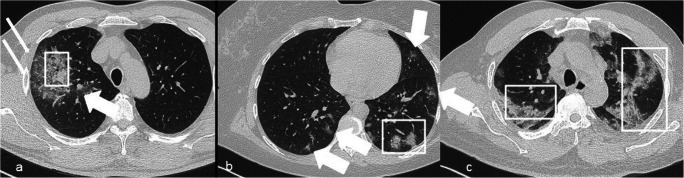
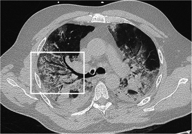
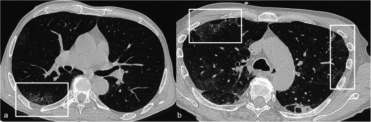
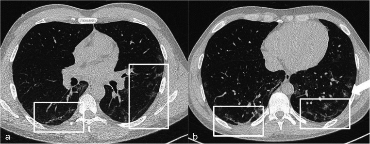
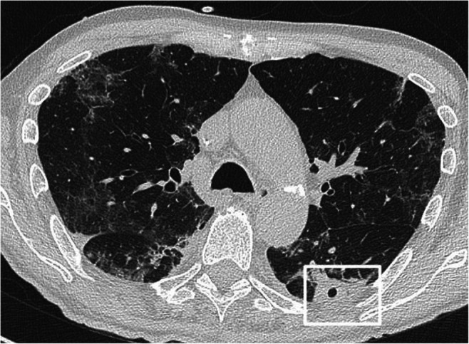
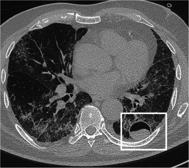
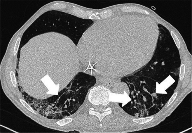
Ultrasound, chest X-ray, and computed tomography (CT) have been used with excellent results in diagnosis, first assessment, and follow-up of COVID-19 confirmed and suspected patients. Ultrasound and chest X-ray have the advantages of the wide availability and acquisition at the patient's bed; CT showed high sensitivity in COVID-19 diagnosis. Ground-glass opacities and consolidation are the main CT and X-ray features; the distribution of lung abnormalities is typically bilateral and peripheral. Less typical findings, including pleural effusion, mediastinal lymphadenopathies, the bubble air sign, and cavitation, can also be visible on chest CT. Radiologists should be aware of the advantages and limitations of the available imaging techniques and of the different pulmonary aspects of COVID-19 infection.

摘要

超声、胸部 X 射线和计算机断层扫描 (CT) 在 COVID-19 确诊和疑似患者的诊断、初步评估和随访中取得了极好的效果。超声和胸部 X 射线具有广泛可用性和可在患者床边采集的优势;CT 显示出在 COVID-19 诊断方面的高灵敏度。磨玻璃影和实变是 CT 和 X 射线的主要特征;肺部异常的分布通常是双侧和外周性的。在胸部 CT 上还可以看到不太典型的表现,包括胸腔积液、纵隔淋巴结病、气泡空气征和空洞。放射科医生应该了解可用成像技术的优缺点,以及 COVID-19 感染的不同肺部表现。

https://cdn.ncbi.nlm.nih.gov/pmc/blobs/6526/7847301/b14e3821b3ec/10140_2021_1905_Fig14_HTML.jpg
https://cdn.ncbi.nlm.nih.gov/pmc/blobs/6526/7847301/dd8da81f884a/10140_2021_1905_Fig1_HTML.jpg
https://cdn.ncbi.nlm.nih.gov/pmc/blobs/6526/7847301/6cc0ed7c1544/10140_2021_1905_Fig2_HTML.jpg
https://cdn.ncbi.nlm.nih.gov/pmc/blobs/6526/7847301/bf26f400016c/10140_2021_1905_Fig3_HTML.jpg
https://cdn.ncbi.nlm.nih.gov/pmc/blobs/6526/7847301/dd8dc1ba6117/10140_2021_1905_Fig4_HTML.jpg
https://cdn.ncbi.nlm.nih.gov/pmc/blobs/6526/7847301/371c7b7b7e58/10140_2021_1905_Fig5_HTML.jpg
https://cdn.ncbi.nlm.nih.gov/pmc/blobs/6526/7847301/f1b10eb9eeb0/10140_2021_1905_Fig6_HTML.jpg
https://cdn.ncbi.nlm.nih.gov/pmc/blobs/6526/7847301/9a5961e88146/10140_2021_1905_Fig7_HTML.jpg
https://cdn.ncbi.nlm.nih.gov/pmc/blobs/6526/7847301/d287892b4766/10140_2021_1905_Fig8_HTML.jpg
https://cdn.ncbi.nlm.nih.gov/pmc/blobs/6526/7847301/2c1ac4f73e0c/10140_2021_1905_Fig9_HTML.jpg
https://cdn.ncbi.nlm.nih.gov/pmc/blobs/6526/7847301/91cd19d692c7/10140_2021_1905_Fig10_HTML.jpg
https://cdn.ncbi.nlm.nih.gov/pmc/blobs/6526/7847301/c71fc1c05b7a/10140_2021_1905_Fig11_HTML.jpg
https://cdn.ncbi.nlm.nih.gov/pmc/blobs/6526/7847301/05bdd25488d0/10140_2021_1905_Fig12_HTML.jpg
https://cdn.ncbi.nlm.nih.gov/pmc/blobs/6526/7847301/7f7dc49ca161/10140_2021_1905_Fig13_HTML.jpg
https://cdn.ncbi.nlm.nih.gov/pmc/blobs/6526/7847301/b14e3821b3ec/10140_2021_1905_Fig14_HTML.jpg

相似文献

1
COVID-19 pneumonia-ultrasound, radiographic, and computed tomography findings: a comprehensive pictorial essay.COVID-19 肺炎的超声、X 线和 CT 表现:全面的影像学图片综述。
Emerg Radiol. 2021 Jun;28(3):519-526. doi: 10.1007/s10140-021-01905-6. Epub 2021 Jan 30.
2
Chest computed tomography findings of COVID-19 pneumonia: pictorial essay with literature review.新型冠状病毒肺炎的胸部计算机断层扫描表现:文献综述配图文章
Jpn J Radiol. 2020 Nov;38(11):1012-1019. doi: 10.1007/s11604-020-01010-7. Epub 2020 Jun 25.
3
Correlation between Chest Computed Tomography and Lung Ultrasonography in Patients with Coronavirus Disease 2019 (COVID-19).新型冠状病毒肺炎(COVID-19)患者的胸部计算机断层扫描与肺部超声的相关性。
Ultrasound Med Biol. 2020 Nov;46(11):2918-2926. doi: 10.1016/j.ultrasmedbio.2020.07.003. Epub 2020 Jul 13.
4
Thoracic imaging tests for the diagnosis of COVID-19.用于诊断新型冠状病毒肺炎的胸部影像学检查
Cochrane Database Syst Rev. 2020 Sep 30;9:CD013639. doi: 10.1002/14651858.CD013639.pub2.
5
Imaging and clinical features of patients with 2019 novel coronavirus SARS-CoV-2.新型冠状病毒 2019 年 SARS-CoV-2 患者的影像学和临床特征。
Eur J Nucl Med Mol Imaging. 2020 May;47(5):1275-1280. doi: 10.1007/s00259-020-04735-9. Epub 2020 Feb 28.
6
Radiology of COVID-19 - Imaging the pulmonary damage.新型冠状病毒肺炎的放射学——肺部损伤成像
J Pak Med Assoc. 2020 May;70(Suppl 3)(5):S60-S63. doi: 10.5455/JPMA.21.
7
COVID-19 and its Mimics: What the Radiologist Needs to Know.COVID-19 及其类似疾病:放射科医生需要了解的知识。
J Thorac Imaging. 2021 Jan;36(1):W1-W10. doi: 10.1097/RTI.0000000000000554.
8
Frequency and Distribution of Chest Radiographic Findings in Patients Positive for COVID-19.COVID-19 阳性患者的胸部 X 线表现的频率和分布。
Radiology. 2020 Aug;296(2):E72-E78. doi: 10.1148/radiol.2020201160. Epub 2020 Mar 27.
9
Similarities and Differences of Early Pulmonary CT Features of Pneumonia Caused by SARS-CoV-2, SARS-CoV and MERS-CoV: Comparison Based on a Systemic Review.新型冠状病毒、严重急性呼吸综合征冠状病毒和中东呼吸综合征冠状病毒所致肺炎早期肺部CT特征的异同:基于系统评价的比较
Chin Med Sci J. 2020 Sep 30;35(3):254-261. doi: 10.24920/003727.
10
Chest computed tomography findings of COVID-19 in children younger than 1 year: a systematic review.儿童 COVID-19 的胸部计算机断层扫描表现:系统综述。
World J Pediatr. 2021 Jun;17(3):234-241. doi: 10.1007/s12519-021-00424-1. Epub 2021 May 7.

引用本文的文献

1
The diagnostic performance of automatic B-lines detection for evaluating pulmonary edema in the emergency department among novice point-of-care ultrasound practitioners.急诊科新手床旁超声从业者中自动B线检测在评估肺水肿方面的诊断性能。
Emerg Radiol. 2025 Apr;32(2):241-246. doi: 10.1007/s10140-025-02319-4. Epub 2025 Feb 14.
2
COVID-19 and Lung Cavitation: A Clue to Pathogenesis?新冠病毒与肺空洞:发病机制的线索?
HCA Healthc J Med. 2022 Aug 29;3(4):253-256. doi: 10.36518/2689-0216.1361. eCollection 2022.
3
Chest x-ray imaging score is associated with severity of COVID-19 pneumonia: the MBrixia score.

本文引用的文献

1
Qualitative and quantitative chest CT parameters as predictors of specific mortality in COVID-19 patients.定性和定量胸部CT参数作为COVID-19患者特定死亡率的预测指标
Emerg Radiol. 2020 Dec;27(6):701-710. doi: 10.1007/s10140-020-01867-1. Epub 2020 Oct 29.
2
COVID-19 pneumonia in the emergency department: correlation of initial chest CT findings with short-term outcome.急诊科的新型冠状病毒肺炎:初始胸部CT表现与短期预后的相关性
Emerg Radiol. 2020 Dec;27(6):691-699. doi: 10.1007/s10140-020-01863-5. Epub 2020 Oct 15.
3
Lung Ultrasound May Support Diagnosis and Monitoring of COVID-19 Pneumonia.
胸部 X 射线成像评分与 COVID-19 肺炎的严重程度相关:MBrixia 评分。
Sci Rep. 2022 Dec 5;12(1):21019. doi: 10.1038/s41598-022-25397-7.
4
Pixel-based analysis of pulmonary changes on CT lung images due to COVID-19 pneumonia.基于像素的新冠病毒肺炎CT肺部图像肺部变化分析
J Clin Imaging Sci. 2022 Feb 19;12:6. doi: 10.25259/JCIS_172_2021. eCollection 2022.
5
Role of Artificial Intelligence in COVID-19 Detection.人工智能在 COVID-19 检测中的作用。
Sensors (Basel). 2021 Dec 1;21(23):8045. doi: 10.3390/s21238045.
6
Deep CNN models for predicting COVID-19 in CT and x-ray images.用于在CT和X光图像中预测新冠肺炎的深度卷积神经网络模型。
J Med Imaging (Bellingham). 2021 Jan;8(Suppl 1):014502. doi: 10.1117/1.JMI.8.S1.014502. Epub 2021 Apr 21.
肺部超声检查可能有助于 COVID-19 肺炎的诊断和监测。
Ultrasound Med Biol. 2020 Nov;46(11):2908-2917. doi: 10.1016/j.ultrasmedbio.2020.07.018. Epub 2020 Jul 20.
4
High-resolution computed tomography finding in 552 patients with symptomatic COVID-19: first report from north of Iran.552例有症状的新型冠状病毒肺炎患者的高分辨率计算机断层扫描结果:来自伊朗北部的首次报告
Emerg Radiol. 2020 Dec;27(6):633-639. doi: 10.1007/s10140-020-01819-9. Epub 2020 Jul 13.
5
Radiographic and chest CT imaging presentation and follow-up of COVID-19 pneumonia: a multicenter experience from an endemic area.新型冠状病毒肺炎的影像学及胸部CT表现与随访:来自疫区的多中心经验
Emerg Radiol. 2020 Dec;27(6):623-632. doi: 10.1007/s10140-020-01817-x. Epub 2020 Jul 11.
6
Lung cavitation due to COVID-19 pneumonia.新型冠状病毒肺炎所致的肺空洞形成。
BMJ Case Rep. 2020 Jul 6;13(7):e237245. doi: 10.1136/bcr-2020-237245.
7
Feasibility, Reproducibility, and Clinical Validity of a Quantitative Chest X-Ray Assessment for COVID-19.COVID-19 的定量胸部 X 射线评估的可行性、可重复性和临床有效性。
Am J Trop Med Hyg. 2020 Aug;103(2):822-827. doi: 10.4269/ajtmh.20-0535. Epub 2020 Jul 2.
8
The role of initial chest X-ray in triaging patients with suspected COVID-19 during the pandemic.在疫情期间,初始胸部X光在对疑似COVID-19患者进行分诊中的作用。
Emerg Radiol. 2020 Dec;27(6):617-621. doi: 10.1007/s10140-020-01808-y. Epub 2020 Jun 22.
9
Pneumomediastinum and spontaneous pneumothorax as an extrapulmonary complication of COVID-19 disease.纵隔气肿和自发性气胸作为新型冠状病毒肺炎的一种肺外并发症。
Emerg Radiol. 2020 Dec;27(6):727-730. doi: 10.1007/s10140-020-01806-0. Epub 2020 Jun 11.
10
Chest Radiography Features Help to Predict a Favorable Outcome in Patients with Coronavirus Disease 2019.胸部X线特征有助于预测2019冠状病毒病患者的良好预后。
Radiology. 2020 Oct;297(1):E238. doi: 10.1148/radiol.2020202326. Epub 2020 Jun 2.