Schreiter Jeannine Susanne, Beescho Christian, Kang Jagdip, Kursawe Laura, Moter Annette, Kikhney Judith, Langer Stefan, Osla Fredrik, Wellner Eric, Kurow Olga
Department of Orthopedics, Trauma and Plastic Surgery, University Hospital Leipzig, Germany.
Leipzig Heart Centre, Leipzig, Germany.
GMS Interdiscip Plast Reconstr Surg DGPW. 2020 Dec 23;9:Doc06. doi: 10.3205/iprs000150. eCollection 2020.
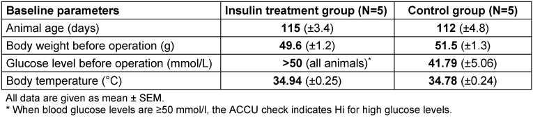
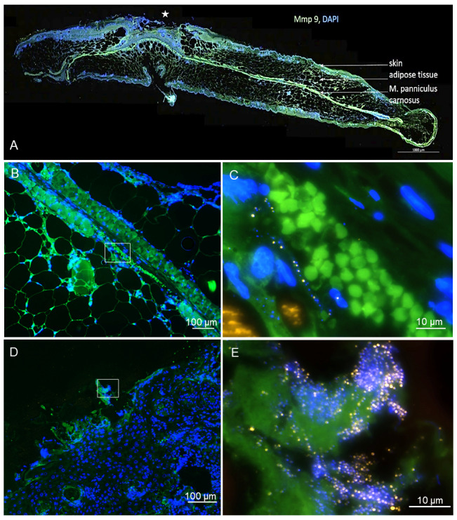
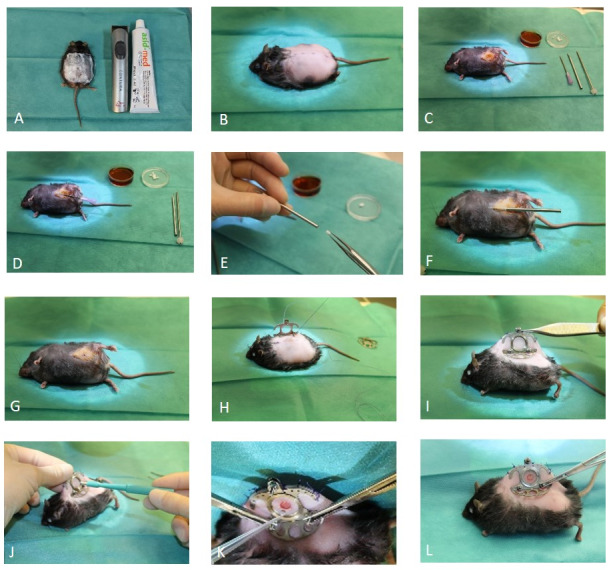
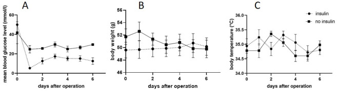
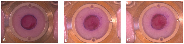
Diabetic patients suffer more frequently from biofilm-associated infections than normoglycemic patients. Well described in the literature is a relationship between elevated blood glucose levels in patients and the occurrence of biofilm-associated wound infections. Nevertheless, the underlying pathophysiological pathways leading to this increased infection vulnerability and its effects on biofilm development still need to be elucidated. We developed in our laboratory a model to allow the investigation of a biofilm-associated wound infection in diabetic mice under controlled insulin treatment. A dorsal skinfold chamber was used on 16 weeks old BKS.Cg-Dock7 +/+ Lepr/J mice and a wound within the observation field of the dorsal skinfold chamber was created. These wounds were infected with ATCC 49230 (10 cells/mL). Simultaneously, we implanted implants for sustained insulin release into the ventral subcutaneous tissue (N=5 mice). Mice of the control group (N=5) were treated with sham implants. Serum glucose levels were registered before intervention and daily after the operation. Densitometrical analysis of the wound size was performed at day 0, 3, and 6 after intervention. Mice were sacrificed on day 6 and wound tissue was submitted to fluorescence hybridization (FISH) and colony forming unit (CFU) analysis in addition to immunohistochemical staining to observe wound healing. Experiments were carried out in accordance with the National Institute of Health Guidelines for the Care and Use of Laboratory Animals (protocol number 05/19). The insulin implants were able to reduce blood glucose levels in the mice. Hence, the diabetic mice in the intervention group were normoglycemic after the implantation. The combination with the dorsal skinfold chamber allowed for continuous, in vivo measurements of the infection development. Implantation of the insulin implant and the dorsal skinfold chamber was a tolerable condition for the diabetic mice. We succeeded to realize reproducible biofilm infections in the animals. We developed a novel model to assess interactions between blood glucose level and -induced biofilm-associated wound infections. The combination of the dorsal skinfold chamber model with a sustained insulin treatment has not been described so far. It allows a broad field of glucose and insulin dependent studies of infection.
糖尿病患者比血糖正常的患者更频繁地遭受生物膜相关感染。患者血糖水平升高与生物膜相关伤口感染的发生之间的关系在文献中有充分描述。然而,导致这种感染易感性增加的潜在病理生理途径及其对生物膜形成的影响仍有待阐明。我们在实验室中开发了一个模型,用于在可控胰岛素治疗下研究糖尿病小鼠的生物膜相关伤口感染。对16周龄的BKS.Cg-Dock7 +/+ Lepr/J小鼠使用背部皮褶小室,并在背部皮褶小室的观察视野内制造一个伤口。这些伤口用ATCC 49230(10个细胞/毫升)感染。同时,我们将用于持续胰岛素释放的植入物植入腹部皮下组织(N = 5只小鼠)。对照组的小鼠(N = 5)接受假植入物治疗。在干预前和术后每天记录血清葡萄糖水平。在干预后第0、3和6天对伤口大小进行光密度分析。在第6天处死小鼠,除了进行免疫组织化学染色以观察伤口愈合外,还对伤口组织进行荧光杂交(FISH)和菌落形成单位(CFU)分析。实验按照美国国立卫生研究院实验室动物护理和使用指南(方案编号05/19)进行。胰岛素植入物能够降低小鼠的血糖水平。因此,干预组的糖尿病小鼠在植入后血糖正常。与背部皮褶小室相结合,可以对感染发展进行连续的体内测量。胰岛素植入物和背部皮褶小室的植入对糖尿病小鼠来说是可以耐受的。我们成功地在动物中实现了可重复的生物膜感染。我们开发了一种新模型来评估血糖水平与诱导的生物膜相关伤口感染之间的相互作用。到目前为止,尚未描述背部皮褶小室模型与持续胰岛素治疗的结合。它允许进行广泛的葡萄糖和胰岛素依赖性感染研究。