• 文献检索
  • 文档翻译
  • 深度研究
  • 学术资讯
  • Suppr Zotero 插件Zotero 插件
  • 邀请有礼
  • 套餐&价格
  • 历史记录
应用&插件
Suppr Zotero 插件Zotero 插件浏览器插件Mac 客户端Windows 客户端微信小程序
定价
高级版会员购买积分包购买API积分包
服务
文献检索文档翻译深度研究API 文档MCP 服务
关于我们
关于 Suppr公司介绍联系我们用户协议隐私条款
关注我们

Suppr 超能文献

核心技术专利:CN118964589B侵权必究
粤ICP备2023148730 号-1Suppr @ 2026

文献检索

告别复杂PubMed语法,用中文像聊天一样搜索,搜遍4000万医学文献。AI智能推荐,让科研检索更轻松。

立即免费搜索

文件翻译

保留排版,准确专业,支持PDF/Word/PPT等文件格式,支持 12+语言互译。

免费翻译文档

深度研究

AI帮你快速写综述,25分钟生成高质量综述,智能提取关键信息,辅助科研写作。

立即免费体验

心脏细胞类型对损伤的特异性反应及对心脏再生的贡献。

Cardiac cell type-specific responses to injury and contributions to heart regeneration.

作者信息

Zhang Weijia, Liang Jinxiu, Han Peidong

机构信息

Division of Medical Genetics and Genomics, Children's Hospital, Zhejiang University School of Medicine and National Clinical Research Center for Child Health, Hangzhou, China.

出版信息

Cell Regen. 2021 Feb 2;10(1):4. doi: 10.1186/s13619-020-00065-1.

DOI:10.1186/s13619-020-00065-1
PMID:33527149
原文链接:https://pmc.ncbi.nlm.nih.gov/articles/PMC7851195/
Abstract

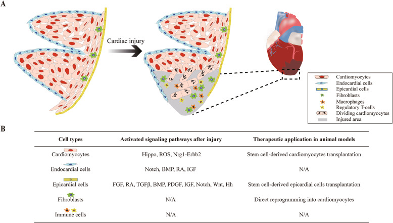
Heart disease is the leading cause of mortality worldwide. Due to the limited proliferation rate of mature cardiomyocytes, adult mammalian hearts are unable to regenerate damaged cardiac muscle following injury. Instead, injured area is replaced by fibrotic scar tissue, which may lead to irreversible cardiac remodeling and organ failure. In contrast, adult zebrafish and neonatal mammalian possess the capacity for heart regeneration and have been widely used as experimental models. Recent studies have shown that multiple types of cells within the heart can respond to injury with the activation of distinct signaling pathways. Determining the specific contributions of each cell type is essential for our understanding of the regeneration network organization throughout the heart. In this review, we provide an overview of the distinct functions and coordinated cell behaviors of several major cell types including cardiomyocytes, endocardial cells, epicardial cells, fibroblasts, and immune cells. The topic focuses on their specific responses and cellular plasticity after injury, and potential therapeutic applications.

摘要

心脏病是全球范围内导致死亡的主要原因。由于成熟心肌细胞的增殖速率有限,成年哺乳动物心脏在受伤后无法再生受损的心肌。相反,受伤区域会被纤维化瘢痕组织取代,这可能导致不可逆的心脏重塑和器官衰竭。相比之下,成年斑马鱼和新生哺乳动物具有心脏再生能力,并已被广泛用作实验模型。最近的研究表明,心脏内的多种细胞类型可通过激活不同的信号通路对损伤做出反应。确定每种细胞类型的具体作用对于我们理解整个心脏的再生网络组织至关重要。在本综述中,我们概述了几种主要细胞类型(包括心肌细胞、心内膜细胞、心外膜细胞、成纤维细胞和免疫细胞)的不同功能和协调的细胞行为。主题聚焦于它们在受伤后的特定反应和细胞可塑性,以及潜在的治疗应用。

https://cdn.ncbi.nlm.nih.gov/pmc/blobs/89a5/7851195/8c0844c1ce03/13619_2020_65_Fig1_HTML.jpg

相似文献

1
Cardiac cell type-specific responses to injury and contributions to heart regeneration.心脏细胞类型对损伤的特异性反应及对心脏再生的贡献。
Cell Regen. 2021 Feb 2;10(1):4. doi: 10.1186/s13619-020-00065-1.
2
Signaling Enhances Epicardial Cell Expansion during Neonatal Mouse Heart Repair.信号传导增强新生小鼠心脏修复过程中心外膜细胞的扩张。
J Cardiovasc Dis Diagn. 2013 Mar;1(1). doi: 10.4172/2329-9517.1000101.
3
Is zebrafish heart regeneration "complete"? Lineage-restricted cardiomyocytes proliferate to pre-injury numbers but some fail to differentiate in fibrotic hearts.斑马鱼的心脏再生是“完全的”吗?谱系限制的心肌细胞增殖到损伤前的数量,但在纤维化的心脏中有些未能分化。
Dev Biol. 2021 Mar;471:106-118. doi: 10.1016/j.ydbio.2020.12.004. Epub 2020 Dec 10.
4
Zebrafish as a Smart Model to Understand Regeneration After Heart Injury: How Fish Could Help Humans.斑马鱼:理解心脏损伤后再生的智能模型——鱼类如何助力人类
Front Cardiovasc Med. 2019 Aug 6;6:107. doi: 10.3389/fcvm.2019.00107. eCollection 2019.
5
Primary contribution to zebrafish heart regeneration by gata4(+) cardiomyocytes.Gata4(+) 心肌细胞对斑马鱼心脏再生的主要贡献。
Nature. 2010 Mar 25;464(7288):601-5. doi: 10.1038/nature08804.
6
The regenerative capacity of the zebrafish heart is dependent on TGFβ signaling.斑马鱼心脏的再生能力依赖于 TGFβ 信号通路。
Development. 2012 Jun;139(11):1921-30. doi: 10.1242/dev.078543. Epub 2012 Apr 18.
7
Wnt/β-catenin signaling in heart regeneration.心脏再生中的Wnt/β-连环蛋白信号通路。
Cell Regen. 2015 Jul 8;4(1):3. doi: 10.1186/s13619-015-0017-8. eCollection 2015.
8
Metabolic Regulation of Cardiac Regeneration.心脏再生的代谢调控
Front Cardiovasc Med. 2022 Jul 8;9:933060. doi: 10.3389/fcvm.2022.933060. eCollection 2022.
9
Extensive scar formation and regression during heart regeneration after cryoinjury in zebrafish.斑马鱼心脏冷冻损伤后再生过程中的广泛瘢痕形成和消退。
Development. 2011 May;138(9):1663-74. doi: 10.1242/dev.060897. Epub 2011 Mar 23.
10
Telomerase Is Essential for Zebrafish Heart Regeneration.端粒酶对斑马鱼心脏再生至关重要。
Cell Rep. 2015 Sep 8;12(10):1691-703. doi: 10.1016/j.celrep.2015.07.064. Epub 2015 Aug 28.

引用本文的文献

1
Contraction-induced endocardial plays a dual role in regulating myocardial contractility and valve formation.收缩诱导的心内膜在调节心肌收缩力和瓣膜形成中起双重作用。
Elife. 2025 Jul 24;13:RP101151. doi: 10.7554/eLife.101151.
2
Increased Left Ventricular Myocardial Extracellular Volume Assessed by Cardiac Computed Tomography as a Consequence of Aortic Stenosis and Coexisting Cardiovascular Risk Factors.心脏计算机断层扫描评估发现,主动脉瓣狭窄及并存心血管危险因素导致左心室心肌细胞外容积增加。
J Clin Med. 2025 Jun 22;14(13):4435. doi: 10.3390/jcm14134435.
3
Deciphering myocardial fibrosis: a comprehensive bibliometric analysis of mechanism over the period 1992-2023.

本文引用的文献

1
An emerging role of regulatory T-cells in cardiovascular repair and regeneration.调节性 T 细胞在心血管修复和再生中的新作用。
Theranostics. 2020 Jul 11;10(20):8924-8938. doi: 10.7150/thno.47118. eCollection 2020.
2
Specific ablation of CD4 T-cells promotes heart regeneration in juvenile mice.特异性消融 CD4 T 细胞促进幼年小鼠的心脏再生。
Theranostics. 2020 Jun 29;10(18):8018-8035. doi: 10.7150/thno.42943. eCollection 2020.
3
Natural Heart Regeneration in a Neonatal Rat Myocardial Infarction Model.新生大鼠心肌梗死模型中的自然心脏再生。
解读心肌纤维化:1992 - 2023年期间机制的全面文献计量分析
J Cardiothorac Surg. 2025 Mar 29;20(1):170. doi: 10.1186/s13019-025-03404-y.
4
Vitamin C Regulates the Profibrotic Activity of Fibroblasts in In Vitro Replica Settings of Myocardial Infarction.维生素 C 调节心肌梗死体外复制模型中成纤维细胞的促纤维化活性。
Int J Mol Sci. 2023 May 6;24(9):8379. doi: 10.3390/ijms24098379.
5
Characterization of diverse populations of sinoatrial node cells and their proliferation potential at single nucleus resolution.在单核分辨率下对窦房结细胞的不同群体及其增殖潜力进行表征。
Heliyon. 2022 Dec 29;9(1):e12708. doi: 10.1016/j.heliyon.2022.e12708. eCollection 2023 Jan.
6
A DUSP6 inhibitor suppresses inflammatory cardiac remodeling and improves heart function after myocardial infarction.一种 DUSP6 抑制剂可抑制心肌梗死后的炎症性心脏重构,改善心脏功能。
Dis Model Mech. 2023 May 1;16(5). doi: 10.1242/dmm.049662. Epub 2022 Dec 7.
7
Heart failure with preserved ejection fraction (HFpEF) in type 2 diabetes mellitus: from pathophysiology to therapeutics.2型糖尿病中射血分数保留的心力衰竭(HFpEF):从病理生理学到治疗方法
J Mol Cell Biol. 2022 Sep 12;14(5). doi: 10.1093/jmcb/mjac028.
8
Endothelial-Mesenchymal Transition or Functional Tissue Regeneration - Two Outcomes of Heart Remodeling.内皮-间充质转化或功能性组织再生——心脏重构的两种结局。
Physiol Res. 2021 Nov 30;70(Suppl 1):S13-S20. doi: 10.33549/physiolres.934780.
9
Advances in Cardiac Development and Regeneration Using Zebrafish as a Model System for High-Throughput Research.以斑马鱼为高通量研究模型系统在心脏发育与再生方面的进展
J Dev Biol. 2021 Sep 25;9(4):40. doi: 10.3390/jdb9040040.
10
Control of cardiomyocyte differentiation timing by intercellular signaling pathways.细胞间信号通路对心肌细胞分化时机的控制。
Semin Cell Dev Biol. 2021 Oct;118:94-106. doi: 10.1016/j.semcdb.2021.06.002. Epub 2021 Jun 16.
Cells. 2020 Jan 16;9(1):229. doi: 10.3390/cells9010229.
4
Single-cell analysis uncovers that metabolic reprogramming by ErbB2 signaling is essential for cardiomyocyte proliferation in the regenerating heart.单细胞分析揭示了 ErbB2 信号转导的代谢重编程对于心脏再生过程中心肌细胞的增殖是必不可少的。
Elife. 2019 Dec 23;8:e50163. doi: 10.7554/eLife.50163.
5
Macrophages in cardiac repair: Environmental cues and therapeutic strategies.心肌修复中的巨噬细胞:环境线索和治疗策略。
Exp Mol Med. 2019 Dec 19;51(12):1-10. doi: 10.1038/s12276-019-0269-4.
6
An acute immune response underlies the benefit of cardiac stem cell therapy.急性免疫反应是心脏干细胞治疗获益的基础。
Nature. 2020 Jan;577(7790):405-409. doi: 10.1038/s41586-019-1802-2. Epub 2019 Nov 27.
7
Cardiac Fibroblasts and the Extracellular Matrix in Regenerative and Nonregenerative Hearts.再生与非再生心脏中的心肌成纤维细胞与细胞外基质
J Cardiovasc Dev Dis. 2019 Aug 20;6(3):29. doi: 10.3390/jcdd6030029.
8
Epicardial cells derived from human embryonic stem cells augment cardiomyocyte-driven heart regeneration.人心肌细胞衍生的人心外膜细胞增强心肌细胞驱动的心脏再生。
Nat Biotechnol. 2019 Aug;37(8):895-906. doi: 10.1038/s41587-019-0197-9. Epub 2019 Aug 2.
9
Regulatory T-cells regulate neonatal heart regeneration by potentiating cardiomyocyte proliferation in a paracrine manner.调节性 T 细胞通过旁分泌方式增强心肌细胞增殖来调节新生儿心脏再生。
Theranostics. 2019 Jun 9;9(15):4324-4341. doi: 10.7150/thno.32734. eCollection 2019.
10
Hemodynamic-mediated endocardial signaling controls in vivo myocardial reprogramming.血流动力学介导的心内膜信号控制体内心肌重编程。
Elife. 2019 Jun 25;8:e44816. doi: 10.7554/eLife.44816.