• 文献检索
  • 文档翻译
  • 深度研究
  • 学术资讯
  • Suppr Zotero 插件Zotero 插件
  • 邀请有礼
  • 套餐&价格
  • 历史记录
应用&插件
Suppr Zotero 插件Zotero 插件浏览器插件Mac 客户端Windows 客户端微信小程序
定价
高级版会员购买积分包购买API积分包
服务
文献检索文档翻译深度研究API 文档MCP 服务
关于我们
关于 Suppr公司介绍联系我们用户协议隐私条款
关注我们

Suppr 超能文献

核心技术专利:CN118964589B侵权必究
粤ICP备2023148730 号-1Suppr @ 2026

文献检索

告别复杂PubMed语法,用中文像聊天一样搜索,搜遍4000万医学文献。AI智能推荐,让科研检索更轻松。

立即免费搜索

文件翻译

保留排版,准确专业,支持PDF/Word/PPT等文件格式,支持 12+语言互译。

免费翻译文档

深度研究

AI帮你快速写综述,25分钟生成高质量综述,智能提取关键信息,辅助科研写作。

立即免费体验

采用基于扩散放射组学的机器学习模型开发和外部验证方法,区分复发性脑胶质瘤和放射性坏死。

Differentiation of recurrent glioblastoma from radiation necrosis using diffusion radiomics with machine learning model development and external validation.

机构信息

Department of Radiology and Research Institute of Radiological Science and Center for Clinical Image Data Science, Yonsei University College of Medicine, 50-1 Yonsei-ro, Seodaemun-gu, Seoul, 120-752, South Korea.

Department of Computer Science, Yonsei University, Seoul, South Korea.

出版信息

Sci Rep. 2021 Feb 3;11(1):2913. doi: 10.1038/s41598-021-82467-y.

DOI:10.1038/s41598-021-82467-y
PMID:33536499
原文链接:https://pmc.ncbi.nlm.nih.gov/articles/PMC7858615/
Abstract

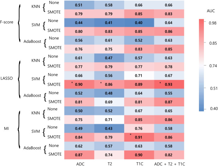
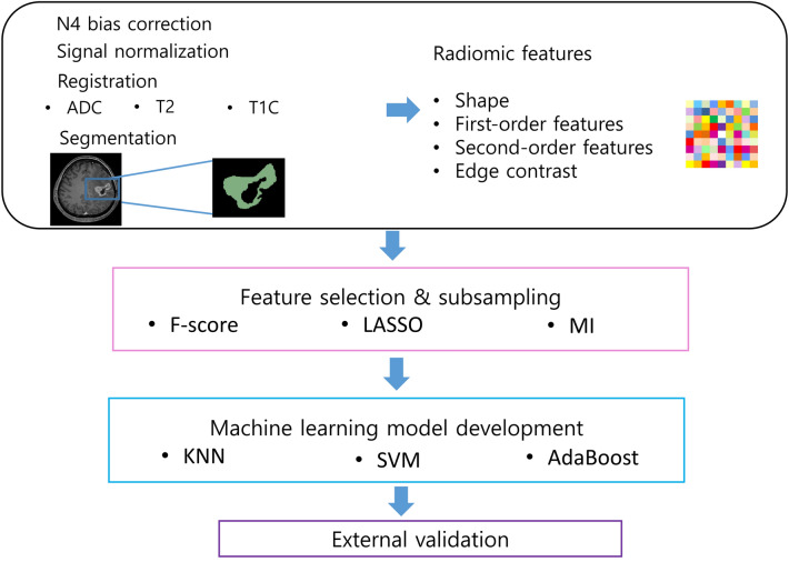
The purpose of this study was to establish a high-performing radiomics strategy with machine learning from conventional and diffusion MRI to differentiate recurrent glioblastoma (GBM) from radiation necrosis (RN) after concurrent chemoradiotherapy (CCRT) or radiotherapy. Eighty-six patients with GBM were enrolled in the training set after they underwent CCRT or radiotherapy and presented with new or enlarging contrast enhancement within the radiation field on follow-up MRI. A diagnosis was established either pathologically or clinicoradiologically (63 recurrent GBM and 23 RN). Another 41 patients (23 recurrent GBM and 18 RN) from a different institution were enrolled in the test set. Conventional MRI sequences (T2-weighted and postcontrast T1-weighted images) and ADC were analyzed to extract 263 radiomic features. After feature selection, various machine learning models with oversampling methods were trained with combinations of MRI sequences and subsequently validated in the test set. In the independent test set, the model using ADC sequence showed the best diagnostic performance, with an AUC, accuracy, sensitivity, specificity of 0.80, 78%, 66.7%, and 87%, respectively. In conclusion, the radiomics models models using other MRI sequences showed AUCs ranging from 0.65 to 0.66 in the test set. The diffusion radiomics may be helpful in differentiating recurrent GBM from RN..

摘要

本研究旨在建立一种基于机器学习的高性能放射组学策略,通过常规和弥散磁共振成像(MRI)来区分同步放化疗(CCRT)或放疗后复发性胶质母细胞瘤(GBM)与放射性坏死(RN)。86 例 GBM 患者在 CCRT 或放疗后出现放射性区域内新的或扩大的对比增强,并在随访 MRI 上表现为复发性 GBM(63 例)或放射性坏死(23 例)。另一组 41 例患者(23 例复发性 GBM 和 18 例放射性坏死)来自不同机构,纳入验证集。对常规 MRI 序列(T2 加权和增强后 T1 加权图像)和 ADC 进行分析,提取 263 个放射组学特征。经过特征选择,使用 MRI 序列和过采样方法对各种机器学习模型进行训练,并在验证集进行验证。在独立验证集中,使用 ADC 序列的模型显示出最佳的诊断性能,AUC、准确性、敏感度和特异度分别为 0.80、78%、66.7%和 87%。总之,其他 MRI 序列的放射组学模型在验证集中的 AUC 值范围为 0.65 至 0.66。扩散放射组学可能有助于区分复发性 GBM 与放射性坏死。

https://cdn.ncbi.nlm.nih.gov/pmc/blobs/e69c/7858615/98df3c07f7ab/41598_2021_82467_Fig2_HTML.jpg
https://cdn.ncbi.nlm.nih.gov/pmc/blobs/e69c/7858615/d11d00880166/41598_2021_82467_Fig1_HTML.jpg
https://cdn.ncbi.nlm.nih.gov/pmc/blobs/e69c/7858615/98df3c07f7ab/41598_2021_82467_Fig2_HTML.jpg
https://cdn.ncbi.nlm.nih.gov/pmc/blobs/e69c/7858615/d11d00880166/41598_2021_82467_Fig1_HTML.jpg
https://cdn.ncbi.nlm.nih.gov/pmc/blobs/e69c/7858615/98df3c07f7ab/41598_2021_82467_Fig2_HTML.jpg

相似文献

1
Differentiation of recurrent glioblastoma from radiation necrosis using diffusion radiomics with machine learning model development and external validation.采用基于扩散放射组学的机器学习模型开发和外部验证方法,区分复发性脑胶质瘤和放射性坏死。
Sci Rep. 2021 Feb 3;11(1):2913. doi: 10.1038/s41598-021-82467-y.
2
Support vector machine multiparametric MRI identification of pseudoprogression from tumor recurrence in patients with resected glioblastoma.支持向量机多参数 MRI 鉴别切除后的胶质母细胞瘤患者肿瘤复发生物假进展。
J Magn Reson Imaging. 2011 Feb;33(2):296-305. doi: 10.1002/jmri.22432.
3
Leveraging radiomics and machine learning to differentiate radiation necrosis from recurrence in patients with brain metastases.利用放射组学和机器学习区分脑转移瘤患者的放射性坏死与复发。
J Neurooncol. 2024 Jun;168(2):307-316. doi: 10.1007/s11060-024-04669-4. Epub 2024 Apr 30.
4
Differentiation of recurrent glioblastoma multiforme from radiation necrosis after external beam radiation therapy with dynamic susceptibility-weighted contrast-enhanced perfusion MR imaging.采用动态磁敏感加权对比增强灌注磁共振成像鉴别多形性胶质母细胞瘤复发与外照射放疗后放射性坏死
Radiology. 2009 Nov;253(2):486-96. doi: 10.1148/radiol.2532090007. Epub 2009 Sep 29.
5
Incorporating diffusion- and perfusion-weighted MRI into a radiomics model improves diagnostic performance for pseudoprogression in glioblastoma patients.将弥散加权和灌注加权 MRI 纳入放射组学模型可提高胶质母细胞瘤患者假性进展的诊断性能。
Neuro Oncol. 2019 Feb 19;21(3):404-414. doi: 10.1093/neuonc/noy133.
6
Primary central nervous system lymphoma and atypical glioblastoma: Differentiation using radiomics approach.原发性中枢神经系统淋巴瘤和非典型性脑胶质瘤:基于放射组学方法的鉴别诊断。
Eur Radiol. 2018 Sep;28(9):3832-3839. doi: 10.1007/s00330-018-5368-4. Epub 2018 Apr 6.
7
Differentiation of Recurrent Glioblastoma from Delayed Radiation Necrosis by Using Voxel-based Multiparametric Analysis of MR Imaging Data.基于体素的磁共振成像数据分析对复发性脑胶质母细胞瘤与迟发性放射性坏死的鉴别诊断。
Radiology. 2017 Oct;285(1):206-213. doi: 10.1148/radiol.2017161588. Epub 2017 May 23.
8
Role of circulating tumor cell detection in differentiating tumor recurrence from treatment necrosis of brain gliomas.循环肿瘤细胞检测在区分脑胶质瘤肿瘤复发与治疗性坏死中的作用。
Biosci Trends. 2021 May 11;15(2):107-117. doi: 10.5582/bst.2021.01017. Epub 2021 Apr 29.
9
Differentiation of Pseudoprogression from True Progressionin Glioblastoma Patients after Standard Treatment: A Machine Learning Strategy Combinedwith Radiomics Features from T-weighted Contrast-enhanced Imaging.基于 T1 加权对比增强成像的放射组学特征与机器学习策略鉴别胶质母细胞瘤患者标准治疗后假性进展与真性进展。
BMC Med Imaging. 2021 Feb 3;21(1):17. doi: 10.1186/s12880-020-00545-5.
10
Radiomics-based differentiation between glioblastoma and primary central nervous system lymphoma: a comparison of diagnostic performance across different MRI sequences and machine learning techniques.基于放射组学的脑胶质母细胞瘤与原发性中枢神经系统淋巴瘤鉴别:不同 MRI 序列和机器学习技术的诊断效能比较。
Eur Radiol. 2021 Nov;31(11):8703-8713. doi: 10.1007/s00330-021-07845-6. Epub 2021 Apr 23.

引用本文的文献

1
Radiomics and Radiogenomics in Differentiating Progression, Pseudoprogression, and Radiation Necrosis in Gliomas.放射组学与放射基因组学在鉴别胶质瘤进展、假性进展和放射性坏死中的应用
Biomedicines. 2025 Jul 21;13(7):1778. doi: 10.3390/biomedicines13071778.
2
Radiomic Profiling of Orthotopic Mouse Models of Glioblastoma Reveals Histopathological Correlations Associated with Tumour Response to Ionising Radiation.胶质母细胞瘤原位小鼠模型的放射组学分析揭示了与肿瘤对电离辐射反应相关的组织病理学相关性。
Cancers (Basel). 2025 Apr 8;17(8):1258. doi: 10.3390/cancers17081258.
3
Radiomics for differentiating radiation-induced brain injury from recurrence in gliomas: systematic review, meta-analysis, and methodological quality evaluation using METRICS and RQS.

本文引用的文献

1
Differentiation of recurrent diffuse glioma from treatment-induced change using amide proton transfer imaging: incremental value to diffusion and perfusion parameters.酰胺质子转移成像在鉴别复发性弥漫性神经胶质瘤与治疗后改变中的作用:对弥散和灌注参数的增量价值。
Neuroradiology. 2021 Mar;63(3):363-372. doi: 10.1007/s00234-020-02542-5. Epub 2020 Sep 2.
2
Radiomics model predicts granulation pattern in growth hormone-secreting pituitary adenomas.放射组学模型可预测生长激素分泌型垂体腺瘤的肉芽形成模式。
Pituitary. 2020 Dec;23(6):691-700. doi: 10.1007/s11102-020-01077-5.
3
Diffusion tensor and postcontrast T1-weighted imaging radiomics to differentiate the epidermal growth factor receptor mutation status of brain metastases from non-small cell lung cancer.
用于鉴别胶质瘤放疗后脑损伤与复发的影像组学:使用METRICS和RQS的系统评价、Meta分析及方法学质量评估
Eur Radiol. 2025 Feb 12. doi: 10.1007/s00330-025-11401-x.
4
Artificial Intelligence for Response Assessment in Neuro Oncology (AI-RANO), part 1: review of current advancements.神经肿瘤学中的人工智能反应评估(AI-RANO),第 1 部分:当前进展综述。
Lancet Oncol. 2024 Nov;25(11):e581-e588. doi: 10.1016/S1470-2045(24)00316-4.
5
Magnetic Resonance-Guided Cancer Therapy Radiomics and Machine Learning Models for Response Prediction.磁共振引导癌症治疗放射组学和机器学习模型用于预测反应。
Tomography. 2024 Sep 2;10(9):1439-1454. doi: 10.3390/tomography10090107.
6
Brain Tumor Recurrence vs. Radiation Necrosis Classification and Patient Survivability Prediction.脑肿瘤复发与放射性坏死的分类及患者生存能力预测
IEEE J Biomed Health Inform. 2024 Oct;28(10):5685-5695. doi: 10.1109/JBHI.2024.3406256. Epub 2024 Oct 3.
7
Diagnostic performance of a deep-learning model using F-FDG PET/CT for evaluating recurrence after radiation therapy in patients with lung cancer.深度学习模型结合 F-FDG PET/CT 对肺癌放疗后复发的评估诊断性能。
Ann Nucl Med. 2024 Jul;38(7):516-524. doi: 10.1007/s12149-024-01925-5. Epub 2024 Apr 8.
8
Recent Outcomes and Challenges of Artificial Intelligence, Machine Learning, and Deep Learning in Neurosurgery.人工智能、机器学习和深度学习在神经外科领域的近期成果与挑战
World Neurosurg X. 2024 Mar 8;23:100301. doi: 10.1016/j.wnsx.2024.100301. eCollection 2024 Jul.
9
A large open access dataset of brain metastasis 3D segmentations on MRI with clinical and imaging information.一个带有临床和影像信息的大型脑转移瘤 MRI 3D 分割的开放获取数据集。
Sci Data. 2024 Feb 29;11(1):254. doi: 10.1038/s41597-024-03021-9.
10
Artificial Intelligence Imaging for Predicting High-risk Molecular Markers of Gliomas.人工智能成像预测脑胶质瘤的高风险分子标志物。
Clin Neuroradiol. 2024 Mar;34(1):33-43. doi: 10.1007/s00062-023-01375-y. Epub 2024 Jan 26.
扩散张量和增强后 T1 加权成像的放射组学在区分表皮生长因子受体突变状态脑转移瘤非小细胞肺癌。
Neuroradiology. 2021 Mar;63(3):343-352. doi: 10.1007/s00234-020-02529-2. Epub 2020 Aug 21.
4
Consensus recommendations for a standardized brain tumor imaging protocol for clinical trials in brain metastases.脑转移瘤临床试验中标准化脑肿瘤成像方案的共识建议。
Neuro Oncol. 2020 Jun 9;22(6):757-772. doi: 10.1093/neuonc/noaa030.
5
Differentiation of Recurrence from Radiation Necrosis in Gliomas Based on the Radiomics of Combinational Features and Multimodality MRI Images.基于组合特征放射组学和多模态 MRI 图像对脑胶质瘤复发与放射性坏死的鉴别诊断。
Comput Math Methods Med. 2019 Dec 1;2019:2893043. doi: 10.1155/2019/2893043. eCollection 2019.
6
Diffusion tensor imaging radiomics in lower-grade glioma: improving subtyping of isocitrate dehydrogenase mutation status.弥散张量成像放射组学在低级别胶质瘤中的应用:提高异柠檬酸脱氢酶突变状态的亚型分类。
Neuroradiology. 2020 Mar;62(3):319-326. doi: 10.1007/s00234-019-02312-y. Epub 2019 Dec 9.
7
Individualized discrimination of tumor recurrence from radiation necrosis in glioma patients using an integrated radiomics-based model.使用基于综合影像组学的模型对胶质瘤患者的肿瘤复发与放射性坏死进行个体化鉴别。
Eur J Nucl Med Mol Imaging. 2020 Jun;47(6):1400-1411. doi: 10.1007/s00259-019-04604-0. Epub 2019 Nov 26.
8
Radiomics MRI Phenotyping with Machine Learning to Predict the Grade of Lower-Grade Gliomas: A Study Focused on Nonenhancing Tumors.基于机器学习的放射组学 MRI 表型预测低级别胶质瘤分级:一项聚焦于非增强肿瘤的研究。
Korean J Radiol. 2019 Sep;20(9):1381-1389. doi: 10.3348/kjr.2018.0814.
9
Radiomics and machine learning may accurately predict the grade and histological subtype in meningiomas using conventional and diffusion tensor imaging.影像组学和机器学习可以使用常规和弥散张量成像准确预测脑膜瘤的分级和组织学亚型。
Eur Radiol. 2019 Aug;29(8):4068-4076. doi: 10.1007/s00330-018-5830-3. Epub 2018 Nov 15.
10
Distinguishing True Progression From Radionecrosis After Stereotactic Radiation Therapy for Brain Metastases With Machine Learning and Radiomics.利用机器学习和放射组学区分脑转移立体定向放射治疗后的真性进展与放射性坏死。
Int J Radiat Oncol Biol Phys. 2018 Nov 15;102(4):1236-1243. doi: 10.1016/j.ijrobp.2018.05.041. Epub 2018 May 24.