Zhao Hongwei, Shi Yuanyuan, Qiu Changyu, Zhao Jun, Gong Yubo, Nie Chuang, Wu Bin, Yang Yanyan, Wang Fei, Luo Ling
Department of Ophthalmology, The PLA Strategic Support Force Characteristic Medical Center, Beijing, China.
China Astronaut Research and Training Center, Beijing, China.
Front Physiol. 2021 Jan 18;11:577325. doi: 10.3389/fphys.2020.577325. eCollection 2020.
It was confirmed that simulated microgravity (SMG) led to ultrastructural alterations and apoptosis in many types of microvascular endothelial cells. However, whether SMG would also affect choroidal vascular endothelial cells (CVECs) remains unknown. This study was designed to investigate the effects of SMG on ultrastructure and apoptosis of CVECs.
The rotary cell culture system (RCCS) was utilized to simulate microgravity condition. Human CVECs were cultured under normal gravity (NG) or SMG condition for 3 days. The ultrastructure was viewed under transmission electron microscopy, and the organization of F-actin was observed by immunofluorescence staining. Additionally, the apoptosis percentage was calculated using flow cytometry. Moreover, the mRNA and protein expression of BAX, Bcl-2, Caspase3, Cytochrome C, p-AKT, and p-PI3K were detected with quantitative PCR and Western blot at different exposure time.
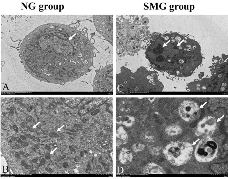
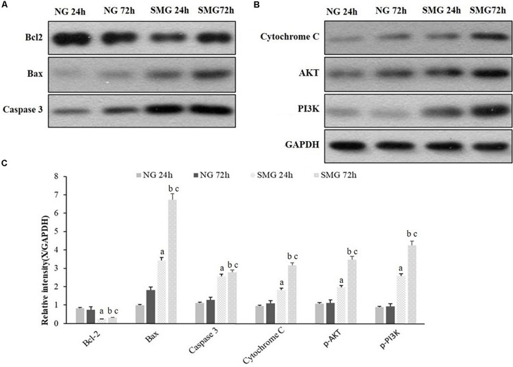
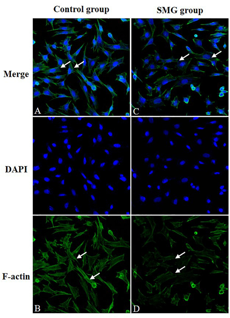
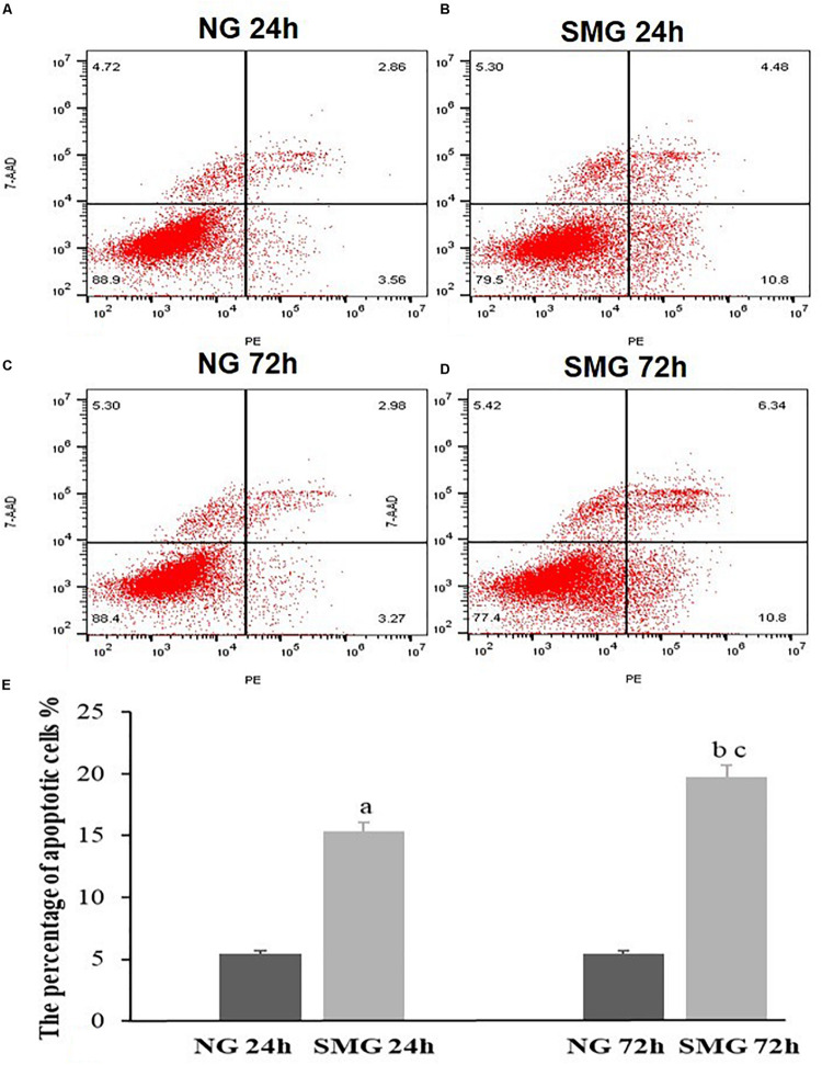
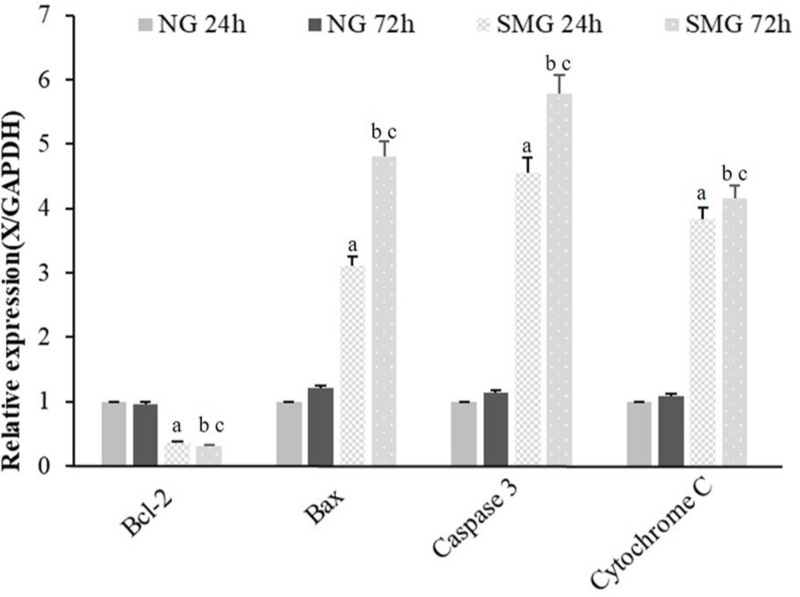
In the SMG group, CVECs presented with a shrunk cell body, chromatin condensation and margination, mitochondria vacuolization, and apoptotic bodies. The amount of F-actin decreased, and the filaments of F-actin were sparse or even partly discontinuous after cultivation under SMG for 72 h. The proportions of apoptotic CVECs in SMG groups at 24 and 72 h were significantly higher than those in the NG group ( < 0.001). The mRNA and protein expression of Bax, Caspase3, and Cytochrome C of CVECs in SMG groups at 24 and 72 h significantly increased than those of the NG group, respectively ( < 0.001). The alterations of p-AKT and p-PI3K protein expression possessed similar trends. On the contrary, the mRNA and protein expression of Bcl-2 in CVECs under SMG at 24 and 72 h were significantly less than that of the NG group, respectively ( < 0.001).
Simulated microgravity conditions can lead the alterations of the F-actin structure and apoptosis of CVECs. The Bcl-2 apoptosis pathway and PI3K/AKT pathway may participate in the damage of CVECs caused by SMG.
已证实模拟微重力(SMG)会导致多种类型的微血管内皮细胞发生超微结构改变和凋亡。然而,SMG是否也会影响脉络膜血管内皮细胞(CVECs)仍不清楚。本研究旨在探讨SMG对CVECs超微结构和凋亡的影响。
利用旋转细胞培养系统(RCCS)模拟微重力条件。将人CVECs在正常重力(NG)或SMG条件下培养3天。在透射电子显微镜下观察超微结构,通过免疫荧光染色观察F-肌动蛋白的组织情况。此外,使用流式细胞术计算凋亡百分比。而且,在不同暴露时间用定量PCR和蛋白质印迹法检测BAX、Bcl-2、Caspase3、细胞色素C、p-AKT和p-PI3K的mRNA和蛋白质表达。
在SMG组中,CVECs呈现细胞体缩小、染色质浓缩和边缘化、线粒体空泡化以及凋亡小体。F-肌动蛋白的量减少,在SMG条件下培养72小时后,F-肌动蛋白的细丝稀疏甚至部分不连续。SMG组在24小时和72小时时凋亡CVECs的比例显著高于NG组(<0.001)。SMG组CVECs在24小时和72小时时Bax、Caspase3和细胞色素C的mRNA和蛋白质表达分别比NG组显著增加(<0.001)。p-AKT和p-PI3K蛋白质表达的变化呈现相似趋势。相反,SMG条件下CVECs在24小时和72小时时Bcl-2的mRNA和蛋白质表达分别显著低于NG组(<0.001)。
模拟微重力条件可导致CVECs的F-肌动蛋白结构改变和凋亡。Bcl-2凋亡途径和PI3K/AKT途径可能参与了SMG引起的CVECs损伤。