Marinescu Răzvan V, Eshaghi Arman, Alexander Daniel C, Golland Polina
Computer Science and Artificial Intelligence Laboratory, Massachusetts Institute of Technology, Cambridge, USA, MA 02139.
Centre for Medical Image Computing, University College London, Gower Street, London, United Kingdom, WC1E 6BT.
Multimodal Brain Image Anal Math Found Comput Anat (2019). 2019 Oct;11846:112-120. doi: 10.1007/978-3-030-33226-6_13. Epub 2019 Oct 10.
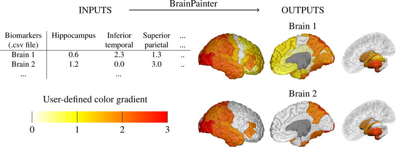
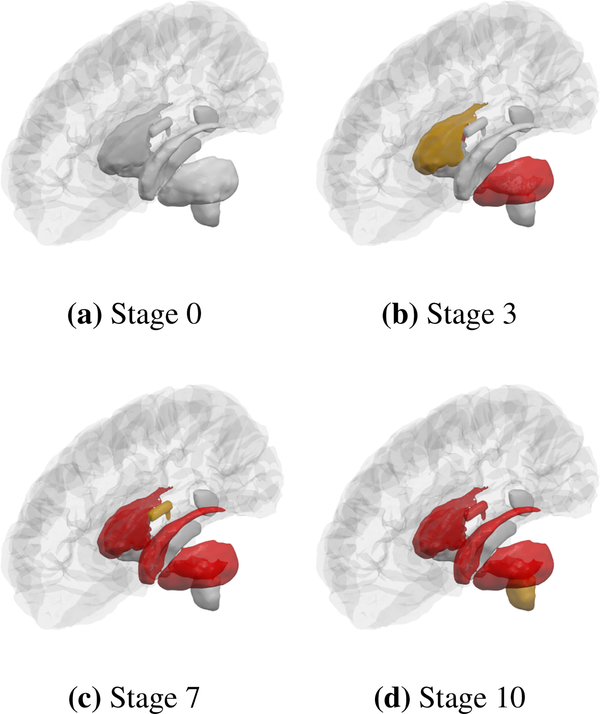
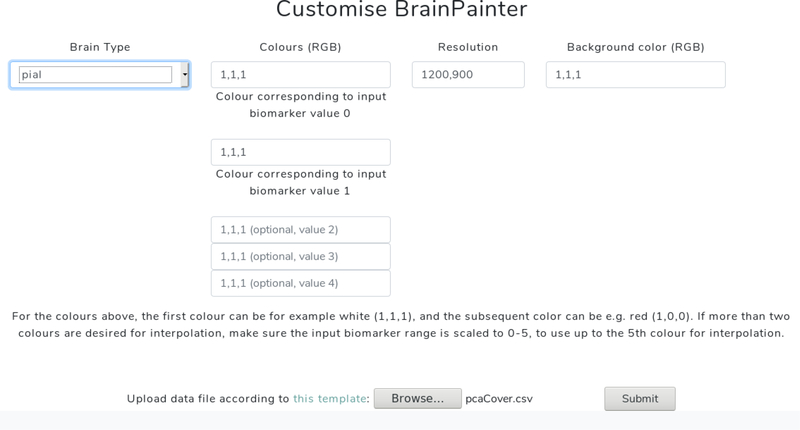
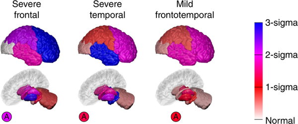
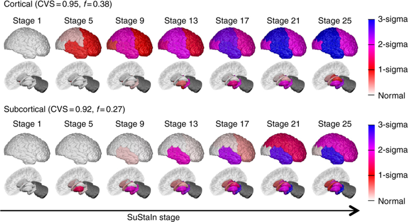
We present BrainPainter, a software that automatically generates images of highlighted brain structures given a list of numbers corresponding to the output colours of each region. Compared to existing visualisation software (i.e. Freesurfer, SPM, 3D Slicer), BrainPainter has three key advantages: (1) it does not require the input data to be in a specialised format, allowing BrainPainter to be used in combination with any neuroimaging analysis tools, (2) it can visualise both cortical and subcortical structures and (3) it can be used to generate movies showing dynamic processes, e.g. propagation of pathology on the brain. We highlight three use cases where BrainPainter was used in existing neuroimaging studies: (1) visualisation of the degree of atrophy through interpolation along a user-defined gradient of colours, (2) visualisation of the progression of pathology in Alzheimer's disease as well as (3) visualisation of pathology in subcortical regions in Huntington's disease. Moreover, through the design of BrainPainter we demonstrate the possibility of using a powerful 3D computer graphics engine such as Blender to generate brain visualisations for the neuroscience community. Blender's capabilities, e.g. particle simulations, motion graphics, UV unwrapping, raster graphics editing, raytracing and illumination effects, open a wealth of possibilities for brain visualisation not available in current neuroimaging software. BrainPainter is customisable, easy to use, and can run straight from the web browser: https://brainpainter.csail.mit.edu, as well as from source-code packaged in a docker container: https://github.com/mrazvan22/brain-coloring. It can be used to visualise biomarker data from any brain imaging modality, or simply to highlight a particular brain structure for e.g. anatomy courses.
我们介绍了BrainPainter,这是一款软件,它能根据与每个区域输出颜色对应的数字列表自动生成突出显示的脑结构图像。与现有的可视化软件(即FreeSurfer、SPM、3D Slicer)相比,BrainPainter具有三个关键优势:(1)它不要求输入数据为特定格式,这使得BrainPainter可与任何神经影像分析工具结合使用;(2)它能可视化皮质和皮质下结构;(3)它可用于生成展示动态过程的影片,例如脑部病变的传播。我们重点介绍了BrainPainter在现有神经影像研究中的三个应用案例:(1)通过沿用户定义的颜色梯度进行插值来可视化萎缩程度;(2)可视化阿尔茨海默病中病理变化的进展;以及(3)可视化亨廷顿病皮质下区域的病变。此外,通过BrainPainter的设计,我们展示了使用强大的3D计算机图形引擎(如Blender)为神经科学界生成脑可视化图像的可能性。Blender的功能,如粒子模拟、动态图形、UV展开、光栅图形编辑、光线追踪和光照效果,为当前神经影像软件所没有的脑可视化开辟了丰富的可能性。BrainPainter可定制、易于使用,并且可以直接从网页浏览器(https://brainpainter.csail.mit.edu)运行,也可以从打包在docker容器中的源代码(https://github.com/mrazvan22/brain-coloring)运行。它可用于可视化来自任何脑成像模态的生物标志物数据,或者仅仅用于突出显示特定的脑结构,例如用于解剖学课程。