• 文献检索
  • 文档翻译
  • 深度研究
  • 学术资讯
  • Suppr Zotero 插件Zotero 插件
  • 邀请有礼
  • 套餐&价格
  • 历史记录
应用&插件
Suppr Zotero 插件Zotero 插件浏览器插件Mac 客户端Windows 客户端微信小程序
定价
高级版会员购买积分包购买API积分包
服务
文献检索文档翻译深度研究API 文档MCP 服务
关于我们
关于 Suppr公司介绍联系我们用户协议隐私条款
关注我们

Suppr 超能文献

核心技术专利:CN118964589B侵权必究
粤ICP备2023148730 号-1Suppr @ 2026

文献检索

告别复杂PubMed语法,用中文像聊天一样搜索,搜遍4000万医学文献。AI智能推荐,让科研检索更轻松。

立即免费搜索

文件翻译

保留排版,准确专业,支持PDF/Word/PPT等文件格式,支持 12+语言互译。

免费翻译文档

深度研究

AI帮你快速写综述,25分钟生成高质量综述,智能提取关键信息,辅助科研写作。

立即免费体验

单侧唇腭裂患者地标识别的可靠性和可重复性:数字侧位片与CBCT衍生的三维头影测量片对比

Reliability and Reproducibility of Landmark Identification in Unilateral Cleft Lip and Palate Patients: Digital Lateral Vis-A-Vis CBCT-Derived 3D Cephalograms.

作者信息

Kochhar Anuraj Singh, Nucci Ludovica, Sidhu Maninder Singh, Prabhakar Mona, Grassia Vincenzo, Perillo Letizia, Kochhar Gulsheen Kaur, Bhasin Ritasha, Dadlani Himanshu, d'Apuzzo Fabrizia

机构信息

Former Consultant Orthodontist Max Hospital Gurgaon, Haryana 122001, India.

Multidisciplinary Department of Medical-Surgical and Dental Specialties, University of Campania Luigi Vanvitelli, 80138 Naples, Italy.

出版信息

J Clin Med. 2021 Feb 2;10(3):535. doi: 10.3390/jcm10030535.

DOI:10.3390/jcm10030535
PMID:33540549
原文链接:https://pmc.ncbi.nlm.nih.gov/articles/PMC7867146/
Abstract

BACKGROUND

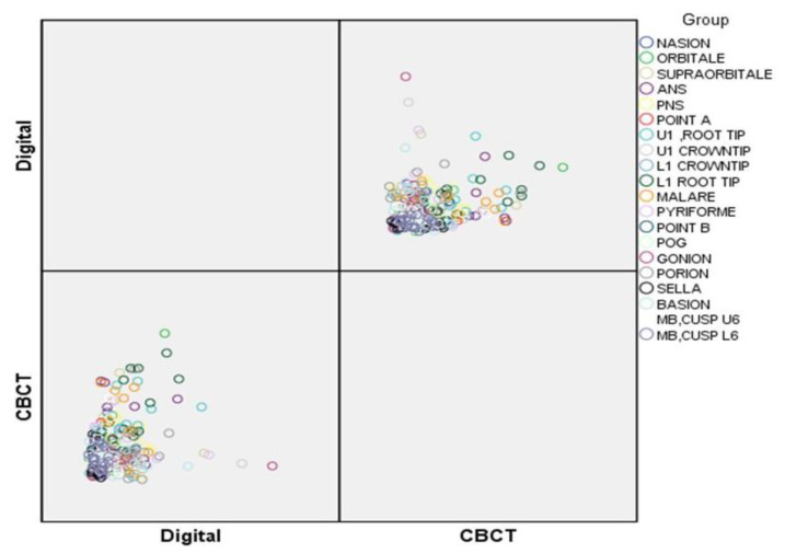
The aim of the retrospective observational study was to compare the precision of landmark identification and its reproducibility using cone beam computed tomography-derived 3D cephalograms and digital lateral cephalograms in unilateral cleft lip and palate patients.

METHODS

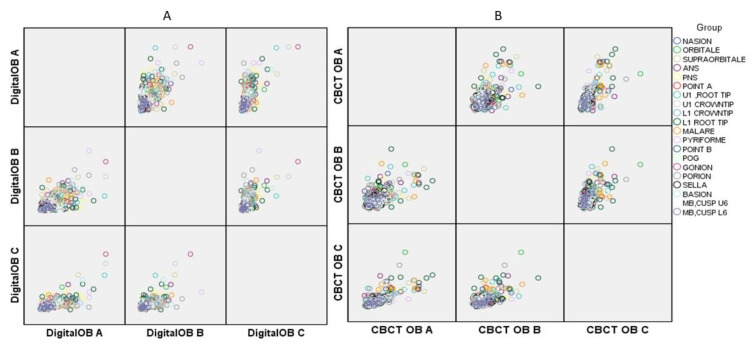
Cephalograms of thirty-one (31) North Indian children (18 boys and 13 girls) with a unilateral cleft lip and palate, who were recommended for orthodontic treatment, were selected. After a thorough analysis of peer-reviewed articles, 20 difficult-to-trace landmarks were selected, and their reliability and reproducibility were studied. These were subjected to landmark identification to evaluate interobserver variability; the coordinates for each point were traced separately by three different orthodontists (OB, OB, OB). Statistical analysis was performed using descriptive and inferential statistics with paired -tests to compare the differences measured by the two methods. Real-scale data are presented in mean ± SD. A -value less than 0.05 was considered as significant at a 95% confidence level.

RESULTS

When comparing, the plotting of points posterior nasal spine (PNS) ( < 0.05), anterior nasal spine (ANS) ( < 0.01), upper 1 root tip ( < 0.05), lower 1 root tip ( < 0.05), malare ( < 0.05), pyriforme ( < 0.05), porion ( < 0.01), and basion ( < 0.05) was statistically significant.

CONCLUSION

In patients with a cleft lip and palate, the interobserver identification of cephalometric landmarks was significantly more precise and reproducible with cone beam computed tomography -derived cephalograms vis-a-vis digital lateral cephalograms.

摘要

背景

这项回顾性观察研究的目的是比较在单侧唇腭裂患者中,使用锥形束计算机断层扫描(CBCT)衍生的三维头影测量片和数字化侧位头影测量片进行标志点识别的精度及其可重复性。

方法

选取了31名北印度单侧唇腭裂儿童(18名男孩和13名女孩)的头影测量片,这些儿童均被建议进行正畸治疗。在对同行评审文章进行全面分析后,选取了20个难以追踪的标志点,并研究了它们的可靠性和可重复性。对这些标志点进行识别以评估观察者间的差异;每个点的坐标由三位不同的正畸医生(OB、OB、OB)分别描绘。使用描述性和推断性统计方法以及配对t检验进行统计分析,以比较两种方法测量的差异。实际数据以平均值±标准差表示。在95%置信水平下,p值小于0.05被认为具有统计学意义。

结果

比较时,后鼻棘(PNS)(p<0.05)、前鼻棘(ANS)(p<0.01)、上1根尖(p<0.05)、下1根尖(p<0.05)、颧骨点(p<0.05)、梨状孔(p<0.05)、耳点(p<0.01)和颅底点(p<0.05)的点描绘具有统计学意义。

结论

在唇腭裂患者中,相对于数字化侧位头影测量片,使用CBCT衍生的头影测量片进行头影测量标志点的观察者间识别明显更精确且可重复。

https://cdn.ncbi.nlm.nih.gov/pmc/blobs/1e68/7867146/71b3be94d552/jcm-10-00535-g002.jpg
https://cdn.ncbi.nlm.nih.gov/pmc/blobs/1e68/7867146/ea3f61ae7b1d/jcm-10-00535-g001.jpg
https://cdn.ncbi.nlm.nih.gov/pmc/blobs/1e68/7867146/71b3be94d552/jcm-10-00535-g002.jpg
https://cdn.ncbi.nlm.nih.gov/pmc/blobs/1e68/7867146/ea3f61ae7b1d/jcm-10-00535-g001.jpg
https://cdn.ncbi.nlm.nih.gov/pmc/blobs/1e68/7867146/71b3be94d552/jcm-10-00535-g002.jpg

相似文献

1
Reliability and Reproducibility of Landmark Identification in Unilateral Cleft Lip and Palate Patients: Digital Lateral Vis-A-Vis CBCT-Derived 3D Cephalograms.单侧唇腭裂患者地标识别的可靠性和可重复性:数字侧位片与CBCT衍生的三维头影测量片对比
J Clin Med. 2021 Feb 2;10(3):535. doi: 10.3390/jcm10030535.
2
The reliability of cephalometric measurements in oral and maxillofacial imaging: Cone beam computed tomography versus two-dimensional digital cephalograms.口腔颌面影像学中头影测量的可靠性:锥形束计算机断层扫描与二维数字化头颅侧位片的比较
Indian J Dent Res. 2016 Jul-Aug;27(4):370-377. doi: 10.4103/0970-9290.191884.
3
Frontal and Axial Evaluation of Craniofacial Morphology in Repaired Unilateral Cleft Lip and Palate Patients Utilizing Cone Beam Computed Tomography; An Observational Study.利用锥形束计算机断层扫描评估单侧唇腭裂修复患者的颅面形态:一项观察性研究。
Int J Environ Res Public Health. 2020 Oct 24;17(21):7786. doi: 10.3390/ijerph17217786.
4
Comparison of reliability in anatomical landmark identification using two-dimensional digital cephalometrics and three-dimensional cone beam computed tomography in vivo.在体使用二维数字头影测量法和三维锥形束计算机断层扫描术进行解剖标志点识别的可靠性比较。
Dentomaxillofac Radiol. 2009 Jul;38(5):262-73. doi: 10.1259/dmfr/81889955.
5
Intraexaminer and interexaminer reliabilities of landmark identification on digitized lateral cephalograms and formatted 3-dimensional cone-beam computerized tomography images.数字化侧位头颅影像和格式化三维锥形束计算机断层扫描影像上标志点识别的观察者内和观察者间可靠性。
Am J Orthod Dentofacial Orthop. 2010 May;137(5):598-604. doi: 10.1016/j.ajodo.2008.07.018.
6
Landmark identification errors on cone-beam computed tomography-derived cephalograms and conventional digital cephalograms.锥形束计算机断层扫描头颅定位误差与传统数字头颅定位误差。
Am J Orthod Dentofacial Orthop. 2011 Dec;140(6):e289-97. doi: 10.1016/j.ajodo.2011.06.024.
7
Evaluation of the accuracy of linear measurements on lateral cephalograms obtained from cone-beam computed tomography scans with digital lateral cephalometric radiography: an in vitro study.锥束计算机断层扫描与数字化侧位头影测量X线摄影所得头颅侧位片上线性测量准确性的评估:一项体外研究
J Craniofac Surg. 2014 Sep;25(5):1710-3. doi: 10.1097/SCS.0000000000000908.
8
A cephalometric study of facial growth in van der Woude syndrome.范德伍德综合征面部生长的头影测量研究。
Cleft Palate Craniofac J. 2002 Mar;39(2):219-25. doi: 10.1597/1545-1569_2002_039_0219_acsofg_2.0.co_2.
9
A comparative study of the reproducibility of landmark identification on posteroanterior and anteroposterior cephalograms generated from cone-beam computed tomography scans.一项关于锥形束计算机断层扫描生成的正位和侧位头颅侧位片上标志点识别可重复性的比较研究。
Korean J Orthod. 2019 Jan;49(1):41-48. doi: 10.4041/kjod.2019.49.1.41. Epub 2018 Dec 19.
10
Identification of cephalometric landmarks in unilateral cleft lip and palate patients: are there alternatives for point A, ANS, and PNS?单侧唇腭裂患者头影测量标志点的确定:对于A点、前鼻棘点和后鼻棘点是否有替代方法?
Cleft Palate Craniofac J. 2008 Jan;45(1):81-6. doi: 10.1597/06-236.1.

引用本文的文献

1
Finite element analysis of maxillary orthodontic therapies with variable alveolar bone grafts under occlusal forces in patient with unilateral cleft lip and palate.单侧唇腭裂患者在咬合力作用下采用不同牙槽骨移植的上颌正畸治疗的有限元分析
Front Bioeng Biotechnol. 2024 Nov 5;12:1448286. doi: 10.3389/fbioe.2024.1448286. eCollection 2024.
2
New Approaches and Technologies in Orthodontics.正畸学的新方法与新技术
J Clin Med. 2024 Apr 24;13(9):2470. doi: 10.3390/jcm13092470.
3
Unilateral Cleft Lip and Palate Has Asymmetry of Bony Orbits: A Retrospective Study.

本文引用的文献

1
Intra- and Interobserver Reliability of Bone Volume Estimation Using OsiriX Software in Patients with Cleft Lip and Palate Using Cone Beam Computed Tomography.使用锥形束计算机断层扫描对唇腭裂患者采用OsiriX软件进行骨体积估计的观察者内和观察者间可靠性
Dent J (Basel). 2021 Jan 22;9(2):14. doi: 10.3390/dj9020014.
2
Frontal and Axial Evaluation of Craniofacial Morphology in Repaired Unilateral Cleft Lip and Palate Patients Utilizing Cone Beam Computed Tomography; An Observational Study.利用锥形束计算机断层扫描评估单侧唇腭裂修复患者的颅面形态:一项观察性研究。
Int J Environ Res Public Health. 2020 Oct 24;17(21):7786. doi: 10.3390/ijerph17217786.
3
单侧唇腭裂存在眶骨不对称:一项回顾性研究。
J Pers Med. 2023 Jun 29;13(7):1067. doi: 10.3390/jpm13071067.
4
Craniofacial Cephalometric Morphology in Caucasian Adult Patients with Cleft Palate Only (CPO).仅患有腭裂(CPO)的成年白种患者的颅面头影测量形态学
Diagnostics (Basel). 2023 Jun 14;13(12):2058. doi: 10.3390/diagnostics13122058.
5
Comparison of palatal volume and surface changes between bone-borne and tooth-tissue-borne maxillary expansion on cone beam computed tomography digital cast models.基于锥形束计算机断层扫描数字模型比较骨支持式和牙组织支持式上颌扩弓器对上颌骨体积和表面变化的影响
Angle Orthod. 2023 May 1;93(3):282-288. doi: 10.2319/040922-278.1.
6
Convolutional Neural Network Performance for Sella Turcica Segmentation and Classification Using CBCT Images.使用CBCT图像的蝶鞍分割与分类的卷积神经网络性能
Diagnostics (Basel). 2022 Sep 16;12(9):2244. doi: 10.3390/diagnostics12092244.
7
Evaluation of Articular Eminence Inclination in Normo-Divergent Subjects with Different Skeletal Classes through CBCT.通过 CBCT 评估不同骨骼类型的正常-低角患者的关节突倾斜度。
Int J Environ Res Public Health. 2021 Jun 3;18(11):5992. doi: 10.3390/ijerph18115992.
Precision Analysis of Supraorbital Transcranial Exits Using Three Dimensional Multidetector Computed Tomography.
使用三维多探测器计算机断层扫描对眶上经颅出口进行精确分析
J Craniofac Surg. 2019 Sep;30(6):1894-1897. doi: 10.1097/SCS.0000000000005557.
4
Reliability of 3D dental and skeletal landmarks on CBCT images.CBCT 图像上 3D 牙和骨骼标志点的可靠性。
Angle Orthod. 2019 Sep;89(5):758-767. doi: 10.2319/082018-612.1. Epub 2019 Mar 18.
5
CBCT in orthodontics: a systematic review on justification of CBCT in a paediatric population prior to orthodontic treatment.口腔锥形束 CT 在正畸学中的应用:正畸治疗前对儿童人群进行口腔锥形束 CT 检查的正当性的系统评价。
Eur J Orthod. 2019 Aug 8;41(4):381-389. doi: 10.1093/ejo/cjy066.
6
Accuracy and reliability of landmark-based, surface-based and voxel-based 3D cone-beam computed tomography superimposition methods.基于标志点、基于表面和基于体素的 3D 锥形束计算机断层扫描叠加方法的准确性和可靠性。
Orthod Craniofac Res. 2017 Nov;20(4):227-236. doi: 10.1111/ocr.12205. Epub 2017 Sep 27.
7
Precision of manual landmark identification between as-received and oriented volume-rendered cone-beam computed tomography images.接收时与定向的容积再现锥形束计算机断层扫描图像之间手动标志点识别的精度。
Am J Orthod Dentofacial Orthop. 2017 Jan;151(1):118-131. doi: 10.1016/j.ajodo.2016.06.027.
8
Oral health related quality of life in cleft lip and palate patients rehabilitated with conventional prostheses or dental implants.采用传统修复体或牙种植体修复的唇腭裂患者的口腔健康相关生活质量
J Int Soc Prev Community Dent. 2015 Nov-Dec;5(6):482-7. doi: 10.4103/2231-0762.168645.
9
Cephalometric landmark variability among orthodontists and dentomaxillofacial radiologists: a comparative study.正畸医生与口腔颌面放射科医生之间头影测量标志点的变异性:一项对比研究。
Imaging Sci Dent. 2015 Dec;45(4):213-20. doi: 10.5624/isd.2015.45.4.213. Epub 2015 Dec 17.
10
Evaluation of dehiscence and fenestration in adolescent patients affected by unilateral cleft lip and palate: A retrospective cone beam computed tomography study.单侧唇腭裂青少年患者创口裂开与开窗的评估:一项回顾性锥形束计算机断层扫描研究
Angle Orthod. 2016 May;86(3):431-6. doi: 10.2319/042715-289.1. Epub 2015 Aug 18.