Department of Bioengineering, Rice University, Houston, TX, USA.
Department of Thoracic/Head and Neck Medical Oncology, The University of Texas MD Anderson Cancer Center, Houston, TX, USA.
Sci Rep. 2021 Feb 4;11(1):3171. doi: 10.1038/s41598-021-82102-w.
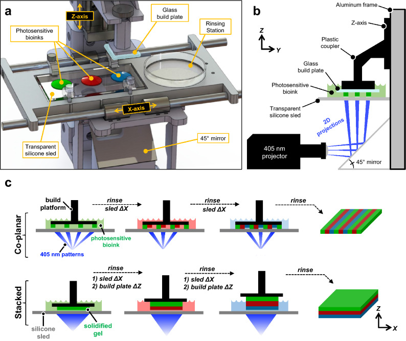
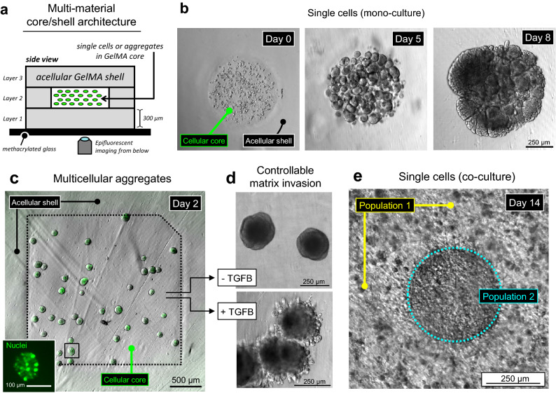
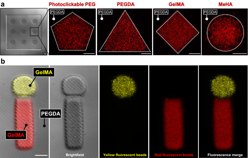
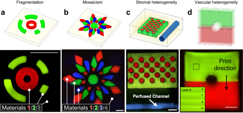
As a 3D bioprinting technique, hydrogel stereolithography has historically been limited in its ability to capture the spatial heterogeneity that permeates mammalian tissues and dictates structure-function relationships. This limitation stems directly from the difficulty of preventing unwanted material mixing when switching between different liquid bioinks. Accordingly, we present the development, characterization, and application of a multi-material stereolithography bioprinter that provides controlled material selection, yields precise regional feature alignment, and minimizes bioink mixing. Fluorescent tracers were first used to highlight the broad design freedoms afforded by this fabrication strategy, complemented by morphometric image analysis to validate architectural fidelity. To evaluate the bioactivity of printed gels, 344SQ lung adenocarcinoma cells were printed in a 3D core/shell architecture. These cells exhibited native phenotypic behavior as evidenced by apparent proliferation and formation of spherical multicellular aggregates. Cells were also printed as pre-formed multicellular aggregates, which appropriately developed invasive protrusions in response to hTGF-β1. Finally, we constructed a simplified model of intratumoral heterogeneity with two separate sub-populations of 344SQ cells, which together grew over 14 days to form a dense regional interface. Together, these studies highlight the potential of multi-material stereolithography to probe heterotypic interactions between distinct cell types in tissue-specific microenvironments.
作为一种 3D 生物打印技术,水凝胶立体光刻技术在捕捉哺乳动物组织中普遍存在的空间异质性以及决定结构-功能关系方面的能力一直受到限制。这种限制直接源于在不同的液体生物墨水之间切换时防止不必要的材料混合的困难。因此,我们提出了一种多材料立体光刻生物打印机的开发、表征和应用,该打印机提供了可控的材料选择、实现了精确的区域特征对准,并最大限度地减少了生物墨水的混合。荧光示踪剂首先被用于突出这种制造策略所提供的广泛设计自由度,并通过形态计量图像分析来验证结构保真度。为了评估打印凝胶的生物活性,将 344SQ 肺腺癌细胞以 3D 核/壳结构打印。这些细胞表现出明显的增殖和形成球形多细胞聚集物,证明了其具有天然表型行为。细胞也被打印为预先形成的多细胞聚集物,这些聚集物在 hTGF-β1 的刺激下适当形成了侵袭性突起。最后,我们构建了一个具有两个独立的 344SQ 细胞亚群的肿瘤内异质性简化模型,这些细胞共同生长了 14 天,形成了一个密集的区域界面。这些研究共同强调了多材料立体光刻在探测组织特异性微环境中不同细胞类型之间异质相互作用的潜力。