State Key Laboratory of Plant Genomics and National Center for Plant Gene Research (Beijing), Institute of Genetics and Developmental Biology, The Innovative Academy of Seed Design, Chinese Academy of Sciences, Beijing 100101, China.
Laboratoire Reproduction et Développement des Plantes, Univ Lyon, ENS de Lyon, UCB Lyon 1, CNRS, INRAE, Inria, 69342 Lyon, France.
STAR Protoc. 2021 Jan 23;2(1):100301. doi: 10.1016/j.xpro.2021.100301. eCollection 2021 Mar 19.
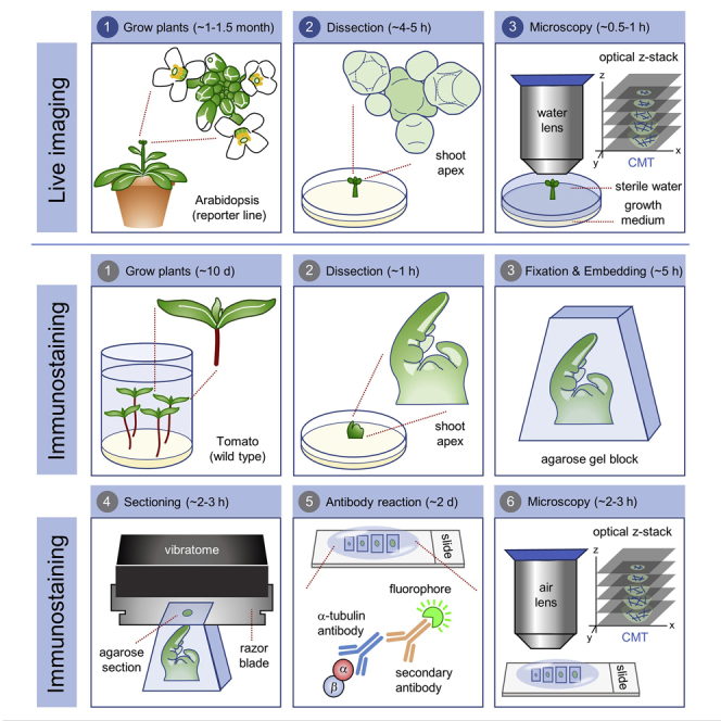
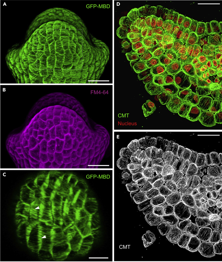
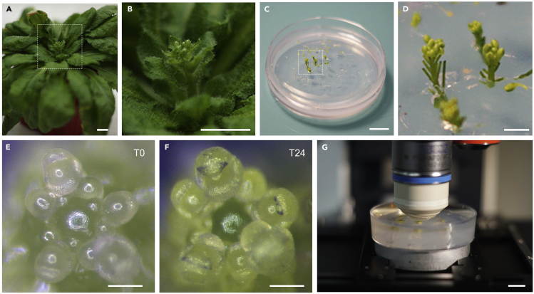
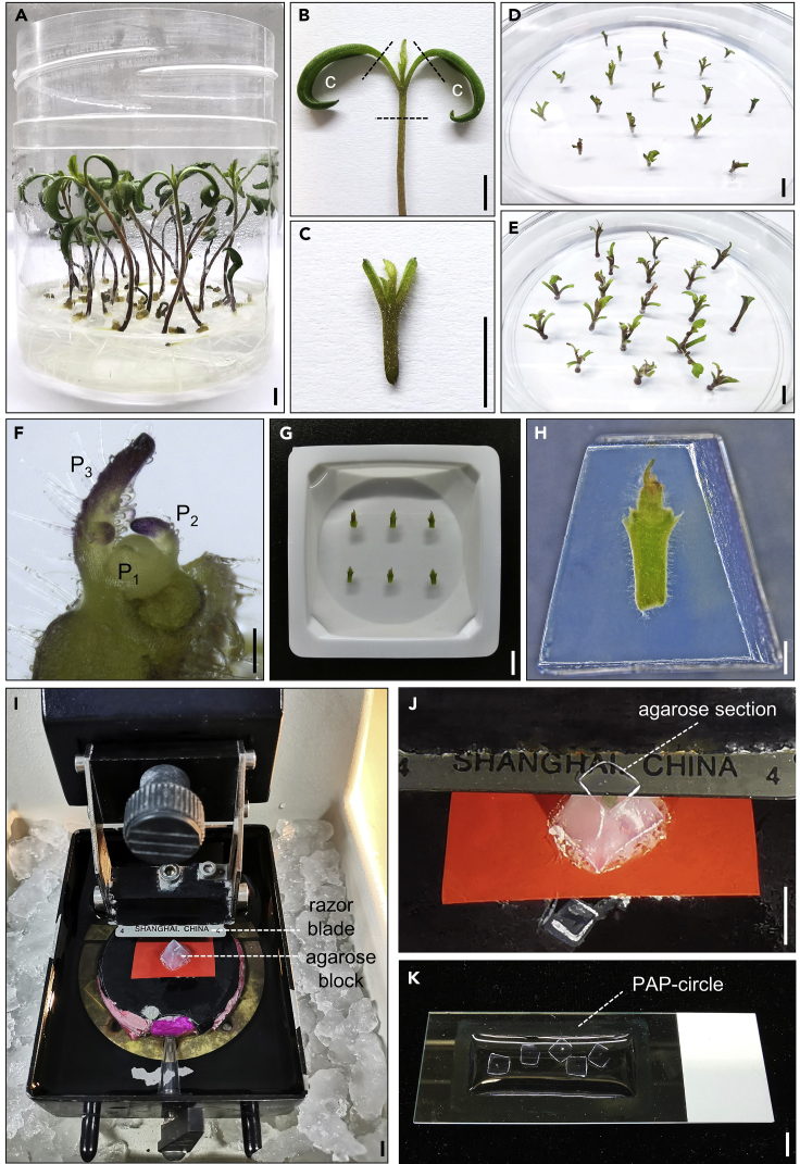
Cortical microtubules (CMTs) play pivotal roles during plant cell growth and division. The organization of CMTs undergoes important changes during different cellular and developmental processes. Here, we describe two methods for the visualization of CMT organization in plant cells using confocal laser scanning microscopy. CMT networks in the outer tissue layers can be directly visualized by live imaging of a fluorescent reporter line, and a protocol combining sectioning and immunostaining is applied for visualization of CMTs throughout tissues. For complete details on the use and execution of this protocol, please refer to Zhao et al. (2020).
皮层微管(CMTs)在植物细胞生长和分裂过程中发挥着关键作用。CMTs 的组织在不同的细胞和发育过程中会发生重要变化。在这里,我们描述了两种使用共聚焦激光扫描显微镜观察植物细胞中 CMT 组织的方法。通过荧光报告线的实时成像,可以直接观察外层组织中的 CMT 网络,并且还应用了一种结合切片和免疫染色的方案来观察整个组织中的 CMT。有关此方案的使用和执行的完整详细信息,请参见 Zhao 等人。(2020 年)。