Department of Quantitative Cell Biology, Institute of Molecular Cell Biology, University of Münster, Münster, Germany.
Group of Molecular Mechanotransduction, Max Planck Institute of Biochemistry, Martinsried, Germany.
Nat Commun. 2021 Feb 10;12(1):919. doi: 10.1038/s41467-021-21142-2.
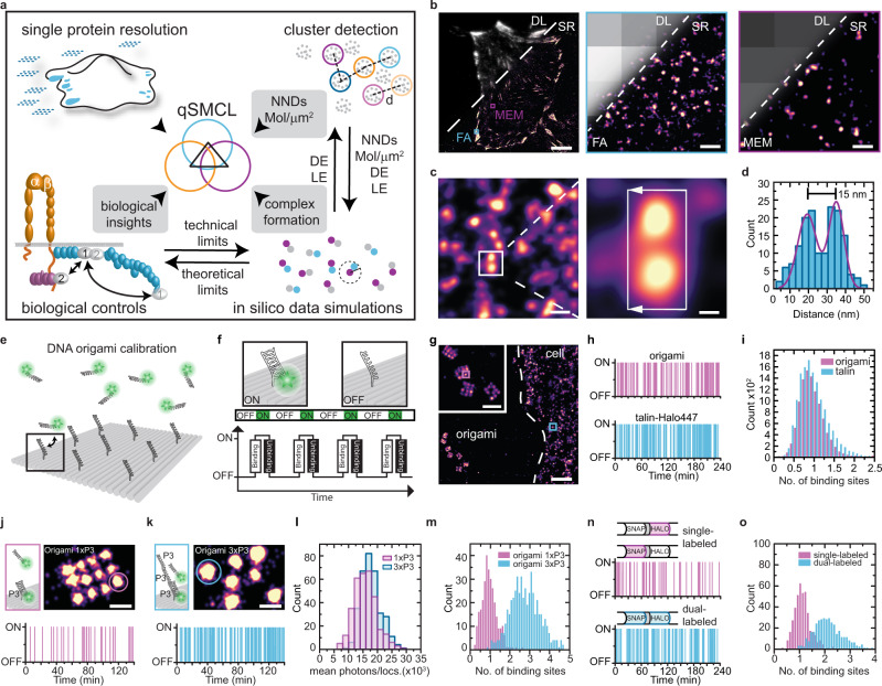
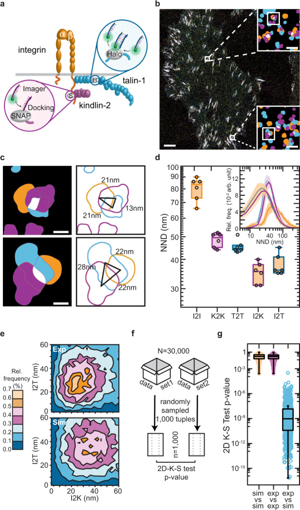
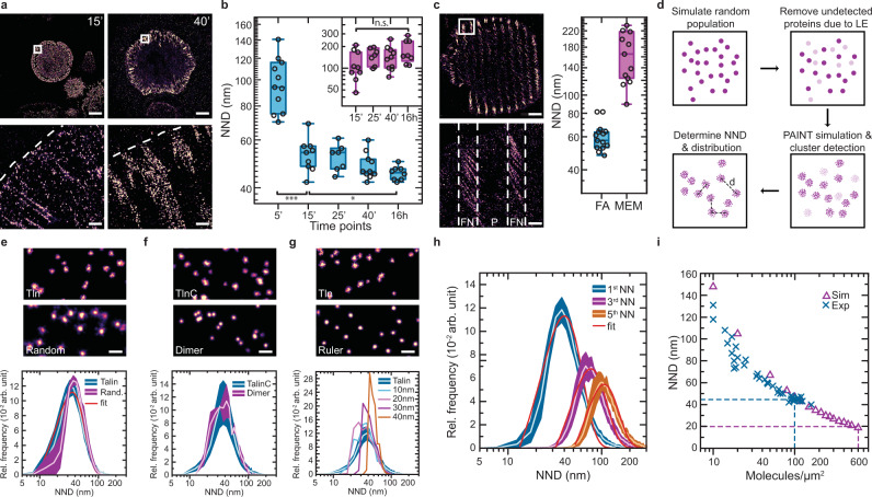
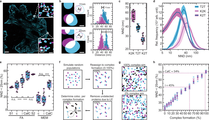
Single-molecule localization microscopy (SMLM) enabling the investigation of individual proteins on molecular scales has revolutionized how biological processes are analysed in cells. However, a major limitation of imaging techniques reaching single-protein resolution is the incomplete and often unknown labeling and detection efficiency of the utilized molecular probes. As a result, fundamental processes such as complex formation of distinct molecular species cannot be reliably quantified. Here, we establish a super-resolution microscopy framework, called quantitative single-molecule colocalization analysis (qSMCL), which permits the identification of absolute molecular quantities and thus the investigation of molecular-scale processes inside cells. The method combines multiplexed single-protein resolution imaging, automated cluster detection, in silico data simulation procedures, and widely applicable experimental controls to determine absolute fractions and spatial coordinates of interacting species on a true molecular level, even in highly crowded subcellular structures. The first application of this framework allowed the identification of a long-sought ternary adhesion complex-consisting of talin, kindlin and active β1-integrin-that specifically forms in cell-matrix adhesion sites. Together, the experiments demonstrate that qSMCL allows an absolute quantification of multiplexed SMLM data and thus should be useful for investigating molecular mechanisms underlying numerous processes in cells.
单分子定位显微镜(SMLM)能够在分子水平上研究单个蛋白质,从而彻底改变了人们在细胞中分析生物过程的方式。然而,达到单蛋白分辨率的成像技术的一个主要局限性是所用分子探针的标记和检测效率不完全,而且通常是未知的。因此,诸如不同分子物种的复杂形成等基本过程无法可靠地定量。在这里,我们建立了一种称为定量单分子共定位分析(qSMCL)的超分辨率显微镜框架,该框架允许确定绝对分子数量,从而可以在细胞内部研究分子尺度的过程。该方法结合了多重单蛋白分辨率成像、自动聚类检测、基于计算机的数据模拟程序以及广泛适用的实验控制,以确定相互作用物种的绝对分数和空间坐标,即使在高度拥挤的亚细胞结构中也是如此。该框架的首次应用允许鉴定一种长期以来备受关注的三元黏附复合物 - 由 talin、kindlin 和活性β1 整合素组成 - 它专门在细胞基质黏附位点形成。总之,这些实验表明,qSMCL 允许对多重 SMLM 数据进行绝对定量,因此对于研究细胞中许多过程的分子机制应该是有用的。