Arangath Anand, Duffy Niamh, Alexandrov Sergey, James Soorya, Neuhaus Kai, Murphy Mary, Leahy Martin
Tissue Optics and Microcirculation Imaging Facility, Physics, School of Natural Sciences, University of Galway, Galway, Ireland.
Regenerative Medicine Institute, University of Galway, Galway, Ireland.
Biomed Opt Express. 2023 Mar 3;14(4):1411-1427. doi: 10.1364/BOE.485082. eCollection 2023 Apr 1.
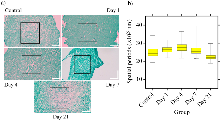
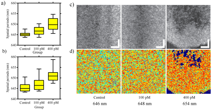
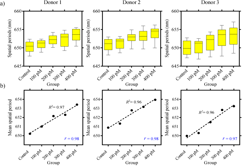
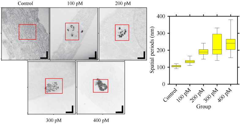
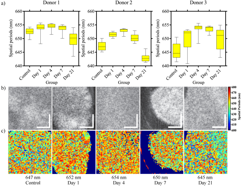
Mesenchymal stromal cells (MSCs) are adult stem cells that have been widely investigated for their potential to regenerate damaged and diseased tissues. Multiple pre-clinical studies and clinical trials have demonstrated a therapeutic response following treatment with MSCs for various pathologies, including cardiovascular, neurological and orthopaedic diseases. The ability to functionally track cells following administration in vivo is pivotal to further elucidating the mechanism of action and safety profile of these cells. Effective monitoring of MSCs and MSC-derived microvesicles requires an imaging modality capable of providing both quantitative and qualitative readouts. Nanosensitive optical coherence tomography (nsOCT) is a recently developed technique that detects nanoscale structural changes within samples. In this study, we demonstrate for the first time, the capability of nsOCT to image MSC pellets following labelling with different concentrations of dual plasmonic gold nanostars. We show that the mean spatial period of MSC pellets increases following the labelling with increasing concentrations of nanostars. Additionally, with the help of extra time points and a more comprehensive analysis, we further improved the understanding of the MSC pellet chondrogenesis model. Despite the limited penetration depth (similar to conventional OCT), the nsOCT is highly sensitive in detecting structural alterations at the nanoscale, which may provide crucial functional information about cell therapies and their modes of action.
间充质基质细胞(MSCs)是成体干细胞,因其具有再生受损和患病组织的潜力而受到广泛研究。多项临床前研究和临床试验表明,用MSCs治疗各种病症(包括心血管、神经和骨科疾病)后会产生治疗反应。在体内给药后对细胞进行功能追踪的能力对于进一步阐明这些细胞的作用机制和安全性至关重要。对MSCs和源自MSCs的微泡进行有效监测需要一种能够提供定量和定性读数的成像方式。纳米敏感光学相干断层扫描(nsOCT)是一种最近开发的技术,可检测样品内的纳米级结构变化。在本研究中,我们首次展示了nsOCT在用不同浓度的双等离子体金纳米星标记后对MSC微球成像的能力。我们表明,随着纳米星浓度的增加,标记后MSC微球的平均空间周期会增加。此外,借助额外的时间点和更全面的分析,我们进一步加深了对MSC微球软骨生成模型的理解。尽管穿透深度有限(类似于传统OCT),但nsOCT在检测纳米级结构变化方面高度敏感,这可能为细胞疗法及其作用方式提供关键的功能信息。