Department of Radiology and Research Institute of Radiology, University of Ulsan College of Medicine, Asan Medical Center, Seoul, Korea.
Biomedical Research Center, Asan Institute for Life Sciences, Asan Medical Center, Seoul, Korea.
Korean J Radiol. 2021 Apr;22(4):624-633. doi: 10.3348/kjr.2020.0914. Epub 2021 Jan 19.
To evaluate the reliability of CT measurements of muscle quantity and quality using variable CT parameters.
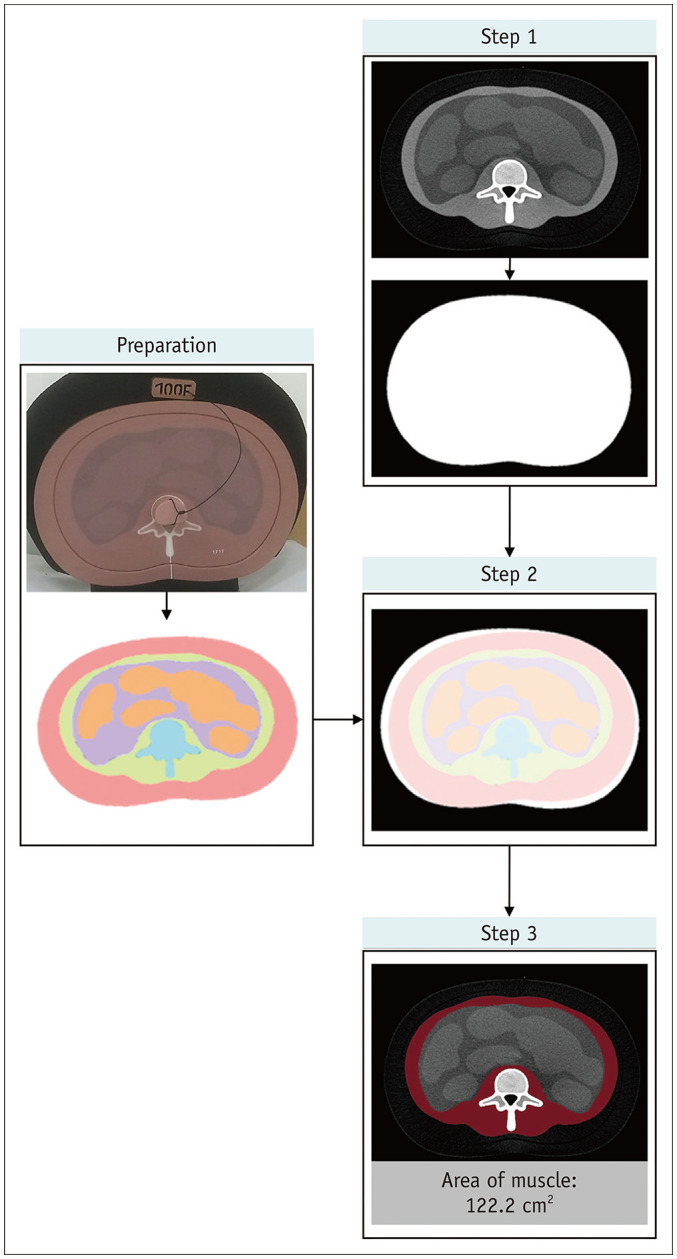
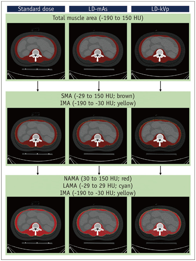
A phantom, simulating the L2-4 vertebral levels, was used for this study. CT images were repeatedly acquired with modulation of tube voltage, tube current, slice thickness, and the image reconstruction algorithm. Reference standard muscle compartments were obtained from the reference maps of the phantom. Cross-sectional area based on the Hounsfield unit (HU) thresholds of muscle and its components, and the mean density of the reference standard muscle compartment, were used to measure the muscle quantity and quality using different CT protocols. Signal-to-noise ratios (SNRs) were calculated in the images acquired with different settings.
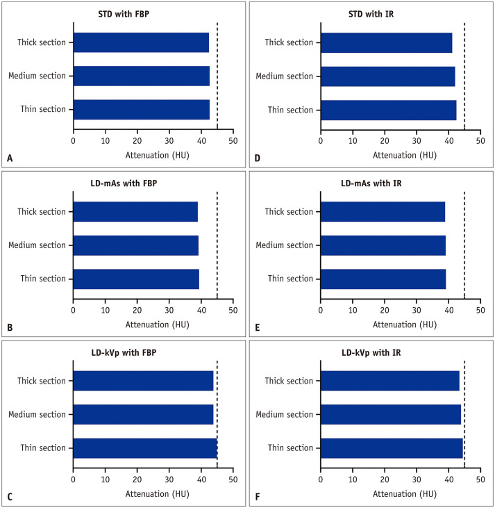
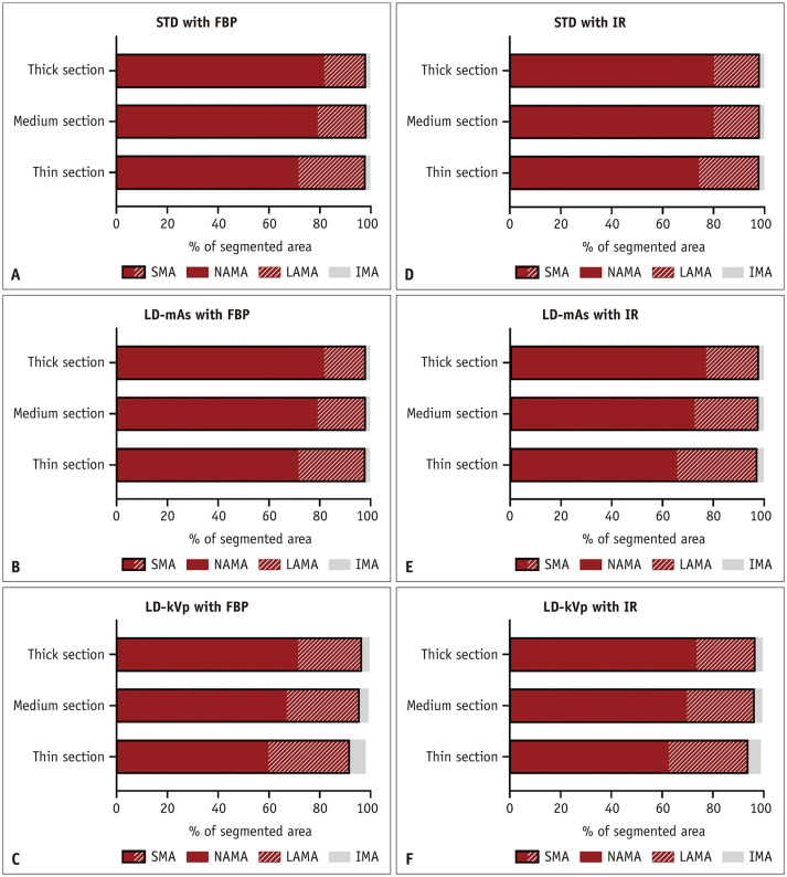
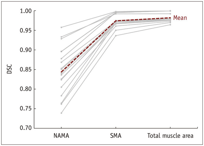
The skeletal muscle area (threshold, ?29 to 150 HU) was constant, regardless of the protocol, occupying at least 91.7% of the reference standard muscle compartment. Conversely, normal attenuation muscle area (30-150 HU) was not constant in the different protocols, varying between 59.7% and 81.7% of the reference standard muscle compartment. The mean density was lower than the target density stated by the manufacturer (45 HU) in all cases (range, 39.0-44.9 HU). The SNR decreased with low tube voltage, low tube current, and in sections with thin slices, whereas it increased when the iterative reconstruction algorithm was used.
Measurement of muscle quantity using HU threshold was reliable, regardless of the CT protocol used. Conversely, the measurement of muscle quality using the mean density and narrow HU thresholds were inconsistent and inaccurate across different CT protocols. Therefore, further studies are warranted in future to determine the optimal CT protocols for reliable measurements of muscle quality.
评估使用可变 CT 参数测量肌肉量和质量的 CT 测量的可靠性。
本研究使用模拟 L2-4 椎体水平的体模。使用管电压、管电流、层厚和图像重建算法调节,对 CT 图像进行重复采集。参考标准肌肉区室从体模的参考图中获得。基于肌肉及其成分的 Hounsfield 单位(HU)阈值的横截面积和参考标准肌肉区室的平均密度,用于使用不同 CT 协议测量肌肉量和质量。在不同设置下采集的图像中计算信噪比(SNR)。
骨骼肌面积(阈值,-29 至 150 HU)不受协议影响,保持不变,至少占据参考标准肌肉区室的 91.7%。相反,不同协议中的正常衰减肌肉面积(30-150 HU)并不恒定,变化范围为参考标准肌肉区室的 59.7%至 81.7%。平均密度均低于制造商规定的目标密度(45 HU)(范围,39.0-44.9 HU)。在所有情况下,SNR 均随管电压降低、管电流降低和薄片层厚降低而降低,而当使用迭代重建算法时则升高。
使用 HU 阈值测量肌肉量是可靠的,无论使用何种 CT 协议。相反,使用平均密度和狭窄 HU 阈值测量肌肉质量在不同 CT 协议中不一致且不准确。因此,未来有必要进行进一步研究,以确定可靠测量肌肉质量的最佳 CT 协议。