• 文献检索
  • 文档翻译
  • 深度研究
  • 学术资讯
  • Suppr Zotero 插件Zotero 插件
  • 邀请有礼
  • 套餐&价格
  • 历史记录
应用&插件
Suppr Zotero 插件Zotero 插件浏览器插件Mac 客户端Windows 客户端微信小程序
定价
高级版会员购买积分包购买API积分包
服务
文献检索文档翻译深度研究API 文档MCP 服务
关于我们
关于 Suppr公司介绍联系我们用户协议隐私条款
关注我们

Suppr 超能文献

核心技术专利:CN118964589B侵权必究
粤ICP备2023148730 号-1Suppr @ 2026

文献检索

告别复杂PubMed语法,用中文像聊天一样搜索,搜遍4000万医学文献。AI智能推荐,让科研检索更轻松。

立即免费搜索

文件翻译

保留排版,准确专业,支持PDF/Word/PPT等文件格式,支持 12+语言互译。

免费翻译文档

深度研究

AI帮你快速写综述,25分钟生成高质量综述,智能提取关键信息,辅助科研写作。

立即免费体验

法国 FISH:基于组织切片荧光原位杂交定量 DNA 拷贝数的鱼类模型。

FrenchFISH: Poisson Models for Quantifying DNA Copy Number From Fluorescence In Situ Hybridization of Tissue Sections.

机构信息

Cancer Research UK, Cambridge Institute, University of Cambridge, Cambridge, UK.

Cavendish Laboratory, Department of Physics, University of Cambridge, Cambridge, UK.

出版信息

JCO Clin Cancer Inform. 2021 Feb;5:176-186. doi: 10.1200/CCI.20.00075.

DOI:10.1200/CCI.20.00075
PMID:33570999
原文链接:https://pmc.ncbi.nlm.nih.gov/articles/PMC8140799/
Abstract

PURPOSE

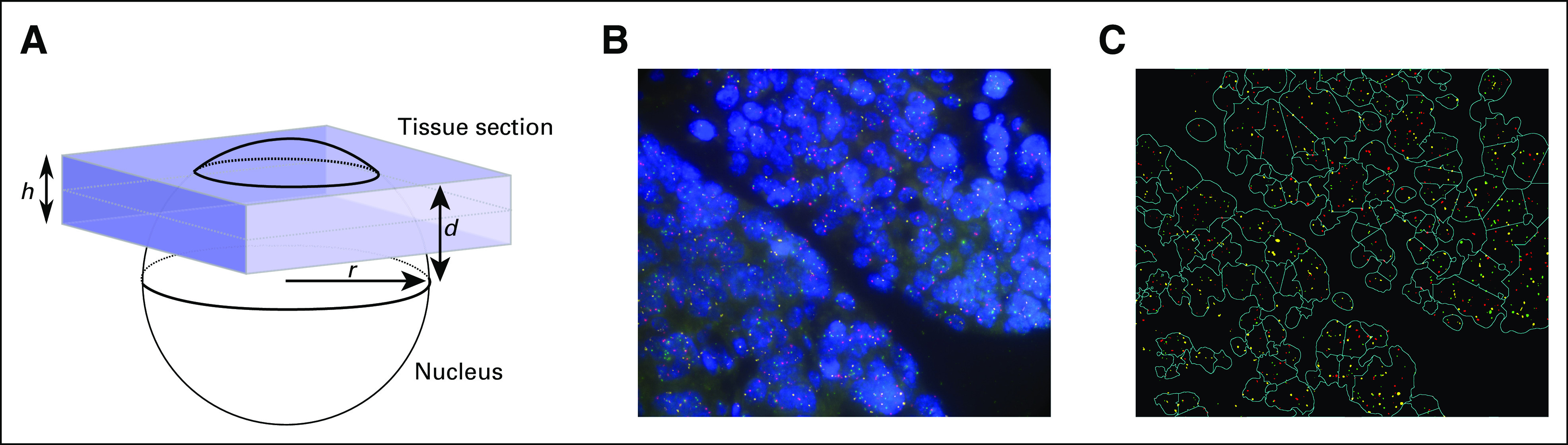
Chromosomal aberration and DNA copy number change are robust hallmarks of cancer. The gold standard for detecting copy number changes in tumor cells is fluorescence in situ hybridization (FISH) using locus-specific probes that are imaged as fluorescent spots. However, spot counting often does not perform well on solid tumor tissue sections due to partially represented or overlapping nuclei.

MATERIALS AND METHODS

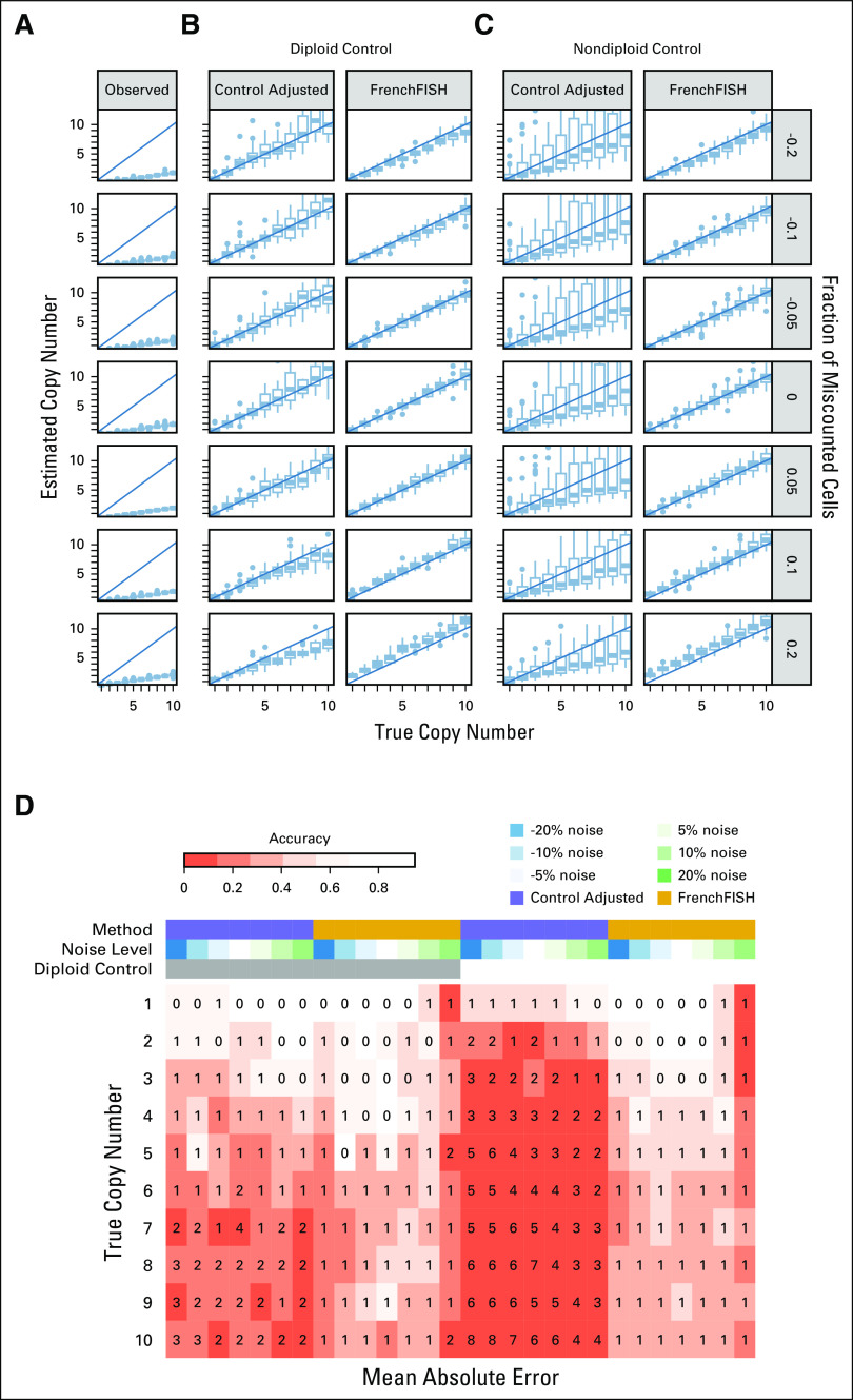
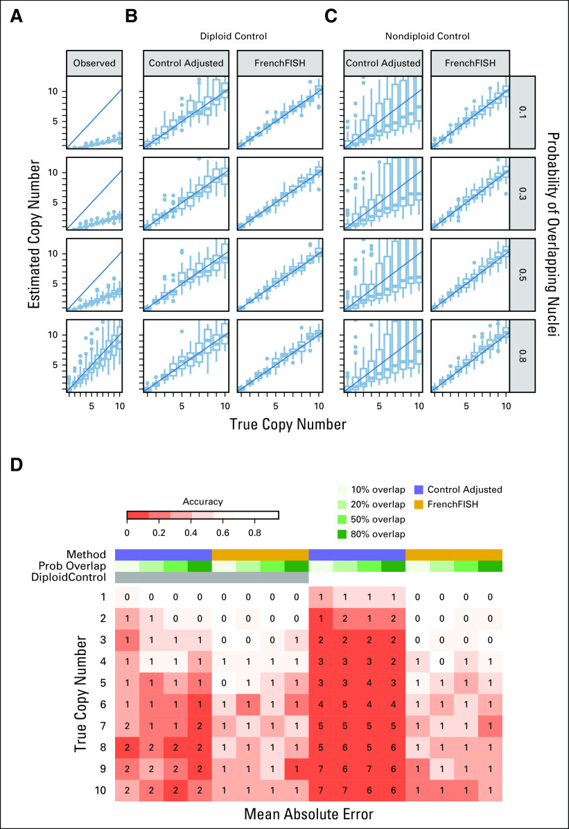
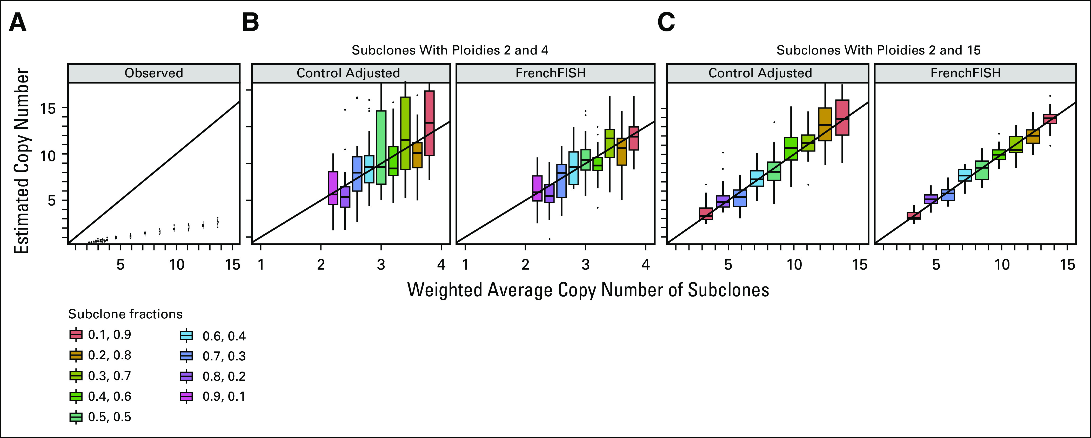
To overcome these challenges, we have developed a computational approach called FrenchFISH, which comprises a nuclear volume correction method coupled with two types of Poisson models: either a Poisson model for improved manual spot counting without the need for control probes or a homogeneous Poisson point process model for automated spot counting.

RESULTS

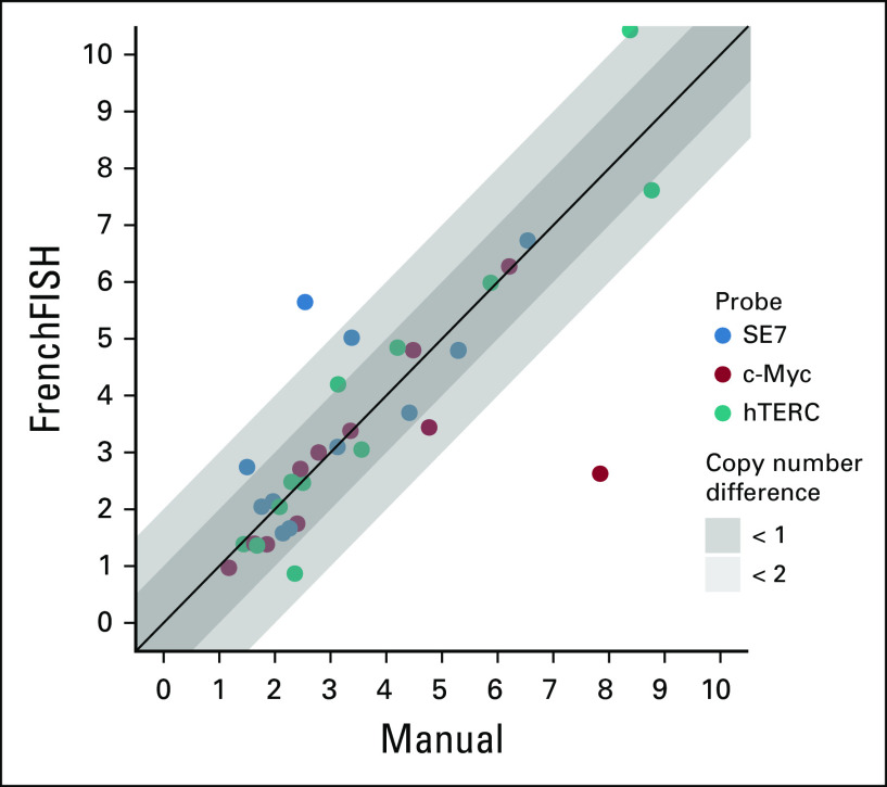
We benchmarked the performance of FrenchFISH against previous approaches using a controlled simulation scenario and tested it experimentally in 12 ovarian carcinoma FFPE-tissue sections for copy number alterations at three loci (c-Myc, hTERC, and SE7). FrenchFISH outperformed standard spot counting with 74% of the automated counts having < 1 copy number difference from the manual counts and 17% having < 2 copy number differences, while taking less than one third of the time of manual counting.

CONCLUSION

FrenchFISH is a general approach that can be used to enhance clinical diagnosis on sections of any tissue by both speeding up and improving the accuracy of spot count estimates.

摘要

目的

染色体畸变和 DNA 拷贝数变化是癌症的显著特征。使用针对特定基因座的探针进行荧光原位杂交(FISH)是检测肿瘤细胞拷贝数变化的金标准,这些探针以荧光点的形式成像。然而,由于部分代表性或重叠的细胞核,点计数在实体肿瘤组织切片上的效果往往不佳。

材料和方法

为了克服这些挑战,我们开发了一种称为 FrenchFISH 的计算方法,该方法包括核体积校正方法和两种泊松模型:一种是改进手动点计数的泊松模型,无需使用对照探针;另一种是用于自动点计数的同质泊松点过程模型。

结果

我们使用受控模拟场景对 FrenchFISH 的性能进行了基准测试,并在 12 个卵巢癌 FFPE 组织切片中针对三个基因座(c-Myc、hTERC 和 SE7)的拷贝数改变进行了实验测试。FrenchFISH 的性能优于标准的点计数,74%的自动计数与手动计数的拷贝数差异小于 1,17%的自动计数与手动计数的拷贝数差异小于 2,而用时不到手动计数的三分之一。

结论

FrenchFISH 是一种通用方法,可通过加快和提高点计数估计的准确性来增强任何组织切片的临床诊断。

https://cdn.ncbi.nlm.nih.gov/pmc/blobs/0ec9/8140799/1959667685d6/cci-5-cci.20.00075-g006.jpg
https://cdn.ncbi.nlm.nih.gov/pmc/blobs/0ec9/8140799/53e5124a6c9e/cci-5-cci.20.00075-g001.jpg
https://cdn.ncbi.nlm.nih.gov/pmc/blobs/0ec9/8140799/47eaed94ae36/cci-5-cci.20.00075-g002.jpg
https://cdn.ncbi.nlm.nih.gov/pmc/blobs/0ec9/8140799/05dc54253ba6/cci-5-cci.20.00075-g003.jpg
https://cdn.ncbi.nlm.nih.gov/pmc/blobs/0ec9/8140799/cab3b5c8ef3f/cci-5-cci.20.00075-g004.jpg
https://cdn.ncbi.nlm.nih.gov/pmc/blobs/0ec9/8140799/bb3b46913613/cci-5-cci.20.00075-g005.jpg
https://cdn.ncbi.nlm.nih.gov/pmc/blobs/0ec9/8140799/1959667685d6/cci-5-cci.20.00075-g006.jpg

相似文献

1
FrenchFISH: Poisson Models for Quantifying DNA Copy Number From Fluorescence In Situ Hybridization of Tissue Sections.法国 FISH:基于组织切片荧光原位杂交定量 DNA 拷贝数的鱼类模型。
JCO Clin Cancer Inform. 2021 Feb;5:176-186. doi: 10.1200/CCI.20.00075.
2
DNA aneuploidy and tissue architecture in oral potentially malignant disorders with epithelial dysplasia assessed by a 10 locus FISH panel.通过10个位点的荧光原位杂交(FISH)检测口腔潜在恶性疾病伴上皮发育异常中的DNA非整倍体和组织结构
Oncol Rep. 2020 Mar;43(3):877-885. doi: 10.3892/or.2020.7461. Epub 2020 Jan 13.
3
Detection of numerical chromosome aberrations using in situ hybridization in paraffin sections of routinely processed bladder cancers.在常规处理的膀胱癌石蜡切片中使用原位杂交检测染色体数目畸变
Mod Pathol. 1991 Jul;4(4):503-13.
4
Fluorescence In Situ Hybridization of Cells, Chromosomes, and Formalin-Fixed Paraffin-Embedded Tissues.细胞、染色体及福尔马林固定石蜡包埋组织的荧光原位杂交
Methods Mol Biol. 2017;1606:265-279. doi: 10.1007/978-1-4939-6990-6_17.
5
Morphological spot counting from stacked images for automated analysis of gene copy numbers by fluorescence in situ hybridization.
J Biomed Opt. 2002 Jan;7(1):109-22. doi: 10.1117/1.1428292.
6
Detection of CCND1 Locus Amplification by Fluorescence In Situ Hybridization.通过荧光原位杂交检测CCND1基因座扩增
Methods Mol Biol. 2018;1726:85-100. doi: 10.1007/978-1-4939-7565-5_9.
7
Comparative FISH analysis of numerical chromosome 7 abnormalities in 5-micron and 15-micron paraffin-embedded tissue sections from prostatic carcinoma.前列腺癌5微米和15微米石蜡包埋组织切片中7号染色体数目异常的比较荧光原位杂交分析
Histochem Cell Biol. 1997 Feb;107(2):121-6. doi: 10.1007/s004180050096.
8
Combination of lamin immunocytochemistry and in situ hybridization for the analysis of chromosome copy numbers in tumor cell areas with high nuclear density.层粘连蛋白免疫细胞化学与原位杂交相结合用于分析高核密度肿瘤细胞区域的染色体拷贝数。
Cytometry. 1996 Jan 1;23(1):1-7. doi: 10.1002/(SICI)1097-0320(19960101)23:1<1::AID-CYTO1>3.0.CO;2-P.
9
MYC Copy Number Detection in Clinical Samples Using a Digital DNA-Hybridization and Detection Method.使用数字 DNA 杂交和检测方法检测临床样本中的 MYC 拷贝数。
Methods Mol Biol. 2021;2318:321-336. doi: 10.1007/978-1-0716-1476-1_18.
10
Analysis of Chromosomal Alterations in Urothelial Carcinoma.尿路上皮癌的染色体改变分析
Methods Mol Biol. 2018;1655:3-17. doi: 10.1007/978-1-4939-7234-0_1.

本文引用的文献

1
Use of Fluorescence In Situ Hybridization (FISH) in Diagnosis and Tailored Therapies in Solid Tumors.荧光原位杂交(FISH)在实体瘤诊断和靶向治疗中的应用。
Molecules. 2020 Apr 17;25(8):1864. doi: 10.3390/molecules25081864.
2
The Driver Mutational Landscape of Ovarian Squamous Cell Carcinomas Arising in Mature Cystic Teratoma.成熟囊性畸胎瘤衍生的卵巢鳞状细胞癌的驱动基因突变图谱。
Clin Cancer Res. 2017 Dec 15;23(24):7633-7640. doi: 10.1158/1078-0432.CCR-17-1789. Epub 2017 Sep 27.
3
Automatic thresholding from the gradients of region boundaries.
基于区域边界梯度的自动阈值处理。
J Microsc. 2017 Feb;265(2):185-195. doi: 10.1111/jmi.12474. Epub 2016 Sep 20.
4
Guidelines for cytogenetic investigations in tumours.肿瘤细胞遗传学检查指南。
Eur J Hum Genet. 2016 Jan;24(1):6-13. doi: 10.1038/ejhg.2015.35. Epub 2015 Mar 25.
5
High fidelity copy number analysis of formalin-fixed and paraffin-embedded tissues using Affymetrix Cytoscan HD chip.使用Affymetrix Cytoscan HD芯片对福尔马林固定石蜡包埋组织进行高保真拷贝数分析。
PLoS One. 2014 Apr 3;9(4):e92820. doi: 10.1371/journal.pone.0092820. eCollection 2014.
6
Lessons from the cancer genome.从癌症基因组中得到的启示。
Cell. 2013 Mar 28;153(1):17-37. doi: 10.1016/j.cell.2013.03.002.
7
State-of-the-art FISHing: automated analysis of cytogenetic aberrations in interphase nuclei.最先进的 FISH 技术:间期细胞核细胞遗传学异常的自动化分析。
Cytometry A. 2012 Aug;81(8):649-63. doi: 10.1002/cyto.a.22082. Epub 2012 Jun 13.
8
Absolute quantification of somatic DNA alterations in human cancer.人类癌症中体细胞 DNA 改变的绝对定量。
Nat Biotechnol. 2012 May;30(5):413-21. doi: 10.1038/nbt.2203.
9
Automated focusing in bright-field microscopy for tuberculosis detection.明场显微镜自动对焦在结核病检测中的应用。
J Microsc. 2010 Nov;240(2):155-63. doi: 10.1111/j.1365-2818.2010.03389.x.
10
Allele-specific copy number analysis of tumors.肿瘤的等位基因特异性拷贝数分析。
Proc Natl Acad Sci U S A. 2010 Sep 28;107(39):16910-5. doi: 10.1073/pnas.1009843107. Epub 2010 Sep 13.