Alamri Ahmad, Nam Jun Yeb, Blancato Jan K
Lombardi Comprehensive Cancer Center, Georgetown University Medical Center, Washington, DC, USA.
Department of Clinical Laboratories Sciences, College of Applied Medical Sciences, King Khalid University, Abha, Saudi Arabia.
Methods Mol Biol. 2017;1606:265-279. doi: 10.1007/978-1-4939-6990-6_17.
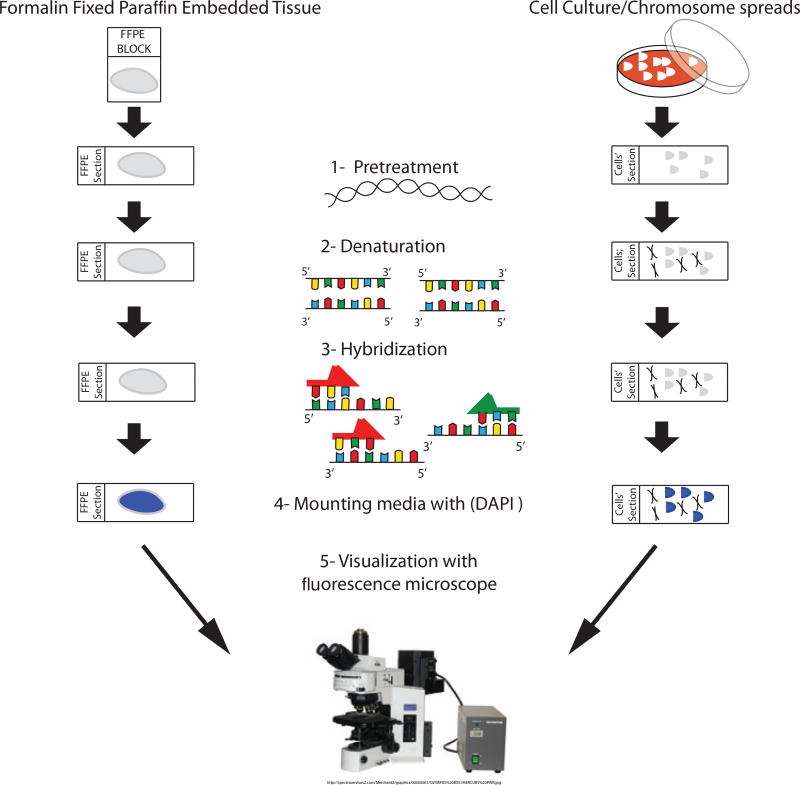
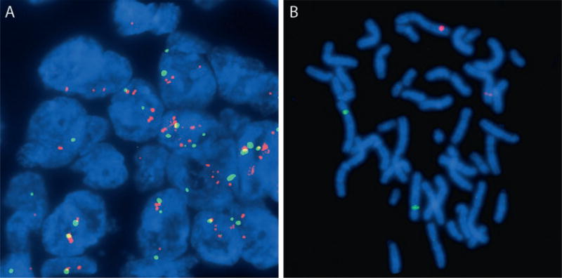
Fluorescence in situ hybridization (FISH) with DNA probes allows the visualization of gene copy number and localization of specific DNA targets with fluorescence microscopy. Cells in culture, metaphase chromosomes, and tissue sections are fixed and prepared on glass slides. Both the DNA in the cells and fluorescently labeled probe are denatured, and the labeled probe is allowed to hybridize to the cellular DNA. The slides are washed, counterstained, and viewed via fluorescence microscopy. We describe the basic method for preparing slides and probes for studies involving DNA copy number changes and structural chromosome rearrangements in formalin-fixed paraffin-embedded (FFPE) tissue sections and cell culture preparations.
使用DNA探针进行荧光原位杂交(FISH)可通过荧光显微镜观察基因拷贝数以及特定DNA靶点的定位。培养的细胞、中期染色体和组织切片经固定后制备在载玻片上。细胞中的DNA和荧光标记的探针均进行变性处理,然后使标记的探针与细胞DNA杂交。将载玻片洗涤、复染,再通过荧光显微镜观察。我们描述了在福尔马林固定石蜡包埋(FFPE)组织切片和细胞培养物中制备载玻片和探针的基本方法,用于研究DNA拷贝数变化和染色体结构重排。