Woo Ha-Na, Park Sujeong, Kim Hae Lin, Jung Min-Kyo, Pack Chan-Gi, Park Jinsu, Cho Yoonsuk, Jo Dong-Gyu, Kim Dong Kyu, Mook-Jung Inhee, Kim Seong Who, Lee Heuiran
Department of Microbiology, University of Ulsan College of Medicine, Seoul 05505, Korea.
Bio-Medical Institute of Technology, Asan Medical Center, Seoul 05505, Korea.
Mol Ther Nucleic Acids. 2020 Dec 23;23:643-656. doi: 10.1016/j.omtn.2020.12.014. eCollection 2021 Mar 5.
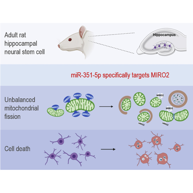
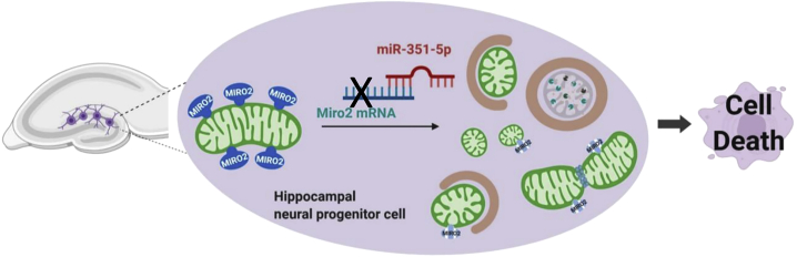
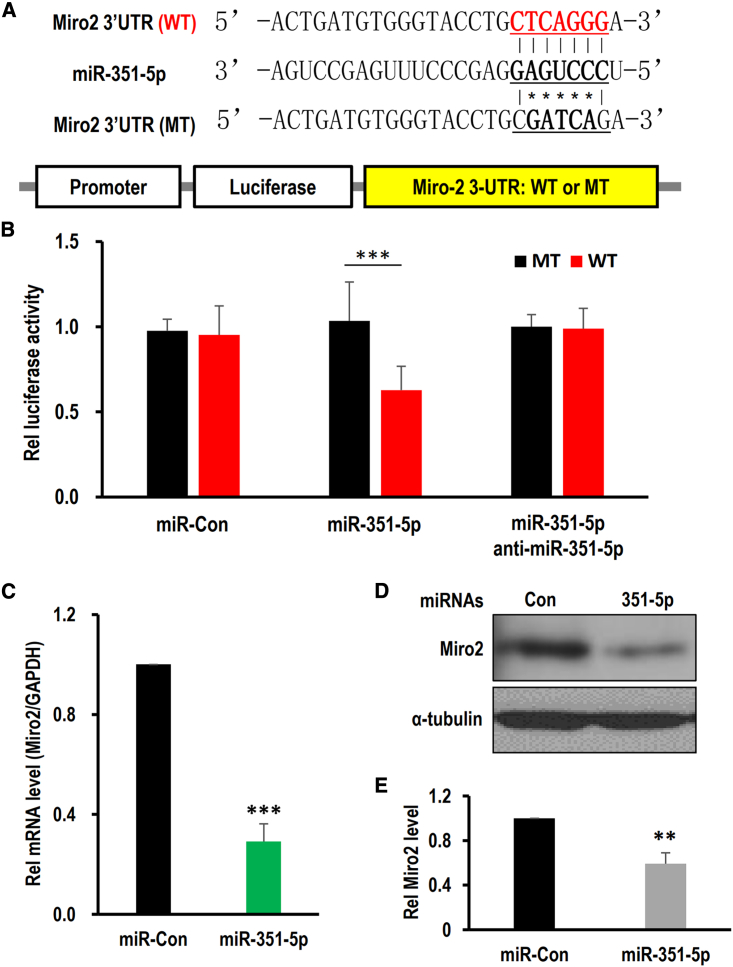
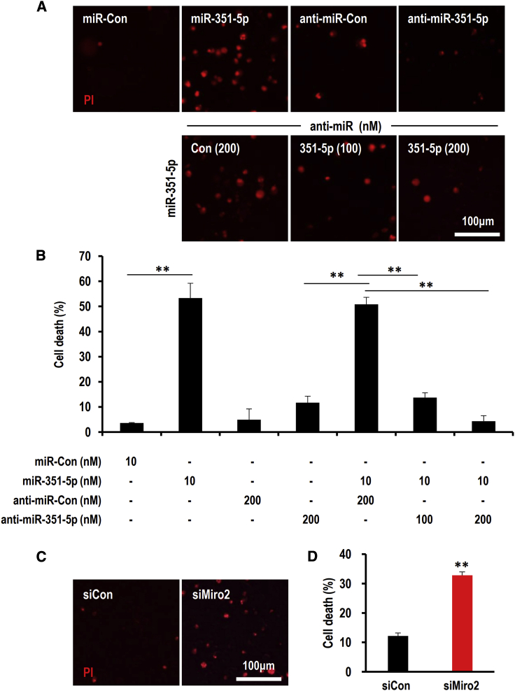
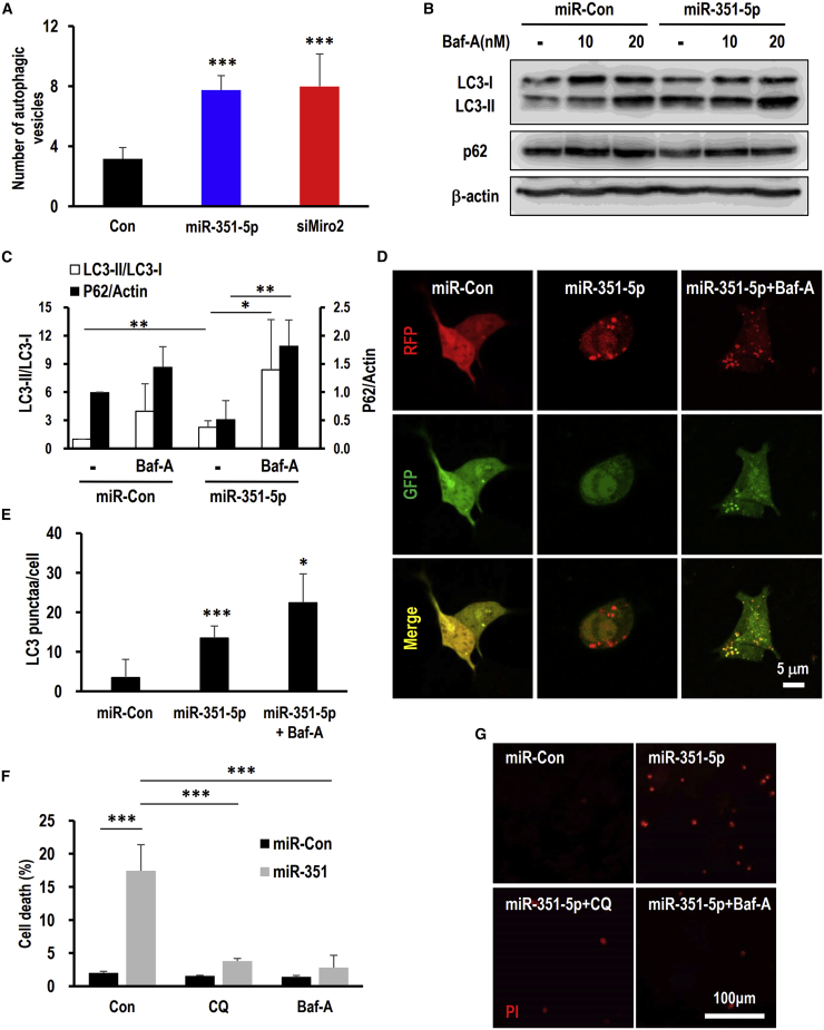
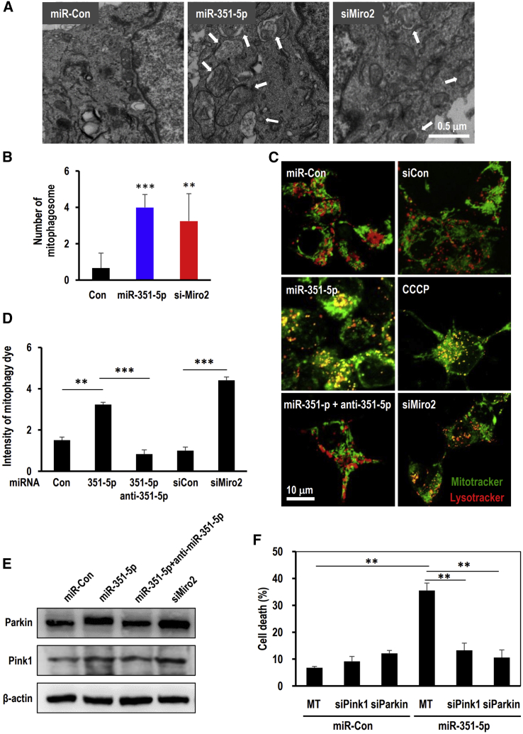
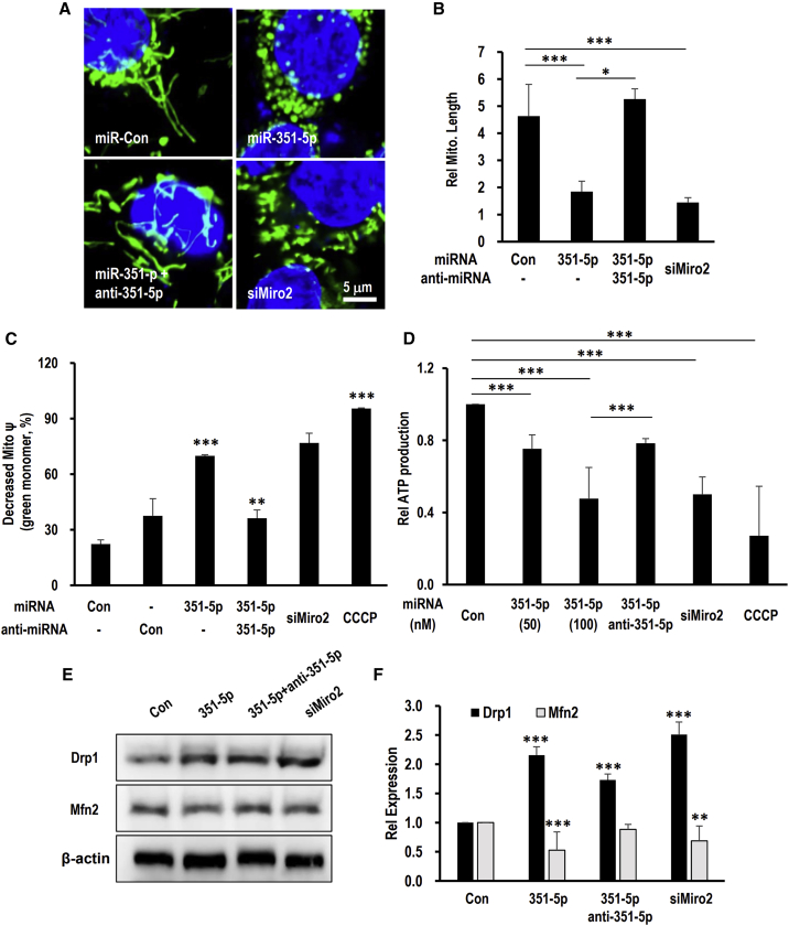
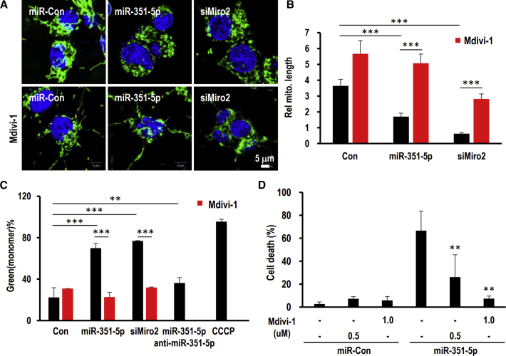
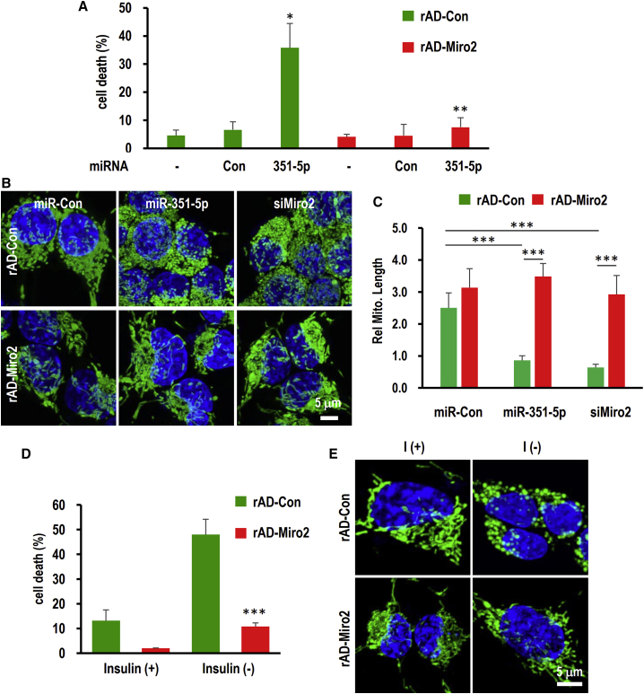
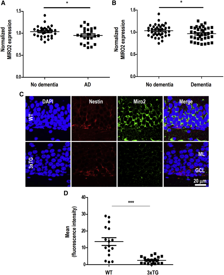
Adult hippocampal neurogenesis supports the structural and functional plasticity of the brain, while its decline is associated with neurodegeneration common in Alzheimer's disease (AD). Although the dysregulation of certain microRNAs (miRNAs) in AD have been observed, the effects of miRNAs on hippocampal neurogenesis are largely unknown. In this study, we demonstrated miR-351-5p as a causative factor in hippocampal neural progenitor cell death through modulation of the mitochondrial guanosine triphosphatase (GTPase), Miro2. Downregulation of Miro2 by siMiro2 induced cell death, similar to miR-351-5p, whereas ectopic Miro2 expression using an adenovirus abolished these effects. Excessively fragmented mitochondria and dysfunctional mitochondria were indexed by decreased mitochondrial potential, and increased reactive oxygen species were identified in miR-351-5p-induced cell death. Moreover, subsequent induction of mitophagy via Pink1 and Parkin was observed in the presence of miR-351-5p and siMiro2. The suppression of mitochondrial fission by Mdivi-1 completely inhibited cell death by miR-351-5p. miR-351-5p expression increased whereas the level of Miro2 decreased in the hippocampus of AD model mice, emulating expression in AD patients. Collectively, the data indicate the mitochondrial fission and accompanying mitophagy by miR-351-5p/Miro2 axis as critical in hippocampal neural progenitor cell death, and a potential therapeutic target in AD.
成体海马神经发生支持大脑的结构和功能可塑性,而其衰退与阿尔茨海默病(AD)中常见的神经退行性变有关。尽管已观察到AD中某些微小RNA(miRNA)的失调,但miRNA对海马神经发生的影响在很大程度上尚不清楚。在本研究中,我们证明miR-351-5p通过调节线粒体鸟苷三磷酸酶(GTP酶)Miro2成为海马神经祖细胞死亡的一个致病因素。与miR-351-5p相似,siMiro2介导的Miro2下调诱导细胞死亡,而使用腺病毒异位表达Miro2可消除这些影响。线粒体电位降低表明线粒体过度碎片化和功能失调,并且在miR-351-5p诱导的细胞死亡中发现活性氧增加。此外,在存在miR-351-5p和siMiro2的情况下,观察到随后通过Pink1和Parkin诱导的线粒体自噬。Mdivi-1对线粒体分裂的抑制完全抑制了miR-351-5p诱导的细胞死亡。在AD模型小鼠的海马中,miR-351-5p表达增加而Miro2水平降低,这与AD患者中的表达情况相似。总体而言,数据表明miR-351-5p/Miro2轴介导的线粒体分裂和伴随的线粒体自噬在海马神经祖细胞死亡中起关键作用,并且是AD的一个潜在治疗靶点。