• 文献检索
  • 文档翻译
  • 深度研究
  • 学术资讯
  • Suppr Zotero 插件Zotero 插件
  • 邀请有礼
  • 套餐&价格
  • 历史记录
应用&插件
Suppr Zotero 插件Zotero 插件浏览器插件Mac 客户端Windows 客户端微信小程序
定价
高级版会员购买积分包购买API积分包
服务
文献检索文档翻译深度研究API 文档MCP 服务
关于我们
关于 Suppr公司介绍联系我们用户协议隐私条款
关注我们

Suppr 超能文献

核心技术专利:CN118964589B侵权必究
粤ICP备2023148730 号-1Suppr @ 2026

文献检索

告别复杂PubMed语法,用中文像聊天一样搜索,搜遍4000万医学文献。AI智能推荐,让科研检索更轻松。

立即免费搜索

文件翻译

保留排版,准确专业,支持PDF/Word/PPT等文件格式,支持 12+语言互译。

免费翻译文档

深度研究

AI帮你快速写综述,25分钟生成高质量综述,智能提取关键信息,辅助科研写作。

立即免费体验

在 COVID-19 肺炎中使用 COVID-19 报告和数据系统(CO-RADS)分类和胸部计算机断层扫描受累评分(CT-IS)。

Use of the COVID-19 Reporting and Data System (CO-RADS) classification and chest computed tomography involvement score (CT-IS) in COVID-19 pneumonia.

机构信息

Department of Emergency Medicine, Diyarbakır Gazi Yasargil Training and Research Hospital, University of Health Sciences, Elazığ Yolu 10. Km Üçkuyular Mevkii 21070, Kayapınar/Diyarbakır, Turkey.

Department of Radiology, Diyarbakır Gazi Yasargil Training and Research Hospital, University of Health Sciences, Diyarbakır, Turkey.

出版信息

Radiol Med. 2021 May;126(5):679-687. doi: 10.1007/s11547-021-01335-x. Epub 2021 Feb 12.

DOI:10.1007/s11547-021-01335-x
PMID:33580449
原文链接:https://pmc.ncbi.nlm.nih.gov/articles/PMC7880207/
Abstract

PURPOSE

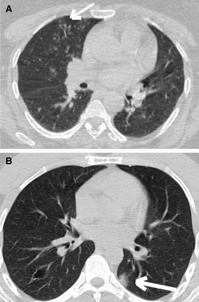
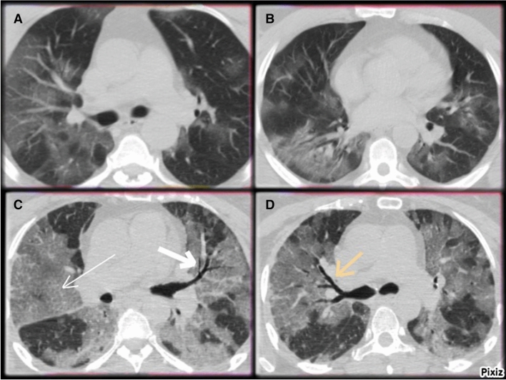
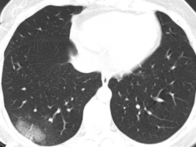
The increasing tendency of chest CT usage throughout the COVID-19 epidemic requires new tools and a systematic scheme for diagnosing and assessing the lung involvement in Coronavirus Disease 2019 (COVID-19). To investigate the use of the COVID-19 Reporting and Data System (CO-RADS) classification and chest CT Involvement Score (CT-IS) in COVID-19 pneumonia.

MATERIAL AND METHODS

This retrospective study enrolled 280 hospitalized patients diagnosed with COVID-19 pneumonia in a tertiary hospital in Turkey. All patients underwent non-contrast CT chest imaging. Two radiologists interpreted all CT images according to CO-RADS classification without knowing the clinical features, laboratory findings. We used CT involvement score (CT-IS) for assessing chest CT images of COVID-19 patients. Also, we examined the relationship between CT-IS and clinical outcomes in COVID-19 patients.

RESULTS

Of the patients, 111(39.6%) had positive real-time reverse transcriptase-polymerase chain reaction (RT-PCR) results. CO-RADS 5 group patients had statistically significant positive RT-PCR results than the other groups (P < 0.001). All of the CO-RADS 2 group patients (30) had negative RT-PCR results. The mean total CT-IS in CO-RADS 2 group was 3.4 ± 2.8. The mean total CT-IS in CO-RADS 5 group was 8.2 ± 4.7. Total CT-IS was statistically significantly different among CO-RADS groups (P < 0.001). The mean total CT-IS was statistically significantly different between survivors and patients died of COVID-19 pneumonia (P < 0.001).

CONCLUSIONS

CO-RADS is useful in detecting COVID-19 disease, even if RT-PCR testing is negative. CT-IS is also helpful as an imaging tool for evaluation of the severity and extent of COVID-19 pneumonia.

摘要

目的

在整个 COVID-19 疫情期间,胸部 CT 使用呈上升趋势,这需要新的工具和系统方案来诊断和评估 2019 年冠状病毒病(COVID-19)肺部受累情况。探讨 COVID-19 报告和数据系统(CO-RADS)分类和胸部 CT 受累评分(CT-IS)在 COVID-19 肺炎中的应用。

材料与方法

本回顾性研究纳入了土耳其一家三级医院 280 例住院 COVID-19 肺炎患者。所有患者均行非对比 CT 胸部成像。两名放射科医生根据 CO-RADS 分类解读所有 CT 图像,而不了解临床特征、实验室结果。我们使用 CT 受累评分(CT-IS)评估 COVID-19 患者的胸部 CT 图像。此外,我们还检查了 COVID-19 患者 CT-IS 与临床结局之间的关系。

结果

患者中,111 例(39.6%)实时逆转录聚合酶链反应(RT-PCR)检测结果阳性。CO-RADS 5 组患者的 RT-PCR 检测结果显著高于其他组(P<0.001)。CO-RADS 2 组所有患者(30 例)的 RT-PCR 检测结果均为阴性。CO-RADS 2 组的平均总 CT-IS 为 3.4±2.8。CO-RADS 5 组的平均总 CT-IS 为 8.2±4.7。CO-RADS 组间的总 CT-IS 差异有统计学意义(P<0.001)。幸存者与 COVID-19 肺炎死亡患者的总 CT-IS 差异有统计学意义(P<0.001)。

结论

即使 RT-PCR 检测结果为阴性,CO-RADS 也有助于检测 COVID-19 疾病。CT-IS 也是评估 COVID-19 肺炎严重程度和范围的有用影像学工具。

https://cdn.ncbi.nlm.nih.gov/pmc/blobs/8e76/7880207/6fb362f5aee6/11547_2021_1335_Fig3_HTML.jpg
https://cdn.ncbi.nlm.nih.gov/pmc/blobs/8e76/7880207/f8e94507f297/11547_2021_1335_Fig1_HTML.jpg
https://cdn.ncbi.nlm.nih.gov/pmc/blobs/8e76/7880207/adacfab7f84e/11547_2021_1335_Fig2_HTML.jpg
https://cdn.ncbi.nlm.nih.gov/pmc/blobs/8e76/7880207/6fb362f5aee6/11547_2021_1335_Fig3_HTML.jpg
https://cdn.ncbi.nlm.nih.gov/pmc/blobs/8e76/7880207/f8e94507f297/11547_2021_1335_Fig1_HTML.jpg
https://cdn.ncbi.nlm.nih.gov/pmc/blobs/8e76/7880207/adacfab7f84e/11547_2021_1335_Fig2_HTML.jpg
https://cdn.ncbi.nlm.nih.gov/pmc/blobs/8e76/7880207/6fb362f5aee6/11547_2021_1335_Fig3_HTML.jpg

相似文献

1
Use of the COVID-19 Reporting and Data System (CO-RADS) classification and chest computed tomography involvement score (CT-IS) in COVID-19 pneumonia.在 COVID-19 肺炎中使用 COVID-19 报告和数据系统(CO-RADS)分类和胸部计算机断层扫描受累评分(CT-IS)。
Radiol Med. 2021 May;126(5):679-687. doi: 10.1007/s11547-021-01335-x. Epub 2021 Feb 12.
2
Automated Assessment of COVID-19 Reporting and Data System and Chest CT Severity Scores in Patients Suspected of Having COVID-19 Using Artificial Intelligence.利用人工智能对疑似 COVID-19 患者的 COVID-19 报告和数据系统及胸部 CT 严重程度评分进行自动评估。
Radiology. 2021 Jan;298(1):E18-E28. doi: 10.1148/radiol.2020202439. Epub 2020 Jul 30.
3
CO-RADS: A Categorical CT Assessment Scheme for Patients Suspected of Having COVID-19-Definition and Evaluation.CO-RADS:一种用于疑似 COVID-19 患者的 CT 分类评估方案——定义和评估。
Radiology. 2020 Aug;296(2):E97-E104. doi: 10.1148/radiol.2020201473. Epub 2020 Apr 27.
4
Chest CT in the Emergency Department for Diagnosis of COVID-19 Pneumonia: Dutch Experience.急诊科胸部 CT 诊断 COVID-19 肺炎:荷兰经验。
Radiology. 2021 Feb;298(2):E98-E106. doi: 10.1148/radiol.2020203465. Epub 2020 Nov 17.
5
Radiological Society of North America (RSNA) Expert Consensus Statement Related to Chest CT Findings in COVID-19 Versus CO-RADS: Comparison of Reporting System Performance Among Chest Radiologists and End-User Preference.北美放射学会(RSNA)关于 COVID-19 与 CO-RADS 胸部 CT 表现的专家共识声明:胸部放射科医生和最终用户偏好之间报告系统性能的比较。
Can Assoc Radiol J. 2021 Nov;72(4):806-813. doi: 10.1177/0846537120968919. Epub 2020 Nov 3.
6
Diagnostic Performance of Chest CT for SARS-CoV-2 Infection in Individuals with or without COVID-19 Symptoms.胸部 CT 对有或无症状 COVID-19 患者 SARS-CoV-2 感染的诊断性能。
Radiology. 2021 Jan;298(1):E30-E37. doi: 10.1148/radiol.2020202708. Epub 2020 Aug 10.
7
Evaluation of the Usefulness of CO-RADS for Chest CT in Patients Suspected of Having COVID-19.CO-RADS在疑似新型冠状病毒肺炎患者胸部CT中的应用价值评估
Diagnostics (Basel). 2020 Aug 19;10(9):608. doi: 10.3390/diagnostics10090608.
8
Comparison of the CO-RADS and the RSNA chest CT classification system concerning sensitivity and reliability for the diagnosis of COVID-19 pneumonia.CO-RADS与RSNA胸部CT分类系统在诊断新型冠状病毒肺炎的敏感性和可靠性方面的比较。
Insights Imaging. 2021 Apr 28;12(1):55. doi: 10.1186/s13244-021-00998-4.
9
Structured reporting of chest CT provides high sensitivity and specificity for early diagnosis of COVID-19 in a clinical routine setting.胸部 CT 的结构化报告在临床常规环境中对 COVID-19 的早期诊断具有较高的敏感性和特异性。
Br J Radiol. 2021 Jan 1;94(1117):20200574. doi: 10.1259/bjr.20200574. Epub 2020 Nov 27.
10
Chest CT for triage during COVID-19 on the emergency department: myth or truth?急诊科在新型冠状病毒肺炎疫情期间使用胸部CT进行分诊:是神话还是事实?
Emerg Radiol. 2020 Dec;27(6):641-651. doi: 10.1007/s10140-020-01821-1. Epub 2020 Jul 20.

引用本文的文献

1
Early Identification of Severe COVID-19 Cases and the Need for ICU Care Based on Clinical and Laboratory Risk Factors.基于临床和实验室风险因素的重症COVID-19病例的早期识别及重症监护需求
Cureus. 2025 Mar 15;17(3):e80611. doi: 10.7759/cureus.80611. eCollection 2025 Mar.
2
Tips and Tricks in Thoracic Radiology for Beginners: A Findings-Based Approach.初学者胸部放射学技巧与窍门:基于发现的方法。
Tomography. 2023 Jun 14;9(3):1153-1186. doi: 10.3390/tomography9030095.
3
Role of Imaging in the Management of Patients with SARS-CoV-2 Lung Involvement Admitted to the Emergency Department: A Systematic Review.

本文引用的文献

1
Chest CT Severity Score: An Imaging Tool for Assessing Severe COVID-19.胸部CT严重程度评分:一种评估重症COVID-19的影像学工具。
Radiol Cardiothorac Imaging. 2020 Mar 30;2(2):e200047. doi: 10.1148/ryct.2020200047. eCollection 2020 Apr.
2
Evaluation of the Usefulness of CO-RADS for Chest CT in Patients Suspected of Having COVID-19.CO-RADS在疑似新型冠状病毒肺炎患者胸部CT中的应用价值评估
Diagnostics (Basel). 2020 Aug 19;10(9):608. doi: 10.3390/diagnostics10090608.
3
Automated Assessment of COVID-19 Reporting and Data System and Chest CT Severity Scores in Patients Suspected of Having COVID-19 Using Artificial Intelligence.
影像检查在急诊科收治的新型冠状病毒肺炎患者管理中的作用:一项系统评价
Diagnostics (Basel). 2023 May 26;13(11):1856. doi: 10.3390/diagnostics13111856.
4
Artificial intelligence and radiation effects on brain tissue in glioblastoma patient: preliminary data using a quantitative tool.人工智能和辐射对胶质母细胞瘤患者脑组织的影响:使用定量工具的初步数据。
Radiol Med. 2023 Jul;128(7):813-827. doi: 10.1007/s11547-023-01655-0. Epub 2023 Jun 8.
5
Qualitative and semi-quantitative ultrasound assessment in delta and Omicron Covid-19 patients: data from high volume reference center.德尔塔和奥密克戎新冠患者的定性和半定量超声评估:来自大容量参考中心的数据。
Infect Agent Cancer. 2023 May 27;18(1):34. doi: 10.1186/s13027-023-00515-w.
6
Diagnostic performance of standardized typical CT findings for COVID-19: a systematic review and meta-analysis.COVID-19标准化典型CT表现的诊断性能:一项系统评价和荟萃分析
Insights Imaging. 2023 May 24;14(1):96. doi: 10.1186/s13244-023-01429-2.
7
Responses to COVID-19, small and medium enterprises' corporate social responsibility and psychological capital of employees: From the mediating perspective of affective commitment.对新冠疫情的应对、中小企业的企业社会责任与员工的心理资本:基于情感承诺的中介视角
Heliyon. 2023 Mar 30;9(4):e15004. doi: 10.1016/j.heliyon.2023.e15004. eCollection 2023 Apr.
8
Colorectal liver metastases patients prognostic assessment: prospects and limits of radiomics and radiogenomics.结直肠癌肝转移患者的预后评估:影像组学和影像基因组学的前景与局限
Infect Agent Cancer. 2023 Mar 16;18(1):18. doi: 10.1186/s13027-023-00495-x.
9
Post-Surgical Imaging Assessment in Rectal Cancer: Normal Findings and Complications.直肠癌术后影像学评估:正常表现与并发症
J Clin Med. 2023 Feb 13;12(4):1489. doi: 10.3390/jcm12041489.
10
Radiation Recall Pneumonitis: The Open Challenge in Differential Diagnosis of Pneumonia Induced by Oncological Treatments.放射性回忆性肺炎:肿瘤治疗所致肺炎鉴别诊断中的公开挑战
J Clin Med. 2023 Feb 10;12(4):1442. doi: 10.3390/jcm12041442.
利用人工智能对疑似 COVID-19 患者的 COVID-19 报告和数据系统及胸部 CT 严重程度评分进行自动评估。
Radiology. 2021 Jan;298(1):E18-E28. doi: 10.1148/radiol.2020202439. Epub 2020 Jul 30.
4
Coronavirus disease 2019 (COVID-19) imaging reporting and data system (COVID-RADS) and common lexicon: a proposal based on the imaging data of 37 studies.2019 年冠状病毒病(COVID-19)影像报告和数据系统(COVID-RADS)及常用词汇:基于 37 项研究的影像数据提出的建议。
Eur Radiol. 2020 Sep;30(9):4930-4942. doi: 10.1007/s00330-020-06863-0. Epub 2020 Apr 28.
5
CO-RADS: A Categorical CT Assessment Scheme for Patients Suspected of Having COVID-19-Definition and Evaluation.CO-RADS:一种用于疑似 COVID-19 患者的 CT 分类评估方案——定义和评估。
Radiology. 2020 Aug;296(2):E97-E104. doi: 10.1148/radiol.2020201473. Epub 2020 Apr 27.
6
Coronavirus Disease 2019 (COVID-19) CT Findings: A Systematic Review and Meta-analysis.新型冠状病毒病 2019(COVID-19)的 CT 表现:系统评价和荟萃分析。
J Am Coll Radiol. 2020 Jun;17(6):701-709. doi: 10.1016/j.jacr.2020.03.006. Epub 2020 Mar 25.
7
The Role of Chest Imaging in Patient Management During the COVID-19 Pandemic: A Multinational Consensus Statement From the Fleischner Society.COVID-19 大流行期间患者管理中的胸部影像学作用: Fleischner 学会的一项多国家共识声明。
Chest. 2020 Jul;158(1):106-116. doi: 10.1016/j.chest.2020.04.003. Epub 2020 Apr 7.
8
CT image visual quantitative evaluation and clinical classification of coronavirus disease (COVID-19).CT 影像视觉定量评估及新型冠状病毒肺炎(COVID-19)临床分类。
Eur Radiol. 2020 Aug;30(8):4407-4416. doi: 10.1007/s00330-020-06817-6. Epub 2020 Mar 25.
9
Relation Between Chest CT Findings and Clinical Conditions of Coronavirus Disease (COVID-19) Pneumonia: A Multicenter Study.新型冠状病毒肺炎的胸部 CT 表现与临床特征的关系:一项多中心研究。
AJR Am J Roentgenol. 2020 May;214(5):1072-1077. doi: 10.2214/AJR.20.22976. Epub 2020 Mar 3.
10
Correlation of Chest CT and RT-PCR Testing for Coronavirus Disease 2019 (COVID-19) in China: A Report of 1014 Cases.中国 2019 年冠状病毒病(COVID-19)的胸部 CT 与 RT-PCR 检测的相关性:1014 例报告。
Radiology. 2020 Aug;296(2):E32-E40. doi: 10.1148/radiol.2020200642. Epub 2020 Feb 26.