Department of Immunology and Microbiology, University of Copenhagen, Copenhagen, Denmark.
Front Immunol. 2021 Feb 2;11:595707. doi: 10.3389/fimmu.2020.595707. eCollection 2020.
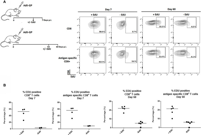
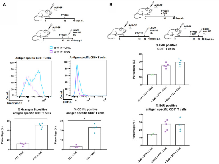
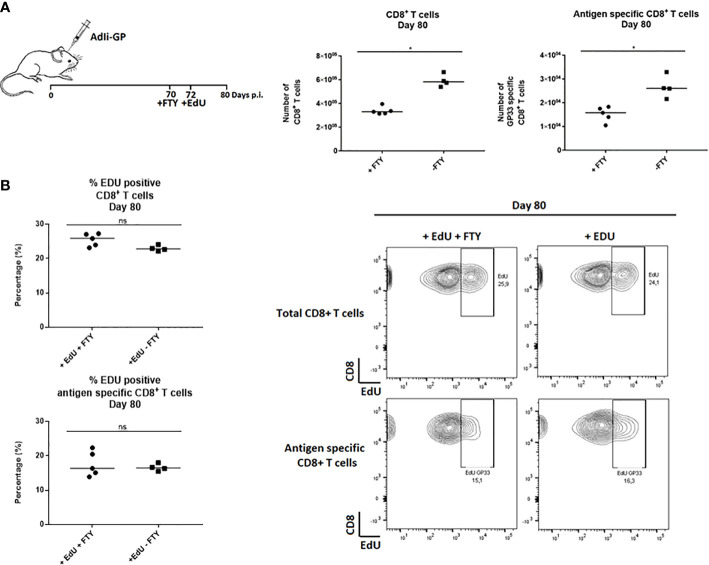
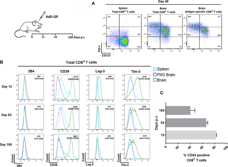
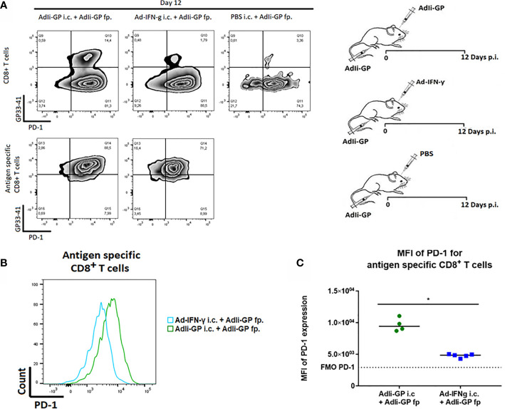
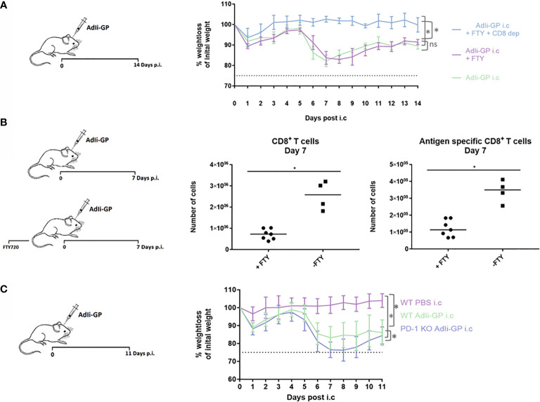
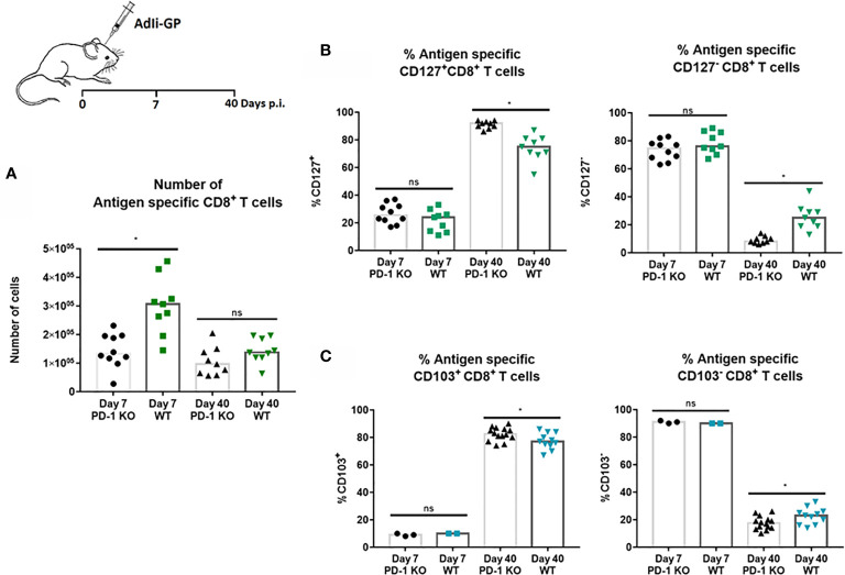
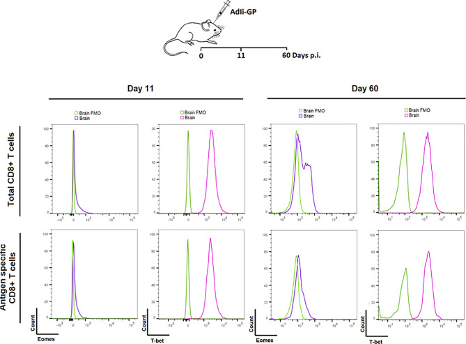
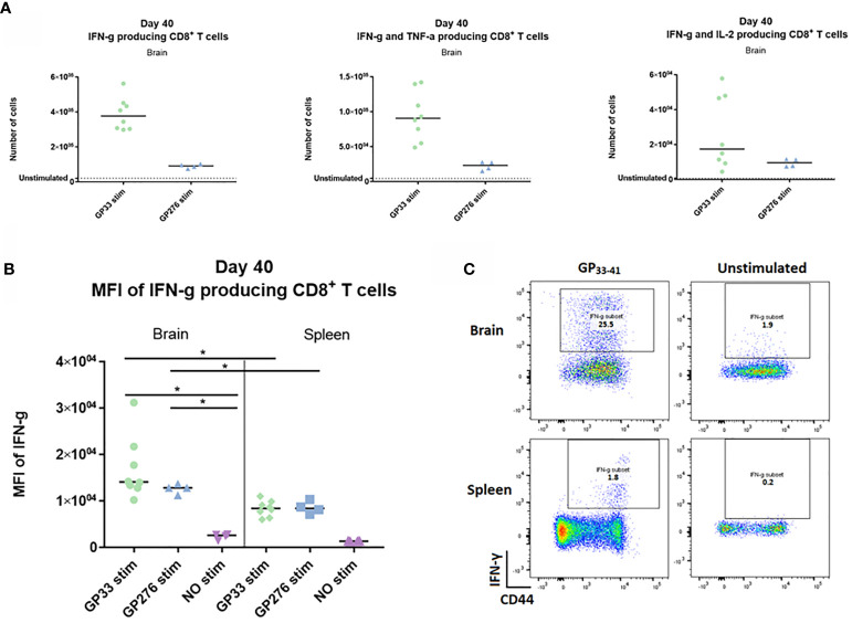
Expression of programmed cell death-1 receptor (PD-1) has traditionally been linked to T-cell exhaustion, as signaling PD-1 dampens the functionality of T-cells upon repetitive antigen exposures during chronic infections. However, resent findings pointing to the involvement of PD-1 both in T-cell survival and in restraining immunopathology, challenge the concept of PD-1 solely as marker for T-cell exhaustion. Tissue resident memory T cells (Trms) hold unique effector qualities, but within a delicate organ like the CNS, these protective abilities could potentially be harmful. In contrast to their counterparts in many other tissues, brain derived CD8 Trms have been found to uniformly and chronically express PD-1. In this study we utilized a recently established model system for generating CNS Trms in order to improve our understanding regarding the role of PD-1 expression by Trms inside the CNS. By intracerebral (i.c.) inoculation with a non-replicating adeno-viral vector, we induced a PD-1 CD8 T cell memory population within the CNS. We found that PD-1 expression lowered the severity of clinical disease associated with the i.c. inoculation. Furthermore, high levels of PD-L1 expression were found on the infiltrating monocytes and macrophages as well as on the resident microglia, oligodendrocytes and astrocytes during the acute phase of the response. Additionally, we showed that the intensity of PD-1 expression correlates with local antigen encounter and found that PD-1 expression was associated with decreased CD8 T cell memory formation in the CNS despite an increased number of infiltrating CD8 T cells. Most importantly, our experiments revealed that despite expression of PD-1 and several additional markers linked to T-cell exhaustion, Tim-3, Lag-3 and CD39, the cells did not show signs of limited effector capacity. Collectively, these results endorse the increasing amount of evidence pointing to an immune-modifying role for PD-1 expression within the CNS, a mechanism we found to correlate with local antigen exposure.
程序性细胞死亡受体 1(PD-1)的表达传统上与 T 细胞耗竭有关,因为在慢性感染期间,PD-1 信号通过重复的抗原暴露抑制 T 细胞的功能。然而,最近的研究结果表明,PD-1 不仅参与 T 细胞存活,还参与抑制免疫病理学,这挑战了 PD-1 仅作为 T 细胞耗竭标志物的概念。组织驻留记忆 T 细胞(Trms)具有独特的效应功能,但在像中枢神经系统这样的精细器官中,这些保护能力可能是有害的。与许多其他组织中的对应物不同,脑源性 CD8 Trms 被发现均匀且慢性地表达 PD-1。在这项研究中,我们利用了最近建立的中枢神经系统 Trms 生成模型系统,以提高我们对中枢神经系统内 Trms 表达 PD-1 的作用的理解。通过脑内(i.c.)接种非复制性腺病毒载体,我们在中枢神经系统内诱导了 PD-1 CD8 T 细胞记忆群体。我们发现 PD-1 表达降低了与 i.c.接种相关的临床疾病的严重程度。此外,在反应的急性期,浸润的单核细胞和巨噬细胞以及驻留的小胶质细胞、少突胶质细胞和星形胶质细胞上发现高水平的 PD-L1 表达。此外,我们表明 PD-1 表达强度与局部抗原接触相关,并且尽管浸润的 CD8 T 细胞数量增加,但 PD-1 表达与中枢神经系统中 CD8 T 细胞记忆形成减少相关。最重要的是,我们的实验表明,尽管表达了 PD-1 和其他几种与 T 细胞耗竭相关的标志物,如 Tim-3、Lag-3 和 CD39,但这些细胞没有表现出效应器功能有限的迹象。总的来说,这些结果支持越来越多的证据表明 PD-1 表达在中枢神经系统中具有免疫调节作用,我们发现这种机制与局部抗原暴露相关。