• 文献检索
  • 文档翻译
  • 深度研究
  • 学术资讯
  • Suppr Zotero 插件Zotero 插件
  • 邀请有礼
  • 套餐&价格
  • 历史记录
应用&插件
Suppr Zotero 插件Zotero 插件浏览器插件Mac 客户端Windows 客户端微信小程序
定价
高级版会员购买积分包购买API积分包
服务
文献检索文档翻译深度研究API 文档MCP 服务
关于我们
关于 Suppr公司介绍联系我们用户协议隐私条款
关注我们

Suppr 超能文献

核心技术专利:CN118964589B侵权必究
粤ICP备2023148730 号-1Suppr @ 2026

文献检索

告别复杂PubMed语法,用中文像聊天一样搜索,搜遍4000万医学文献。AI智能推荐,让科研检索更轻松。

立即免费搜索

文件翻译

保留排版,准确专业,支持PDF/Word/PPT等文件格式,支持 12+语言互译。

免费翻译文档

深度研究

AI帮你快速写综述,25分钟生成高质量综述,智能提取关键信息,辅助科研写作。

立即免费体验

对疑似新型冠状病毒肺炎患者肺部超声与胸部X线摄影比较的描述性分析。

Descriptive analysis of a comparison between lung ultrasound and chest radiography in patients suspected of COVID-19.

作者信息

Volpicelli Giovanni, Cardinale Luciano, Fraccalini Thomas, Calandri Marco, Piatti Clara, Geninatti Carlotta, Stranieri Giuseppe

机构信息

Department of Emergency Medicine, San Luigi Gonzaga University Hospital, Torino, Italy.

Department of Oncology, Radiology Unit, San Luigi Gonzaga University Hospital, Torino, Italy.

出版信息

Ultrasound J. 2021 Feb 26;13(1):11. doi: 10.1186/s13089-021-00215-9.

DOI:10.1186/s13089-021-00215-9
PMID:33635443
原文链接:https://pmc.ncbi.nlm.nih.gov/articles/PMC7907795/
Abstract

BACKGROUND

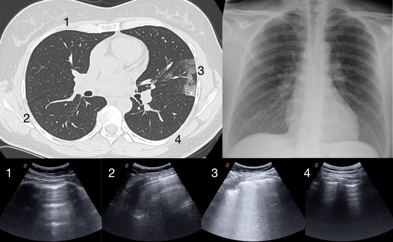
Lung ultrasound (LUS) and chest radiography (CXR) are the most used chest imaging tools in the early diagnosis of COVID-19 associated pneumonia. However, the relationship between LUS and CXR is not clearly defined. The aim of our study was to describe the comparison between LUS interpretation and CXR readings in the first approach to patients suspected of COVID-19.

METHODS

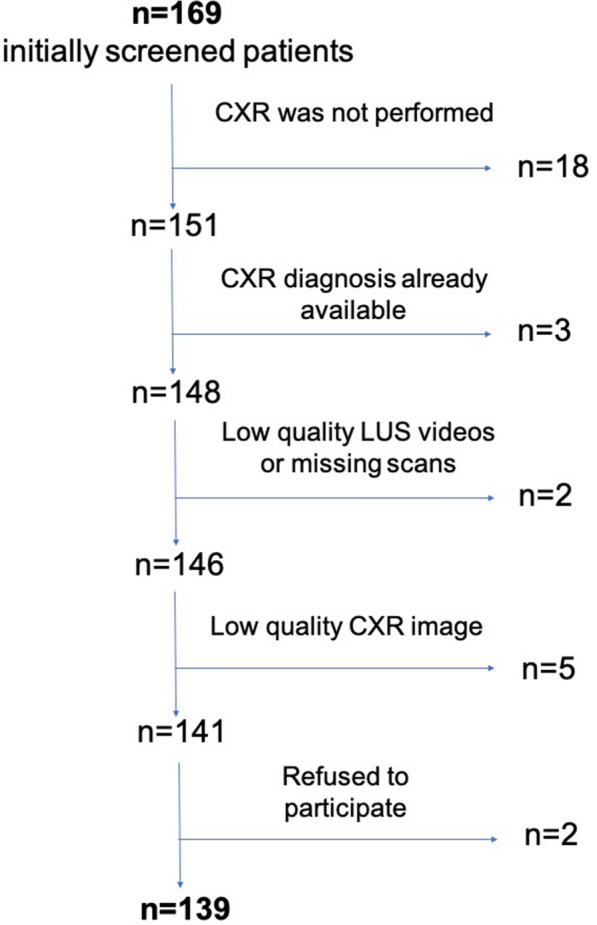
In the time of the first COVID-19 pandemic surge, we prospectively evaluated adult patients presenting to an emergency department complaining of symptoms raising suspicion of COVID-19. Patients were studied by LUS and only those performing also CXR were analyzed. All the patients performed viral reverse transcriptase-polymerase chain reaction (RT-PCR). LUS studies were classified in 4 categories of probabilities, based on the presence of typical or alternative signs of COVID-19-associated interstitial pneumonia. Accordingly, the CXR readings were retrospectively adapted by 2 experts in 4 categories following the standard language that describes the computed tomography (CT) findings. Patients were divided in two groups, based on the agreement of the LUS and CXR categories. Results were also compared to RT-PCR and, when available, to CT studies.

RESULTS

We analyzed 139 cases (55 women, mean age 59.1 ± 15.5 years old). The LUS vs CXR results disagreed in 60 (43.2%) cases. RT-PCR was positive in 88 (63.3%) cases. In 45 cases, a CT scan was also performed and only 4 disagreed with LUS interpretation versus 24 in the comparison between CT and CXR. In 18 cases, LUS detected signs of COVID-19 pneumonia (high and intermediate probabilities) while CXR reading was negative; in 14 of these cases, a CT scan or a RT-PCR-positive result confirmed the LUS interpretation. In 6 cases, LUS detected signs of alternative diagnoses to COVID-19 pneumonia while CXR was negative; in 4 of these cases, CT scan confirmed atypical findings.

CONCLUSION

Our study demonstrated a strong disagreement between LUS interpretation and CXR reading in the early approach to patients suspected of COVID-19. Comparison with CT studies and RT-PCR results seems to confirm the superiority of LUS over a second retrospective reading of CXR.

摘要

背景

肺部超声(LUS)和胸部X线摄影(CXR)是新型冠状病毒肺炎(COVID-19)相关肺炎早期诊断中最常用的胸部成像工具。然而,LUS与CXR之间的关系尚未明确界定。我们研究的目的是描述在对疑似COVID-19患者的初次检查中LUS解读与CXR读数之间的比较。

方法

在首次COVID-19大流行高峰期间,我们前瞻性地评估了因出现引起COVID-19怀疑症状而到急诊科就诊的成年患者。对患者进行LUS检查,仅分析那些也进行了CXR检查的患者。所有患者均进行了病毒逆转录聚合酶链反应(RT-PCR)检测。根据是否存在COVID-19相关间质性肺炎的典型或替代征象,LUS检查结果分为4类概率。相应地,2位专家按照描述计算机断层扫描(CT)结果的标准语言,将CXR读数回顾性地分为4类。根据LUS和CXR分类的一致性,将患者分为两组。结果还与RT-PCR结果进行比较,如有CT检查结果,也与CT检查结果进行比较。

结果

我们分析了139例患者(55名女性,平均年龄59.1±15.5岁)。LUS与CXR结果不一致的有60例(43.2%)。RT-PCR检测阳性的有88例(63.3%)。45例患者还进行了CT扫描,其中只有4例与LUS解读结果不一致,而CT与CXR比较有24例不一致。18例患者中,LUS检测到COVID-19肺炎征象(高概率和中概率),而CXR读数为阴性;其中14例患者,CT扫描或RT-PCR阳性结果证实了LUS解读结果。6例患者中,LUS检测到COVID-19肺炎替代诊断的征象,而CXR为阴性;其中4例患者,CT扫描证实了非典型表现。

结论

我们的研究表明,在对疑似COVID-19患者的早期检查中,LUS解读与CXR读数之间存在很大差异。与CT检查结果和RT-PCR结果的比较似乎证实了LUS优于对CXR的第二次回顾性读数。

https://cdn.ncbi.nlm.nih.gov/pmc/blobs/84be/7910398/96769e9358d2/13089_2021_215_Fig5_HTML.jpg
https://cdn.ncbi.nlm.nih.gov/pmc/blobs/84be/7910398/1e4f565feb9c/13089_2021_215_Fig1_HTML.jpg
https://cdn.ncbi.nlm.nih.gov/pmc/blobs/84be/7910398/50d7314b7492/13089_2021_215_Fig2_HTML.jpg
https://cdn.ncbi.nlm.nih.gov/pmc/blobs/84be/7910398/e3eb8a17fbad/13089_2021_215_Fig3_HTML.jpg
https://cdn.ncbi.nlm.nih.gov/pmc/blobs/84be/7910398/4598b433f6af/13089_2021_215_Fig4_HTML.jpg
https://cdn.ncbi.nlm.nih.gov/pmc/blobs/84be/7910398/96769e9358d2/13089_2021_215_Fig5_HTML.jpg

相似文献

1
Descriptive analysis of a comparison between lung ultrasound and chest radiography in patients suspected of COVID-19.对疑似新型冠状病毒肺炎患者肺部超声与胸部X线摄影比较的描述性分析。
Ultrasound J. 2021 Feb 26;13(1):11. doi: 10.1186/s13089-021-00215-9.
2
Lung Ultrasound vs. Chest X-Ray Study for the Radiographic Diagnosis of COVID-19 Pneumonia in a High-Prevalence Population.肺部超声与 X 线胸片对高流行地区 COVID-19 肺炎的放射学诊断比较。
J Emerg Med. 2021 May;60(5):615-625. doi: 10.1016/j.jemermed.2021.01.041. Epub 2021 Feb 4.
3
Comparison of Lung Ultrasound versus Chest X-ray for Detection of Pulmonary Infiltrates in COVID-19.新型冠状病毒肺炎中肺部超声与胸部X线检查对肺浸润检测的比较
Diagnostics (Basel). 2021 Feb 22;11(2):373. doi: 10.3390/diagnostics11020373.
4
Lung Ultrasonography: A Viable Alternative to Chest Radiography in Children with Suspected Pneumonia?肺部超声检查:对于疑似肺炎的儿童,它是胸部X线摄影的可行替代方法吗?
J Pediatr. 2016 Sep;176:93-98.e7. doi: 10.1016/j.jpeds.2016.05.033. Epub 2016 Jun 16.
5
Correlation between Chest Computed Tomography and Lung Ultrasonography in Patients with Coronavirus Disease 2019 (COVID-19).新型冠状病毒肺炎(COVID-19)患者的胸部计算机断层扫描与肺部超声的相关性。
Ultrasound Med Biol. 2020 Nov;46(11):2918-2926. doi: 10.1016/j.ultrasmedbio.2020.07.003. Epub 2020 Jul 13.
6
Lung ultrasound in the emergency department - a valuable tool in the management of patients presenting with respiratory symptoms during the SARS-CoV-2 pandemic.急诊科肺部超声 - SARS-CoV-2 大流行期间,处理呼吸系统症状患者的有价值工具。
BMC Emerg Med. 2020 Dec 7;20(1):96. doi: 10.1186/s12873-020-00389-w.
7
Appropriateness of lung ultrasound for the diagnosis of COVID-19 pneumonia.肺部超声用于诊断新型冠状病毒肺炎的适用性
Health Sci Rep. 2021 May 24;4(2):e302. doi: 10.1002/hsr2.302. eCollection 2021 Jun.
8
Point-of-care Lung Ultrasound Is More Sensitive than Chest Radiograph for Evaluation of COVID-19.床旁肺部超声比胸部 X 线摄影对 COVID-19 的评估更敏感。
West J Emerg Med. 2020 Jun 19;21(4):771-778. doi: 10.5811/westjem.2020.5.47743.
9
Comparison of admission chest computed tomography and lung ultrasound performance for diagnosis of COVID-19 pneumonia in populations with different disease prevalence.比较不同疾病流行人群中入院时胸部 CT 与肺部超声对 COVID-19 肺炎的诊断性能。
Eur J Radiol. 2020 Dec;133:109344. doi: 10.1016/j.ejrad.2020.109344. Epub 2020 Oct 8.
10
Lung Ultrasound in the Diagnosis of COVID-19 Pneumonia: Not Always and Not Only What Is COVID-19 "Glitters".肺部超声在COVID-19肺炎诊断中的应用:并非总是且不仅仅是COVID-19的“闪光点”。
Front Med (Lausanne). 2021 Jul 19;8:707602. doi: 10.3389/fmed.2021.707602. eCollection 2021.

引用本文的文献

1
Ten Questions on Using Lung Ultrasonography to Diagnose and Manage Pneumonia in the Hospital-at-Home Model: Part I-Techniques and Patterns.关于在居家医院模式下使用肺部超声诊断和管理肺炎的十个问题:第一部分——技术与模式
Diagnostics (Basel). 2024 Dec 13;14(24):2799. doi: 10.3390/diagnostics14242799.
2
[Diagnostic comparison of bedside lung ultrasound and chest radiography in the intensive care unit].[重症监护病房床旁肺部超声与胸部X线摄影的诊断比较]
An Sist Sanit Navar. 2024 Nov 15;47(3):e1088. doi: 10.23938/ASSN.1088.
3
Point-of-care ultrasound use in COVID-19: a narrative review.

本文引用的文献

1
Lung ultrasound for the early diagnosis of COVID-19 pneumonia: an international multicenter study.肺部超声在 COVID-19 肺炎早期诊断中的应用:一项国际多中心研究。
Intensive Care Med. 2021 Apr;47(4):444-454. doi: 10.1007/s00134-021-06373-7. Epub 2021 Mar 20.
2
Lung Ultrasonography for the Diagnosis of SARS-CoV-2 Pneumonia in the Emergency Department.急诊科应用肺部超声诊断新型冠状病毒肺炎
Ann Emerg Med. 2021 Apr;77(4):385-394. doi: 10.1016/j.annemergmed.2020.10.008. Epub 2020 Oct 13.
3
False-negative results of initial RT-PCR assays for COVID-19: A systematic review.
即时超声在2019冠状病毒病中的应用:一项叙述性综述。
Ann Transl Med. 2024 Feb 1;12(1):13. doi: 10.21037/atm-23-1403. Epub 2023 Jul 13.
4
Chest X-rays and Lung Ultrasound Are Not Interchangeable in Intensive Care Practice.在重症监护实践中,胸部X光和肺部超声不可相互替代。
Diagnostics (Basel). 2023 Dec 29;14(1):82. doi: 10.3390/diagnostics14010082.
5
The correlation between point-of-care ultrasound and digital tomosynthesis when used with suspected COVID-19 pneumonia patients in primary care.在基层医疗中,即时超声与数字断层合成技术用于疑似新冠肺炎肺炎患者时的相关性。
Ultrasound J. 2022 Feb 22;14(1):11. doi: 10.1186/s13089-022-00257-7.
6
Retrospective analysis of the application of CT scan in the emergency department to screen clinically asymptomatic COVID-19 before hospital admission.回顾性分析 CT 扫描在急诊入院前对临床无症状 COVID-19 的筛查应用。
Emerg Radiol. 2022 Apr;29(2):235-241. doi: 10.1007/s10140-022-02016-6. Epub 2022 Jan 8.
7
Implementing Lung Ultrasound in the Outpatient Management of COVID-19 Pneumonia: A Pilot Study to Update Local Guidelines.在COVID-19肺炎门诊管理中实施肺部超声检查:一项更新当地指南的初步研究
Front Med (Lausanne). 2021 Nov 26;8:774035. doi: 10.3389/fmed.2021.774035. eCollection 2021.
8
Assessing COVID-19 pneumonia-Clinical extension and risk with point-of-care ultrasound: A multicenter, prospective, observational study.评估新型冠状病毒肺炎-床旁超声检查的临床扩展及风险:一项多中心、前瞻性、观察性研究。
J Am Coll Emerg Physicians Open. 2021 May 1;2(3):e12429. doi: 10.1002/emp2.12429. eCollection 2021 Jun.
COVID-19 初始 RT-PCR 检测的假阴性结果:系统评价。
PLoS One. 2020 Dec 10;15(12):e0242958. doi: 10.1371/journal.pone.0242958. eCollection 2020.
4
Thoracic imaging tests for the diagnosis of COVID-19.用于诊断新型冠状病毒肺炎的胸部影像学检查
Cochrane Database Syst Rev. 2020 Nov 26;11:CD013639. doi: 10.1002/14651858.CD013639.pub3.
5
Chest X-ray findings in a large cohort of 1117 patients with SARS-CoV-2 infection: a multicenter study during COVID-19 outbreak in Italy.1117 例 SARS-CoV-2 感染患者的大队列胸部 X 线检查结果:意大利 COVID-19 爆发期间的一项多中心研究。
Intern Emerg Med. 2021 Aug;16(5):1173-1181. doi: 10.1007/s11739-020-02561-3. Epub 2020 Nov 20.
6
Comparison of Chest Ultrasound and Standard X-Ray Imaging in COVID-19 Patients.COVID-19患者胸部超声与标准X线成像的比较
Ultrasound Int Open. 2020 Sep;6(2):E36-E40. doi: 10.1055/a-1217-1603. Epub 2020 Sep 2.
7
Lung Ultrasound for Patients With Coronavirus Disease 2019 Pulmonary Disease.新冠肺炎肺部疾病患者的肺部超声检查。
Chest. 2021 Jan;159(1):205-211. doi: 10.1016/j.chest.2020.08.2054. Epub 2020 Aug 21.
8
Comparative study of lung ultrasound and chest computed tomography scan in the assessment of severity of confirmed COVID-19 pneumonia.肺超声与胸部 CT 扫描在评估确诊 COVID-19 肺炎严重程度中的对比研究。
Intensive Care Med. 2020 Sep;46(9):1707-1713. doi: 10.1007/s00134-020-06186-0. Epub 2020 Jul 29.
9
Point-of-care Lung Ultrasound Is More Sensitive than Chest Radiograph for Evaluation of COVID-19.床旁肺部超声比胸部 X 线摄影对 COVID-19 的评估更敏感。
West J Emerg Med. 2020 Jun 19;21(4):771-778. doi: 10.5811/westjem.2020.5.47743.
10
Why, when, and how to use lung ultrasound during the COVID-19 pandemic: enthusiasm and caution.在新冠疫情期间何时、为何以及如何使用肺部超声:热情与谨慎并存
Eur Heart J Cardiovasc Imaging. 2020 Sep 1;21(9):941-948. doi: 10.1093/ehjci/jeaa163.