• 文献检索
  • 文档翻译
  • 深度研究
  • 学术资讯
  • Suppr Zotero 插件Zotero 插件
  • 邀请有礼
  • 套餐&价格
  • 历史记录
应用&插件
Suppr Zotero 插件Zotero 插件浏览器插件Mac 客户端Windows 客户端微信小程序
定价
高级版会员购买积分包购买API积分包
服务
文献检索文档翻译深度研究API 文档MCP 服务
关于我们
关于 Suppr公司介绍联系我们用户协议隐私条款
关注我们

Suppr 超能文献

核心技术专利:CN118964589B侵权必究
粤ICP备2023148730 号-1Suppr @ 2026

文献检索

告别复杂PubMed语法,用中文像聊天一样搜索,搜遍4000万医学文献。AI智能推荐,让科研检索更轻松。

立即免费搜索

文件翻译

保留排版,准确专业,支持PDF/Word/PPT等文件格式,支持 12+语言互译。

免费翻译文档

深度研究

AI帮你快速写综述,25分钟生成高质量综述,智能提取关键信息,辅助科研写作。

立即免费体验

1117 例 SARS-CoV-2 感染患者的大队列胸部 X 线检查结果:意大利 COVID-19 爆发期间的一项多中心研究。

Chest X-ray findings in a large cohort of 1117 patients with SARS-CoV-2 infection: a multicenter study during COVID-19 outbreak in Italy.

机构信息

Department of Radiology, Fondazione IRCCS Ca' Granda Ospedale Maggiore Policlinico, University of Milan, Milan, Italy.

Postgraduate School of Diagnostic and Interventional Radiology, University of Milan, Milan, Italy.

出版信息

Intern Emerg Med. 2021 Aug;16(5):1173-1181. doi: 10.1007/s11739-020-02561-3. Epub 2020 Nov 20.

DOI:10.1007/s11739-020-02561-3
PMID:33216258
原文链接:https://pmc.ncbi.nlm.nih.gov/articles/PMC7677441/
Abstract

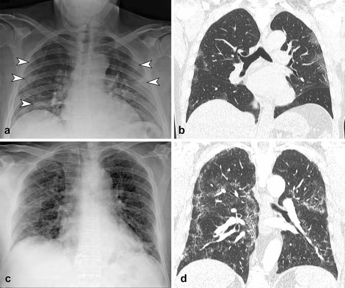
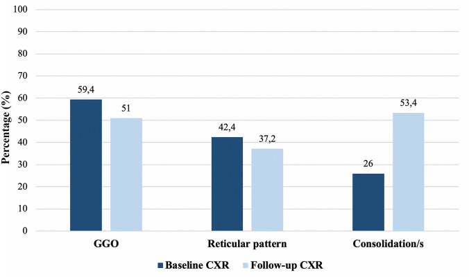
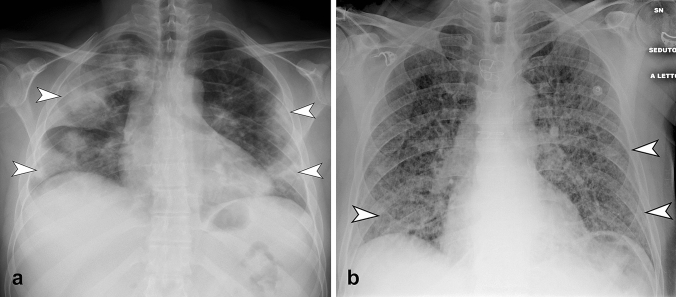
To describe radiographic key patterns on Chest X-ray (CXR) in patients with SARS-CoV-2 infection, assessing the prevalence of radiographic signs of interstitial pneumonia. To evaluate pattern variation between a baseline and a follow-up CXR. 1117 patients tested positive for SARS-CoV-2 infection were retrospectively enrolled from four centers in Lombardy region. All patients underwent a CXR at presentation. Follow-up CXR was performed when clinically indicated. Two radiologists in each center reviewed images and classified them as suggestive or not for interstitial pneumonia, recording the presence of ground-glass opacity (GGO), reticular pattern or consolidation and their distribution. Pearson's χ test for categorical variables and McNemar test (χ for paired data) were performed. Patients mean age 63.3 years, 767 were males (65.5%). The main result is the large proportion of positive CXR in COVID-19 patients. Baseline CXR was positive in 940 patients (80.3%), with significant differences in age and sex distribution between patients with positive and negative CXR. 382 patients underwent a follow-up CXR. The most frequent pattern on baseline CXR was the GGO (66.1%), on follow-up was consolidation (53.4%). The most common distributions were peripheral and middle-lower lung zone. We described key-patterns and their distribution on CXR in a large cohort of COVID-19 patients: GGO was the most frequent finding on baseline CXR, while we found an increase in the proportion of lung consolidation on follow-up CXR. CXR proved to be a reliable tool in our cohort obtaining positive results in 80.3% of the baseline cases.

摘要

描述 SARS-CoV-2 感染患者的胸部 X 线(CXR)的放射学关键模式,评估间质性肺炎的放射学征象的发生率。评估基线和随访 CXR 之间的模式变化。从伦巴第地区的四个中心回顾性招募了 1117 名 SARS-CoV-2 感染检测呈阳性的患者。所有患者在就诊时均进行 CXR。当临床需要时进行随访 CXR。每个中心的两名放射科医生审查图像并将其分类为是否提示间质性肺炎,并记录磨玻璃影(GGO)、网状模式或实变及其分布的存在。对分类变量进行 Pearson χ检验,对配对数据进行 McNemar 检验(χ 检验)。患者的平均年龄为 63.3 岁,767 名男性(65.5%)。主要结果是 COVID-19 患者中 CXR 阳性的比例较大。940 名患者的基线 CXR 阳性,阳性和阴性 CXR 患者的年龄和性别分布存在显著差异。382 名患者进行了随访 CXR。基线 CXR 最常见的模式是 GGO(66.1%),随访时最常见的模式是实变(53.4%)。最常见的分布是外周和中下肺区。我们描述了 COVID-19 患者大队列中 CXR 的关键模式及其分布:GGO 是基线 CXR 最常见的发现,而我们发现随访 CXR 中肺实变的比例增加。CXR 在我们的队列中是一种可靠的工具,在 80.3%的基线病例中获得了阳性结果。

https://cdn.ncbi.nlm.nih.gov/pmc/blobs/22ee/8310506/ddfdaffc985b/11739_2020_2561_Fig3_HTML.jpg
https://cdn.ncbi.nlm.nih.gov/pmc/blobs/22ee/8310506/2db283536b33/11739_2020_2561_Fig1_HTML.jpg
https://cdn.ncbi.nlm.nih.gov/pmc/blobs/22ee/8310506/ec31e2cc0622/11739_2020_2561_Fig2_HTML.jpg
https://cdn.ncbi.nlm.nih.gov/pmc/blobs/22ee/8310506/ddfdaffc985b/11739_2020_2561_Fig3_HTML.jpg
https://cdn.ncbi.nlm.nih.gov/pmc/blobs/22ee/8310506/2db283536b33/11739_2020_2561_Fig1_HTML.jpg
https://cdn.ncbi.nlm.nih.gov/pmc/blobs/22ee/8310506/ec31e2cc0622/11739_2020_2561_Fig2_HTML.jpg
https://cdn.ncbi.nlm.nih.gov/pmc/blobs/22ee/8310506/ddfdaffc985b/11739_2020_2561_Fig3_HTML.jpg

相似文献

1
Chest X-ray findings in a large cohort of 1117 patients with SARS-CoV-2 infection: a multicenter study during COVID-19 outbreak in Italy.1117 例 SARS-CoV-2 感染患者的大队列胸部 X 线检查结果:意大利 COVID-19 爆发期间的一项多中心研究。
Intern Emerg Med. 2021 Aug;16(5):1173-1181. doi: 10.1007/s11739-020-02561-3. Epub 2020 Nov 20.
2
Radiographic findings in 240 patients with COVID-19 pneumonia: time-dependence after the onset of symptoms.240 例新冠肺炎患者的放射学表现:症状出现后的时间依赖性。
Eur Radiol. 2020 Nov;30(11):6161-6169. doi: 10.1007/s00330-020-06967-7. Epub 2020 May 30.
3
Chest X-ray in the emergency department during COVID-19 pandemic descending phase in Italy: correlation with patients' outcome.意大利 COVID-19 大流行下降阶段急诊科的胸部 X 射线:与患者结局的相关性。
Radiol Med. 2021 May;126(5):661-668. doi: 10.1007/s11547-020-01327-3. Epub 2021 Jan 4.
4
Chest X-ray in new Coronavirus Disease 2019 (COVID-19) infection: findings and correlation with clinical outcome.新型冠状病毒病 2019(COVID-19)感染的胸部 X 线表现:特征与临床转归的相关性。
Radiol Med. 2020 Aug;125(8):730-737. doi: 10.1007/s11547-020-01232-9. Epub 2020 Jun 9.
5
Radiographic patterns and severity scoring of COVID-19 pneumonia in children: a retrospective study.儿童 COVID-19 肺炎的影像学表现和严重程度评分:一项回顾性研究。
BMC Med Imaging. 2023 Nov 30;23(1):199. doi: 10.1186/s12880-023-01154-8.
6
Thorax computed tomography (CTX) guided ground truth annotation of CHEST radiographs (CXR) for improved classification and detection of COVID-19.胸部 CT 引导的 CHEST 射线照片(CXR)标注以提高 COVID-19 的分类和检测。
Int J Numer Method Biomed Eng. 2024 Jun;40(6):e3823. doi: 10.1002/cnm.3823. Epub 2024 Apr 8.
7
Lung Ultrasound vs. Chest X-Ray Study for the Radiographic Diagnosis of COVID-19 Pneumonia in a High-Prevalence Population.肺部超声与 X 线胸片对高流行地区 COVID-19 肺炎的放射学诊断比较。
J Emerg Med. 2021 May;60(5):615-625. doi: 10.1016/j.jemermed.2021.01.041. Epub 2021 Feb 4.
8
Progression and Resolution of COVID-19 Pneumonia on Chest Radiograph.COVID-19 肺炎在胸部 X 光片上的进展和转归。
J Coll Physicians Surg Pak. 2021 Mar;31(3):258-261. doi: 10.29271/jcpsp.2021.03.258.
9
Comparison of chest radiography and chest CT for evaluation of pediatric COVID-19 pneumonia: Does CT add diagnostic value?比较胸部 X 线摄影和胸部 CT 对儿童 COVID-19 肺炎的评估:CT 是否增加诊断价值?
Pediatr Pulmonol. 2021 Jun;56(6):1409-1418. doi: 10.1002/ppul.25313. Epub 2021 Feb 25.
10
Baseline chest X-ray in coronavirus disease 19 (COVID-19) patients: association with clinical and laboratory data.新冠肺炎(COVID-19)患者的基线胸部 X 光片:与临床和实验室数据的关联。
Radiol Med. 2020 Dec;125(12):1271-1279. doi: 10.1007/s11547-020-01272-1. Epub 2020 Sep 7.

引用本文的文献

1
COVID-19 and its enduring influence on medical imaging.新型冠状病毒肺炎及其对医学影像学的持久影响。
J Med Radiat Sci. 2022 Sep;69(3):279-281. doi: 10.1002/jmrs.608. Epub 2022 Jul 20.
2
Time course of lung ultrasound findings in patients with COVID-19 pneumonia and cardiac dysfunction.新型冠状病毒肺炎合并心脏功能障碍患者肺部超声检查结果的时间进程
Ultrasound J. 2022 Jul 7;14(1):28. doi: 10.1186/s13089-022-00278-2.
3
Chest radiography findings of COVID-19 pneumonia: a specific pattern for a confident differential diagnosis.COVID-19 肺炎的胸部 X 线摄影表现:一种用于明确鉴别诊断的特定模式。

本文引用的文献

1
Protocol for Prevention and Control of COVID-19 (Edition 6).新型冠状病毒肺炎防控方案(第六版)
China CDC Wkly. 2020 May 8;2(19):321-326. doi: 10.46234/ccdcw2020.082.
2
Imaging Profile of the COVID-19 Infection: Radiologic Findings and Literature Review.新型冠状病毒肺炎感染的影像学表现:放射学发现与文献综述
Radiol Cardiothorac Imaging. 2020 Feb 13;2(1):e200034. doi: 10.1148/ryct.2020200034. eCollection 2020 Feb.
3
Chest CT findings of COVID-19 pneumonia by duration of symptoms.以症状持续时间为特征的 COVID-19 肺炎的胸部 CT 表现。
Acta Radiol. 2022 Dec;63(12):1619-1626. doi: 10.1177/02841851211055163. Epub 2021 Nov 13.
4
A Pictorial Review of the Role of Imaging in the Detection, Management, Histopathological Correlations, and Complications of COVID-19 Pneumonia.关于影像学在新型冠状病毒肺炎检测、管理、组织病理学相关性及并发症中的作用的图文综述
Diagnostics (Basel). 2021 Mar 4;11(3):437. doi: 10.3390/diagnostics11030437.
5
Descriptive analysis of a comparison between lung ultrasound and chest radiography in patients suspected of COVID-19.对疑似新型冠状病毒肺炎患者肺部超声与胸部X线摄影比较的描述性分析。
Ultrasound J. 2021 Feb 26;13(1):11. doi: 10.1186/s13089-021-00215-9.
6
Should CT be used for the diagnosis of RT-PCR-negative suspected COVID-19 patients?CT 用于诊断 RT-PCR 阴性的疑似 COVID-19 患者是否合理?
Clin Respir J. 2021 May;15(5):491-498. doi: 10.1111/crj.13332. Epub 2021 Feb 2.
Eur J Radiol. 2020 Jun;127:109009. doi: 10.1016/j.ejrad.2020.109009. Epub 2020 Apr 18.
4
COVID-19 patients and the radiology department - advice from the European Society of Radiology (ESR) and the European Society of Thoracic Imaging (ESTI).COVID-19 患者与放射科——欧洲放射学会(ESR)和欧洲胸部影像学会(ESTI)的建议。
Eur Radiol. 2020 Sep;30(9):4903-4909. doi: 10.1007/s00330-020-06865-y. Epub 2020 Apr 20.
5
The role of imaging in 2019 novel coronavirus pneumonia (COVID-19).影像学在 2019 新型冠状病毒肺炎(COVID-19)中的作用。
Eur Radiol. 2020 Sep;30(9):4874-4882. doi: 10.1007/s00330-020-06827-4. Epub 2020 Apr 15.
6
The Role of Chest Imaging in Patient Management during the COVID-19 Pandemic: A Multinational Consensus Statement from the Fleischner Society.《COVID-19 大流行期间患者管理中的胸部影像学作用:Fleischner 学会的一项多国共识声明》。
Radiology. 2020 Jul;296(1):172-180. doi: 10.1148/radiol.2020201365. Epub 2020 Apr 7.
7
Frequency and Distribution of Chest Radiographic Findings in Patients Positive for COVID-19.COVID-19 阳性患者的胸部 X 线表现的频率和分布。
Radiology. 2020 Aug;296(2):E72-E78. doi: 10.1148/radiol.2020201160. Epub 2020 Mar 27.
8
Radiology Department Preparedness for COVID-19: Scientific Expert Review Panel.放射科针对2019冠状病毒病的准备工作:科学专家评审小组
Radiology. 2020 Aug;296(2):E106-E112. doi: 10.1148/radiol.2020200988. Epub 2020 Mar 16.
9
CT Features of Coronavirus Disease 2019 (COVID-19) Pneumonia in 62 Patients in Wuhan, China.中国武汉 62 例 2019 年冠状病毒病(COVID-19)肺炎的 CT 特征。
AJR Am J Roentgenol. 2020 Jun;214(6):1287-1294. doi: 10.2214/AJR.20.22975. Epub 2020 Mar 5.
10
The response of Milan's Emergency Medical System to the COVID-19 outbreak in Italy.米兰紧急医疗系统对意大利新冠疫情的应对。
Lancet. 2020 Mar 14;395(10227):e49-e50. doi: 10.1016/S0140-6736(20)30493-1. Epub 2020 Feb 28.