• 文献检索
  • 文档翻译
  • 深度研究
  • 学术资讯
  • Suppr Zotero 插件Zotero 插件
  • 邀请有礼
  • 套餐&价格
  • 历史记录
应用&插件
Suppr Zotero 插件Zotero 插件浏览器插件Mac 客户端Windows 客户端微信小程序
定价
高级版会员购买积分包购买API积分包
服务
文献检索文档翻译深度研究API 文档MCP 服务
关于我们
关于 Suppr公司介绍联系我们用户协议隐私条款
关注我们

Suppr 超能文献

核心技术专利:CN118964589B侵权必究
粤ICP备2023148730 号-1Suppr @ 2026

文献检索

告别复杂PubMed语法,用中文像聊天一样搜索,搜遍4000万医学文献。AI智能推荐,让科研检索更轻松。

立即免费搜索

文件翻译

保留排版,准确专业,支持PDF/Word/PPT等文件格式,支持 12+语言互译。

免费翻译文档

深度研究

AI帮你快速写综述,25分钟生成高质量综述,智能提取关键信息,辅助科研写作。

立即免费体验

影像学在 2019 新型冠状病毒肺炎(COVID-19)中的作用。

The role of imaging in 2019 novel coronavirus pneumonia (COVID-19).

机构信息

Department of Magnetic Resonance Imaging, Fuwai Hospital, National Center for Cardiovascular Diseases, Chinese Academy of Medical Sciences and Peking Union Medical College, #167 Beilishi Road, Xicheng District, Beijing, 100037, China.

Department of Clinical Medicine, Wuhan University, Wuhan, Hubei, 430071, China.

出版信息

Eur Radiol. 2020 Sep;30(9):4874-4882. doi: 10.1007/s00330-020-06827-4. Epub 2020 Apr 15.

DOI:10.1007/s00330-020-06827-4
PMID:32296940
原文链接:https://pmc.ncbi.nlm.nih.gov/articles/PMC7156903/
Abstract

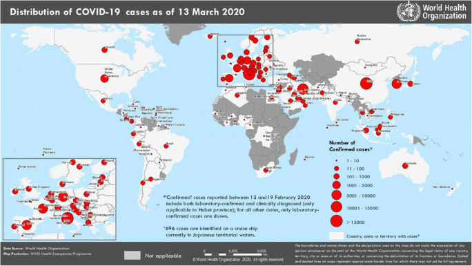
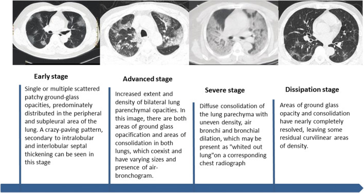
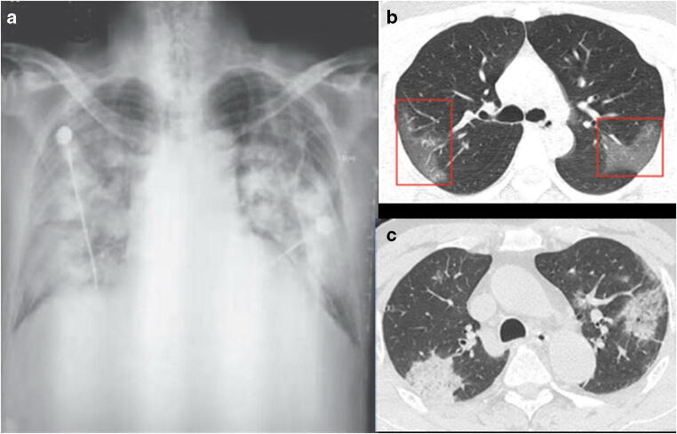
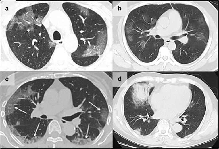
Almost the entire world, not only China, is currently experiencing the outbreak of a novel coronavirus that causes respiratory disease, severe pneumonia, and even death. The outbreak began in Wuhan, China, in December of 2019 and is currently still ongoing. This novel coronavirus is highly contagious and has resulted in a continuously increasing number of infections and deaths that have already surpassed the SARS-CoV outbreak that occurred in China between 2002 and 2003. It is now officially a pandemic, announced by WHO on the 11th of March. Currently, the 2019 novel coronavirus (SARS-CoV-2) can be identified by virus isolation or viral nucleic acid detection; however, false negatives associated with the nucleic acid detection provide a clinical challenge and thus make the imaging examination crucial. Imaging exams have been a main clinical diagnostic criteria for the 2019 novel coronavirus disease (COVID-19) in China. Imaging features of multiple patchy areas of ground glass opacity and consolidation predominately in the periphery of the lungs are characteristic manifestations on chest CT and extremely helpful in the early detection and diagnosis of this disease, which aids prompt diagnosis and the eventual control of this emerging global health emergency. Key Points • In December 2019, China, an outbreak of pneumonia caused by a novel, highly contagious coronavirus raised grave concerns and posed a huge threat to global public health. • Among the infected patients, characteristic findings on CT imaging include multiple, patchy, ground-glass opacity, crazy-paving pattern, and consolidation shadows, mainly distributed in the peripheral and subpleural areas of both lungs, which are very helpful for the frontline clinicians. • Imaging examination has become the indispensable means not only in the early detection and diagnosis but also in monitoring the clinical course, evaluating the disease severity, and may be presented as an important warning signal preceding the negative RT-PCR test results.

摘要

几乎整个世界,不仅是中国,目前都在经历一种新型冠状病毒引发的呼吸道疾病、重症肺炎,甚至死亡。该疫情始于 2019 年 12 月的中国武汉,目前仍在持续。这种新型冠状病毒具有高度传染性,导致感染和死亡人数不断增加,已经超过了 2002 年至 2003 年在中国发生的 SARS-CoV 疫情。现在世界卫生组织于 3 月 11 日正式宣布该疫情为大流行。目前,可以通过病毒分离或病毒核酸检测来识别 2019 年新型冠状病毒(SARS-CoV-2);然而,核酸检测的假阴性结果给临床带来了挑战,因此影像学检查至关重要。影像学检查一直是中国 2019 年新型冠状病毒病(COVID-19)的主要临床诊断标准。胸部 CT 上的主要特征表现为多发斑片状磨玻璃影和实变影,主要位于肺部外周,这是 COVID-19 的特征性表现,有助于早期发现和诊断这种疾病,有助于及时诊断和最终控制这一新兴的全球卫生紧急情况。关键点•2019 年 12 月,中国发生了一种新型、高度传染性冠状病毒引起的肺炎疫情,引起了严重关注,并对全球公共卫生构成了巨大威胁。•在感染患者中,CT 影像学上的特征性表现包括多发斑片状磨玻璃影、铺路石征和实变影,主要分布在双肺的外周和胸膜下区,这对一线临床医生非常有帮助。•影像学检查不仅是早期发现和诊断的不可或缺手段,也是监测临床病程、评估疾病严重程度的手段,并且可能成为 RT-PCR 检测结果转为阴性之前的重要预警信号。

https://cdn.ncbi.nlm.nih.gov/pmc/blobs/28aa/7156903/ed89e212e9ac/330_2020_6827_Fig4_HTML.jpg
https://cdn.ncbi.nlm.nih.gov/pmc/blobs/28aa/7156903/b487bd17eed5/330_2020_6827_Fig1_HTML.jpg
https://cdn.ncbi.nlm.nih.gov/pmc/blobs/28aa/7156903/ceb9d2c4befb/330_2020_6827_Fig2_HTML.jpg
https://cdn.ncbi.nlm.nih.gov/pmc/blobs/28aa/7156903/f43848d03604/330_2020_6827_Fig3_HTML.jpg
https://cdn.ncbi.nlm.nih.gov/pmc/blobs/28aa/7156903/ed89e212e9ac/330_2020_6827_Fig4_HTML.jpg

相似文献

1
The role of imaging in 2019 novel coronavirus pneumonia (COVID-19).影像学在 2019 新型冠状病毒肺炎(COVID-19)中的作用。
Eur Radiol. 2020 Sep;30(9):4874-4882. doi: 10.1007/s00330-020-06827-4. Epub 2020 Apr 15.
2
Imaging and clinical features of patients with 2019 novel coronavirus SARS-CoV-2.新型冠状病毒 2019 年 SARS-CoV-2 患者的影像学和临床特征。
Eur J Nucl Med Mol Imaging. 2020 May;47(5):1275-1280. doi: 10.1007/s00259-020-04735-9. Epub 2020 Feb 28.
3
Similarities and Differences of Early Pulmonary CT Features of Pneumonia Caused by SARS-CoV-2, SARS-CoV and MERS-CoV: Comparison Based on a Systemic Review.新型冠状病毒、严重急性呼吸综合征冠状病毒和中东呼吸综合征冠状病毒所致肺炎早期肺部CT特征的异同:基于系统评价的比较
Chin Med Sci J. 2020 Sep 30;35(3):254-261. doi: 10.24920/003727.
4
Imaging features of the initial chest thin-section CT scans from 110 patients after admission with suspected or confirmed diagnosis of COVID-19.110 例疑似或确诊 COVID-19 患者入院后首次胸部薄层 CT 扫描的影像学特征。
BMC Med Imaging. 2020 Jun 15;20(1):64. doi: 10.1186/s12880-020-00464-5.
5
[Clinical features and high resolution CT imaging evolution of coronavirus disease 2019].新型冠状病毒肺炎的临床特征及高分辨率CT影像学演变
Zhonghua Jie He He Hu Xi Za Zhi. 2020 Jun 12;43(6):509-515. doi: 10.3760/cma.j.cn112147-20200214-00094.
6
Diagnostic value and key features of computed tomography in Coronavirus Disease 2019.计算机断层扫描在 2019 年冠状病毒病中的诊断价值和关键特征。
Emerg Microbes Infect. 2020 Dec;9(1):787-793. doi: 10.1080/22221751.2020.1750307.
7
Clinical and radiological features of novel coronavirus pneumonia.新型冠状病毒肺炎的临床和影像学特征。
J Xray Sci Technol. 2020;28(3):391-404. doi: 10.3233/XST-200687.
8
Chest CT features of coronavirus disease 2019 (COVID-19) pneumonia: key points for radiologists.COVID-19 肺炎的胸部 CT 特征:放射科医生的关键点。
Radiol Med. 2020 Jul;125(7):636-646. doi: 10.1007/s11547-020-01237-4. Epub 2020 Jun 4.
9
Coronavirus Disease 2019 (COVID-19): A Perspective from China.2019 冠状病毒病(COVID-19):来自中国的视角。
Radiology. 2020 Aug;296(2):E15-E25. doi: 10.1148/radiol.2020200490. Epub 2020 Feb 21.
10
Initial CT findings and temporal changes in patients with the novel coronavirus pneumonia (2019-nCoV): a study of 63 patients in Wuhan, China.新型冠状病毒肺炎(2019-nCoV)患者的初始 CT 表现及时间演变:中国武汉 63 例患者研究。
Eur Radiol. 2020 Jun;30(6):3306-3309. doi: 10.1007/s00330-020-06731-x. Epub 2020 Feb 13.

引用本文的文献

1
Differences in chest imaging between Omicron and non-Omicron coronavirus disease 2019 (COVID-19) patients: a systematic review and meta-analysis.奥密克戎与非奥密克戎新型冠状病毒肺炎(COVID-19)患者胸部影像学差异:一项系统评价与荟萃分析
BMC Infect Dis. 2025 Apr 29;25(1):631. doi: 10.1186/s12879-025-11032-z.
2
[Distanciamiento social por COVID-19: efectos en la rutina de estudiantes universitáriosSocial distancing during COVID-19: Effects on daily routine of college students].[新冠疫情期间的社交距离:对大学生日常活动的影响。新冠疫情期间的社交距离:对大学生日常生活的影响]
Rev Cuid. 2022 Aug 20;13(1):e11. doi: 10.15649/cuidarte.2093. eCollection 2022 Jan-Apr.
3

本文引用的文献

1
Radiological findings from 81 patients with COVID-19 pneumonia in Wuhan, China: a descriptive study.中国武汉 81 例新冠肺炎患者的放射学特征:一项描述性研究。
Lancet Infect Dis. 2020 Apr;20(4):425-434. doi: 10.1016/S1473-3099(20)30086-4. Epub 2020 Feb 24.
2
Correlation of Chest CT and RT-PCR Testing for Coronavirus Disease 2019 (COVID-19) in China: A Report of 1014 Cases.中国 2019 年冠状病毒病(COVID-19)的胸部 CT 与 RT-PCR 检测的相关性:1014 例报告。
Radiology. 2020 Aug;296(2):E32-E40. doi: 10.1148/radiol.2020200642. Epub 2020 Feb 26.
3
Chest CT Findings in Coronavirus Disease-19 (COVID-19): Relationship to Duration of Infection.
Enhanced Multi-Model Deep Learning for Rapid and Precise Diagnosis of Pulmonary Diseases Using Chest X-Ray Imaging.
基于胸部X光成像,利用增强多模型深度学习实现肺部疾病的快速精准诊断
Diagnostics (Basel). 2025 Jan 22;15(3):248. doi: 10.3390/diagnostics15030248.
4
Artificial Intelligence-guided Total Opacity Scores and Obstructive Sleep Apnea in Adults with COVID-19 Pneumonia.人工智能指导下的COVID-19肺炎成人患者全肺实变评分与阻塞性睡眠呼吸暂停
Thorac Res Pract. 2025 Apr 30;26(3):107-114. doi: 10.4274/ThoracResPract.2024.24090. Epub 2025 Jan 20.
5
Linking and GenBank to the National Clinical Cohort Collaborative.将Linking和GenBank与国家临床队列协作项目相连接。
Learn Health Syst. 2024 Sep 12;9(1):e10457. doi: 10.1002/lrh2.10457. eCollection 2025 Jan.
6
Role of Imaging in Diagnosis and Management of COVID-19: Evidence-Based Approaches.影像学在 COVID-19 诊断和管理中的作用:循证方法。
Adv Exp Med Biol. 2024;1457:237-246. doi: 10.1007/978-3-031-61939-7_13.
7
Human-in-the-Loop-A Deep Learning Strategy in Combination with a Patient-Specific Gaussian Mixture Model Leads to the Fast Characterization of Volumetric Ground-Glass Opacity and Consolidation in the Computed Tomography Scans of COVID-19 Patients.人在回路中——一种结合患者特异性高斯混合模型的深度学习策略可快速表征COVID-19患者计算机断层扫描中的磨玻璃密度影和实变。
J Clin Med. 2024 Sep 4;13(17):5231. doi: 10.3390/jcm13175231.
8
A Literature Review on the Relative Diagnostic Accuracy of Chest CT Scans versus RT-PCR Testing for COVID-19 Diagnosis.关于胸部 CT 扫描与 RT-PCR 检测用于 COVID-19 诊断的相对诊断准确性的文献综述。
Tomography. 2024 Jun 14;10(6):935-948. doi: 10.3390/tomography10060071.
9
Association between chest X-ray score and clinical outcome in COVID-19 patients: A study on modified radiographic assessment of lung edema score (mRALE) in Indonesia.胸部 X 射线评分与 COVID-19 患者临床结局的关系:印度尼西亚改良放射性肺水肿评分(mRALE)的研究。
Narra J. 2024 Apr;4(1):e691. doi: 10.52225/narra.v4i1.691. Epub 2024 Apr 6.
10
Prevalence of Ear-Related Problems in Individuals Recovered From COVID-19.新冠康复者耳部相关问题的患病率
Iran J Otorhinolaryngol. 2024 May;36(3):489-497. doi: 10.22038/IJORL.2024.71040.3414.
胸部计算机断层扫描在 2019 年冠状病毒病(COVID-19)中的表现:与感染持续时间的关系。
Radiology. 2020 Jun;295(3):200463. doi: 10.1148/radiol.2020200463. Epub 2020 Feb 20.
4
Sensitivity of Chest CT for COVID-19: Comparison to RT-PCR.胸部CT对新型冠状病毒肺炎的敏感性:与逆转录聚合酶链反应的比较。
Radiology. 2020 Aug;296(2):E115-E117. doi: 10.1148/radiol.2020200432. Epub 2020 Feb 19.
5
Pre- and Posttreatment Chest CT Findings: 2019 Novel Coronavirus (2019-nCoV) Pneumonia.治疗前和治疗后胸部CT表现:2019新型冠状病毒(2019-nCoV)肺炎
Radiology. 2020 Apr;295(1):21. doi: 10.1148/radiol.2020200323. Epub 2020 Feb 12.
6
Chest CT for Typical Coronavirus Disease 2019 (COVID-19) Pneumonia: Relationship to Negative RT-PCR Testing.胸部 CT 与典型 2019 冠状病毒病(COVID-19)肺炎:与阴性 RT-PCR 检测的关系。
Radiology. 2020 Aug;296(2):E41-E45. doi: 10.1148/radiol.2020200343. Epub 2020 Feb 12.
7
Use of Chest CT in Combination with Negative RT-PCR Assay for the 2019 Novel Coronavirus but High Clinical Suspicion.胸部CT联合2019新型冠状病毒逆转录聚合酶链反应检测结果为阴性但临床高度怀疑的应用
Radiology. 2020 Apr;295(1):22-23. doi: 10.1148/radiol.2020200330. Epub 2020 Feb 12.
8
Evolution of CT Manifestations in a Patient Recovered from 2019 Novel Coronavirus (2019-nCoV) Pneumonia in Wuhan, China.中国武汉一名从2019新型冠状病毒(2019-nCoV)肺炎康复患者的CT表现演变
Radiology. 2020 Apr;295(1):20. doi: 10.1148/radiol.2020200269. Epub 2020 Feb 7.
9
Clinical Characteristics of 138 Hospitalized Patients With 2019 Novel Coronavirus-Infected Pneumonia in Wuhan, China.《武汉 2019 年新型冠状病毒感染的肺炎 138 例住院患者临床特征分析》
JAMA. 2020 Mar 17;323(11):1061-1069. doi: 10.1001/jama.2020.1585.
10
A rapid advice guideline for the diagnosis and treatment of 2019 novel coronavirus (2019-nCoV) infected pneumonia (standard version).新型冠状病毒(2019-nCoV)感染的肺炎快速诊治指南(标准版)。
Mil Med Res. 2020 Feb 6;7(1):4. doi: 10.1186/s40779-020-0233-6.