Pfaff Johannes A R, Füssel Bianka, Harlan Marcial E, Hubert Alexander, Bendszus Martin
Department of Neuroradiology, Heidelberg University Hospital, Im Neuenheimer Feld 400, 69120, Heidelberg, Germany.
Neurol Res Pract. 2021 Mar 2;3(1):10. doi: 10.1186/s42466-021-00109-0.
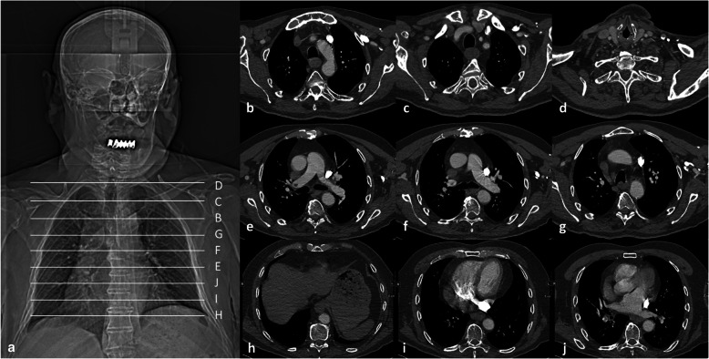
Computed tomography angiography (CTA) of the head and neck during acute ischemic stroke (AIS) usually includes visualization of lung apices. The possibility to evaluate for pulmonary changes, e.g. peripheral ground-glass and consolidative opacities suggestive of coronavirus disease 2019 (COVID-19)-related pneumonia, depends on the area of the lung covered by CTA.
We performed an analysis of a real-world scenario assessing the variability of lung coverage on CTA in patients presenting with AIS to a comprehensive stroke center (CSC) or to one of eight primary stroke centers (PSC) within a teleradiological network covered by the comprehensive stroke center in 2019.
Our final analysis included n = 940 CTA, and in n = 573 (61%) merely lung apices were covered. In 19/940 (2%) of patients no lung tissue was covered by CTA. CTA scanning protocols in the CSC began significantly more frequently at the level of the ascending aorta (CSC: n = 180 (38.2%), PSC: n = 127 (27.1%), p-value < 0.001) and the aortic arch (CSC: n = 140 (29.7%), PSC: n = 83 (17.7%), p-value < 0.001), and by this covered less frequently the lower lobes compared to CTA acquired in one of the PSC.
In our pre-COVID-19 pandemic representative stroke patient cohort, CTA for AIS covered most often only lung apices. In 37% of the patients CTA visualized at least parts of the lower lobes, the lingula or the middle lobe allowing for a more extensive assessment of the lungs.
急性缺血性卒中(AIS)期间的头颈部计算机断层血管造影(CTA)通常包括肺尖的可视化。评估肺部变化的可能性,例如提示2019冠状病毒病(COVID-19)相关肺炎的外周磨玻璃影和实变影,取决于CTA覆盖的肺部区域。
我们对一个真实世界的情况进行了分析,评估了2019年在一个综合卒中中心(CSC)或远程放射学网络中由该综合卒中中心覆盖的八个初级卒中中心(PSC)之一就诊的AIS患者CTA上肺部覆盖范围的变异性。
我们的最终分析包括n = 940次CTA,其中n = 573次(61%)仅覆盖了肺尖。在19/940(2%)的患者中,CTA未覆盖任何肺组织。CSC的CTA扫描方案在升主动脉水平(CSC:n = 180(38.2%),PSC:n = 127(27.1%),p值<0.001)和主动脉弓水平(CSC:n = 140(29.7%),PSC:n = 83(17.7%),p值<0.001)开始的频率明显更高,因此与在PSC之一进行的CTA相比,覆盖下叶的频率更低。
在我们COVID-19大流行前具有代表性的卒中患者队列中,AIS的CTA最常仅覆盖肺尖。在37%的患者中,CTA显示了下叶、舌叶或中叶的至少部分区域,从而可以对肺部进行更广泛的评估。