Stella Martina, Braat Arthur J A T, Lam Marnix G E H, de Jong Hugo W A M, van Rooij Rob
Department of Radiology and Nuclear Medicine, UMC Utrecht, Heidelberglaan 100, 3584 CX, Utrecht, The Netherlands.
EJNMMI Phys. 2021 Mar 2;8(1):22. doi: 10.1186/s40658-021-00372-9.
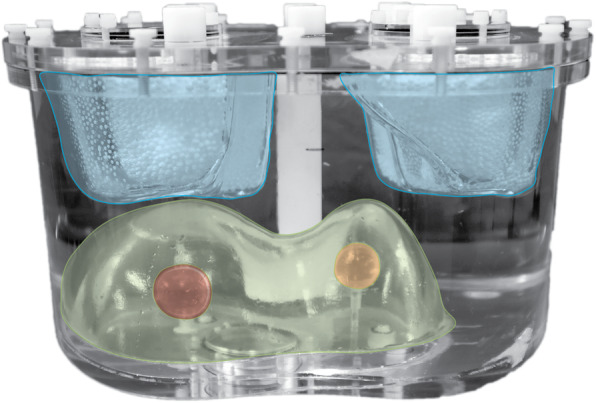
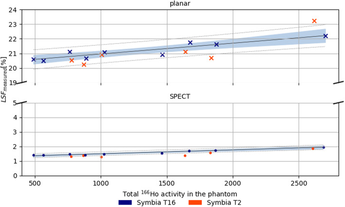
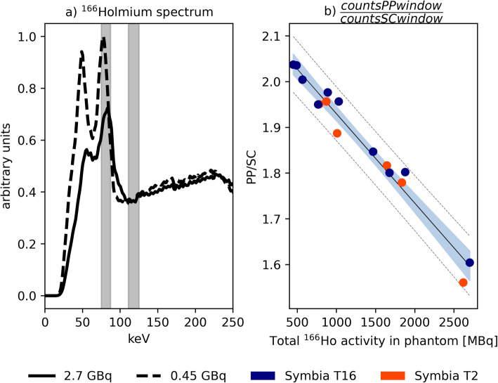
High activities of holmium-166 (Ho)-labeled microspheres are used for therapeutic radioembolization, ideally directly followed by SPECT imaging for dosimetry purposes. The resulting high-count rate potentially impacts dead time, affecting the image quality and dosimetric accuracy. This study assesses gamma camera performance and SPECT image quality at high Ho activities of several GBq. To this purpose, the liver compartment, including two tumors, of an anthropomorphic phantom was filled with Ho-chloride, with a tumor to non-tumorous liver activity concentration ratio of 10:1. Multiple SPECT/CT scans were acquired over a range of activities up to 2.7 GBq. Images were reconstructed using a commercially available protocol incorporating attenuation and scatter correction. Dead time effects were assessed from the observed count rate in the photopeak (81 keV, 15% width) and upper scatter (118 keV, 12% width) window. Post reconstruction, each image was scaled with an individual conversion factor to match the known total activity in the phantom at scanning time. The resulting activity concentration was measured in the tumors and non-tumorous liver. The image quality as a function of activity was assessed by a visual check of the absence of artifacts by a nuclear medicine physician. The apparent lung shunt fraction (nonzero due to scatter) was estimated on planar and SPECT images.
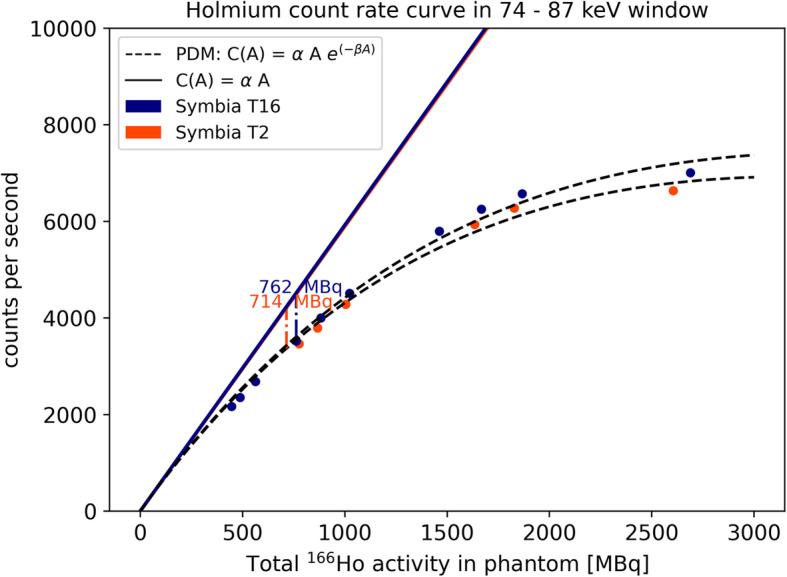
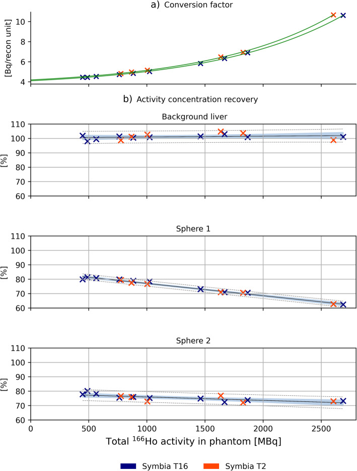
A 20% count loss due to dead time was observed around 0.7 GBq in the photopeak window. Independent of the count losses, the measured activity concentration was up to 100% of the real value for non-tumorous liver, when reconstructions were normalized to the known activity at scanning time. However, for tumor spheres, activity concentration recovery was ~80% at the lowest activity, decreasing with increasing activity in the phantom. Measured lung shunt fractions were relatively constant over the considered activity range.
At high Ho count rate, all images, visually assessed, presented no artifacts, even at considerable dead time losses. A quantitative evaluation revealed the possibility of reliable dosimetry within the healthy liver, as long as a post-reconstruction scaling to scanning activity is applied. Reliable tumor dosimetry, instead, remained hampered by the dead time.
高活度的钬 - 166(Ho)标记微球用于治疗性放射性栓塞,理想情况下紧接着进行SPECT成像以进行剂量测定。由此产生的高计数率可能会影响死时间,进而影响图像质量和剂量测定准确性。本研究评估了在数GBq的高Ho活度下γ相机的性能和SPECT图像质量。为此,在一个仿真人体模型的肝脏区域(包括两个肿瘤)中注入氯化钬,肿瘤与非肿瘤肝脏的活度浓度比为10:1。在高达2.7 GBq的一系列活度下进行了多次SPECT/CT扫描。使用包含衰减和散射校正的商用协议对图像进行重建。通过在光电峰(81 keV,15% 宽度)和上部散射(118 keV,12% 宽度)窗口中观察到的计数率来评估死时间效应。重建后,每个图像用单独的转换因子进行缩放,以匹配扫描时模型中已知的总活度。在肿瘤和非肿瘤肝脏中测量得到的活度浓度。由核医学医师通过目视检查图像是否存在伪影来评估图像质量随活度的变化。在平面图像和SPECT图像上估计表观肺分流分数(由于散射不为零)。
在光电峰窗口中,在约0.7 GBq处观察到由于死时间导致的20% 的计数损失。与计数损失无关,当重建图像根据扫描时的已知活度进行归一化时,非肿瘤肝脏的测量活度浓度高达实际值的100%。然而,对于肿瘤微球,在最低活度时活度浓度恢复率约为80%,随着模型中活度增加而降低。在所考虑的活度范围内,测量的肺分流分数相对恒定。
在高Ho计数率下,所有经目视评估的图像均未出现伪影,即使在死时间损失相当大的情况下。定量评估表明,只要应用重建后按扫描活度进行缩放,在健康肝脏内进行可靠的剂量测定是可能的。相反,死时间仍然阻碍了可靠的肿瘤剂量测定。