Dept. of Imaging Chemistry and Biology, School of Biomedical Engineering and Imaging Sciences, King's College London, St. Thomas' Hospital, London, United Kingdom.
Nanotheranostics. 2021 Feb 13;5(3):256-274. doi: 10.7150/ntno.51676. eCollection 2021.
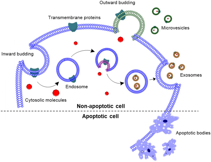
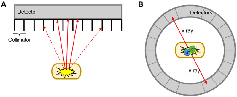
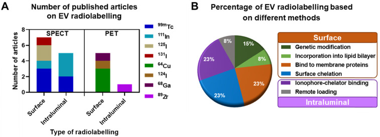
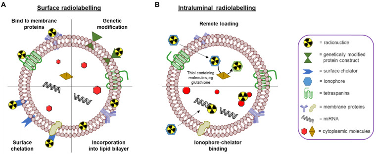
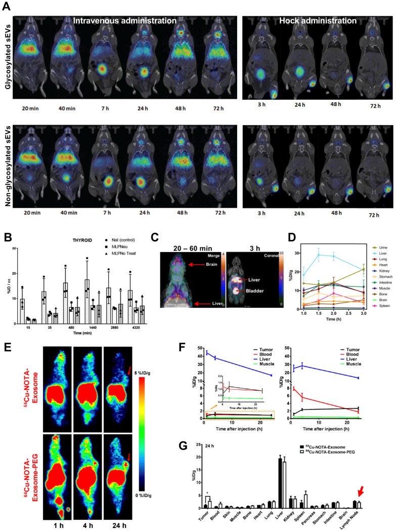
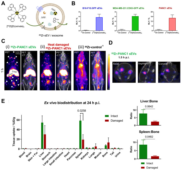
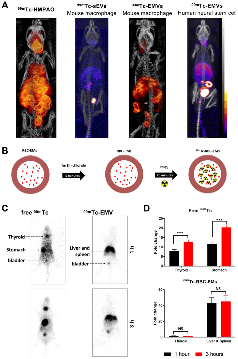
Extracellular vesicles (EVs) such as exosomes and microvesicles have gained recent attention as potential biomarkers of disease as well as nanomedicinal tools, but their behaviour remains mostly unexplored. In order to gain knowledge of their biodistribution it is important to develop imaging tools that allow us to track EVs over time and at the whole-body level. Radionuclide-based imaging (PET and SPECT) have properties that allow us to do so efficiently, mostly due to their high sensitivity, imaging signal tissue penetration, and accurate quantification. Furthermore, they can be easily translated from animals to humans. In this review, we summarise and discuss the different studies that have used PET or SPECT to study the behaviour of EVs . With a focus on the different radiolabelling methods used, we also discuss the advantages and disadvantages of each one, and the challenges of imaging EVs due to their variable stability and heterogeneity.
细胞外囊泡(EVs),如外泌体和微泡,最近作为疾病的潜在生物标志物以及纳米药物工具引起了关注,但它们的行为仍在很大程度上未被探索。为了了解它们的生物分布,开发能够随时间和在全身水平跟踪 EVs 的成像工具非常重要。基于放射性核素的成像(PET 和 SPECT)具有允许我们高效地做到这一点的特性,主要是由于它们的高灵敏度、成像信号组织穿透性和准确的定量。此外,它们可以很容易地从动物转化为人类。在这篇综述中,我们总结和讨论了使用 PET 或 SPECT 研究 EVs 行为的不同研究。我们重点介绍了使用的不同放射性标记方法,还讨论了每种方法的优缺点,以及由于 EVs 的可变稳定性和异质性而导致的成像挑战。