• 文献检索
  • 文档翻译
  • 深度研究
  • 学术资讯
  • Suppr Zotero 插件Zotero 插件
  • 邀请有礼
  • 套餐&价格
  • 历史记录
应用&插件
Suppr Zotero 插件Zotero 插件浏览器插件Mac 客户端Windows 客户端微信小程序
定价
高级版会员购买积分包购买API积分包
服务
文献检索文档翻译深度研究API 文档MCP 服务
关于我们
关于 Suppr公司介绍联系我们用户协议隐私条款
关注我们

Suppr 超能文献

核心技术专利:CN118964589B侵权必究
粤ICP备2023148730 号-1Suppr @ 2026

文献检索

告别复杂PubMed语法,用中文像聊天一样搜索,搜遍4000万医学文献。AI智能推荐,让科研检索更轻松。

立即免费搜索

文件翻译

保留排版,准确专业,支持PDF/Word/PPT等文件格式,支持 12+语言互译。

免费翻译文档

深度研究

AI帮你快速写综述,25分钟生成高质量综述,智能提取关键信息,辅助科研写作。

立即免费体验

痴呆症的神经影像学。

Neuroimaging in dementia.

机构信息

Department of Biomedical Imaging and Image-guided Therapy, Medical University of Vienna, Waehringerguertel 18-20, 1090, Vienna, Austria.

出版信息

Wien Med Wochenschr. 2021 Sep;171(11-12):274-281. doi: 10.1007/s10354-021-00825-x. Epub 2021 Mar 3.

DOI:10.1007/s10354-021-00825-x
PMID:33660199
原文链接:https://pmc.ncbi.nlm.nih.gov/articles/PMC8397686/
Abstract

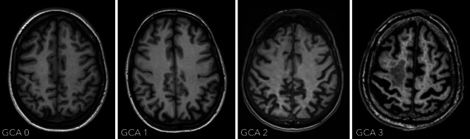
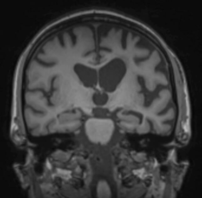
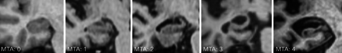
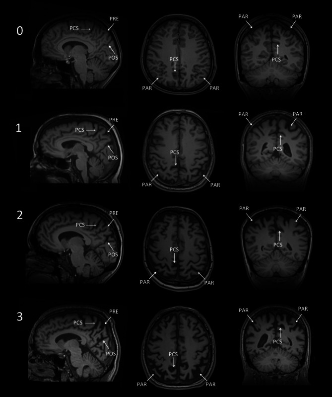
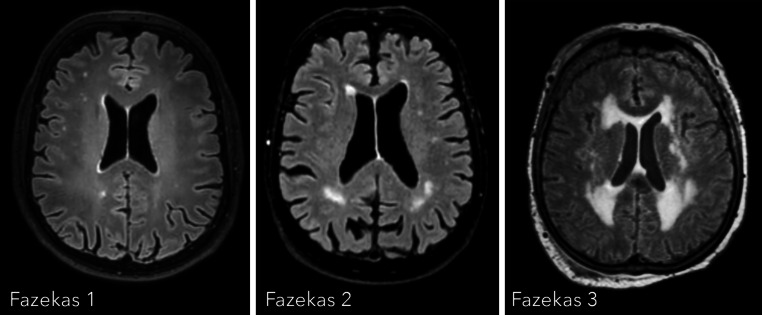
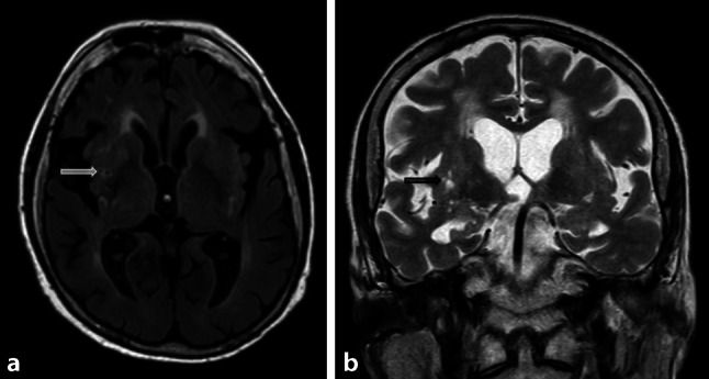
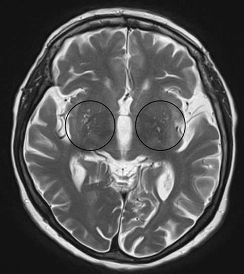
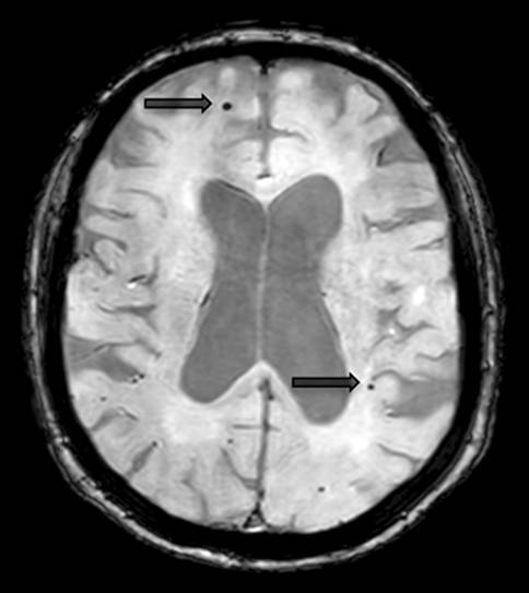
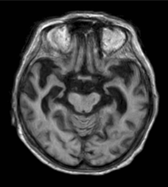
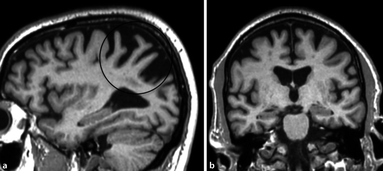
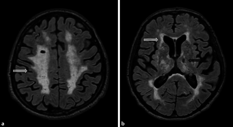
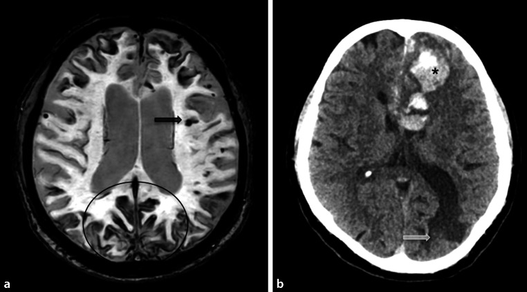
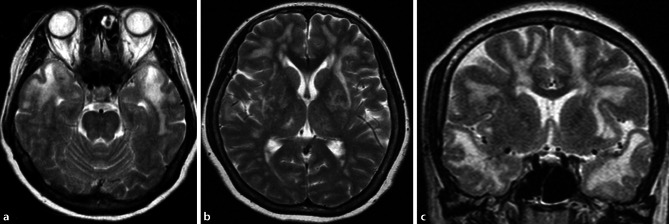
Despite the fact that the diagnosis of dementia is mainly based on clinical criteria, the role of neuroimaging is still expanding. Among other imaging techniques, magnetic resonance imaging (MRI) plays a core role in assisting with the differentiation between various dementia syndromes and excluding other underlying pathologies that cause dementia, such as brain tumors and subdural hemorrhages. This article gives an overview of the standard MRI protocol and of structural radiological reporting systems in patients who suffer from dementia. Moreover, it presents characteristic MRI features of the most common dementia subtypes.

摘要

尽管痴呆症的诊断主要基于临床标准,但神经影像学的作用仍在不断扩大。在其他成像技术中,磁共振成像(MRI)在辅助区分各种痴呆综合征和排除导致痴呆的其他潜在病理方面起着核心作用,如脑肿瘤和硬膜下血肿。本文概述了痴呆患者的标准 MRI 方案和结构放射学报告系统。此外,还介绍了最常见的痴呆亚型的特征性 MRI 特征。

https://cdn.ncbi.nlm.nih.gov/pmc/blobs/f3cc/8397686/3795ca5c9ede/10354_2021_825_Fig13_HTML.jpg
https://cdn.ncbi.nlm.nih.gov/pmc/blobs/f3cc/8397686/191ee554da04/10354_2021_825_Fig1_HTML.jpg
https://cdn.ncbi.nlm.nih.gov/pmc/blobs/f3cc/8397686/cfcdf285cf3a/10354_2021_825_Fig2_HTML.jpg
https://cdn.ncbi.nlm.nih.gov/pmc/blobs/f3cc/8397686/f70205d12ba7/10354_2021_825_Fig3_HTML.jpg
https://cdn.ncbi.nlm.nih.gov/pmc/blobs/f3cc/8397686/66311a499e35/10354_2021_825_Fig4_HTML.jpg
https://cdn.ncbi.nlm.nih.gov/pmc/blobs/f3cc/8397686/9d2fe07ec73a/10354_2021_825_Fig5_HTML.jpg
https://cdn.ncbi.nlm.nih.gov/pmc/blobs/f3cc/8397686/1068413e896e/10354_2021_825_Fig6_HTML.jpg
https://cdn.ncbi.nlm.nih.gov/pmc/blobs/f3cc/8397686/373483edc00f/10354_2021_825_Fig7_HTML.jpg
https://cdn.ncbi.nlm.nih.gov/pmc/blobs/f3cc/8397686/7e4756cefde7/10354_2021_825_Fig8_HTML.jpg
https://cdn.ncbi.nlm.nih.gov/pmc/blobs/f3cc/8397686/07b7ba746dd8/10354_2021_825_Fig9_HTML.jpg
https://cdn.ncbi.nlm.nih.gov/pmc/blobs/f3cc/8397686/a07d161add6e/10354_2021_825_Fig10_HTML.jpg
https://cdn.ncbi.nlm.nih.gov/pmc/blobs/f3cc/8397686/6b39ad1b9f95/10354_2021_825_Fig11_HTML.jpg
https://cdn.ncbi.nlm.nih.gov/pmc/blobs/f3cc/8397686/6977986b62bd/10354_2021_825_Fig12_HTML.jpg
https://cdn.ncbi.nlm.nih.gov/pmc/blobs/f3cc/8397686/3795ca5c9ede/10354_2021_825_Fig13_HTML.jpg

相似文献

1
Neuroimaging in dementia.痴呆症的神经影像学。
Wien Med Wochenschr. 2021 Sep;171(11-12):274-281. doi: 10.1007/s10354-021-00825-x. Epub 2021 Mar 3.
2
Structural MR Imaging in the Diagnosis of Alzheimer's Disease and Other Neurodegenerative Dementia: Current Imaging Approach and Future Perspectives.结构磁共振成像在阿尔茨海默病及其他神经退行性痴呆诊断中的应用:当前成像方法及未来展望
Korean J Radiol. 2016 Nov-Dec;17(6):827-845. doi: 10.3348/kjr.2016.17.6.827. Epub 2016 Oct 31.
3
[Dementia: role of imaging].[痴呆:影像学的作用]
J Radiol. 2003 Nov;84(11 Pt 2):1819-28.
4
Structural Brain Magnetic Resonance Imaging to Rule Out Comorbid Pathology in the Assessment of Alzheimer's Disease Dementia: Findings from the Ontario Neurodegenerative Disease Research Initiative (ONDRI) Study and Clinical Trials Over the Past 10 Years.结构脑磁共振成像排除阿尔茨海默病痴呆评估中的共病病理学:来自安大略神经退行性疾病研究倡议(ONDRI)研究和过去 10 年临床试验的结果。
J Alzheimers Dis. 2020;74(3):747-757. doi: 10.3233/JAD-191097.
5
Methodological problems and clinical relevance of structural neuroimaging in dementia research.痴呆症研究中结构神经影像学的方法学问题及临床相关性
J Neural Transm Gen Sect. 1995;99(1-3):131-43. doi: 10.1007/BF01271474.
6
Imaging in Dementia With Lewy Bodies: An Overview.路易体痴呆的影像学:综述
J Geriatr Psychiatry Neurol. 2016 Sep;29(5):254-60. doi: 10.1177/0891988716654984.
7
Magnetic Resonance Texture Analysis in Alzheimer's disease.磁共振纹理分析在阿尔茨海默病中的应用。
Acad Radiol. 2020 Dec;27(12):1774-1783. doi: 10.1016/j.acra.2020.01.006. Epub 2020 Feb 10.
8
[How Useful is Brain MRI for Early Diagnosis of Alzheimer's Disease?].[脑部磁共振成像对阿尔茨海默病早期诊断有多大用处?]
Brain Nerve. 2023 Aug;75(8):923-932. doi: 10.11477/mf.1416202446.
9
What is the subtype of dementia in patients with fragility hip fracture?脆性髋部骨折患者的痴呆亚型是什么?
PLoS One. 2022 Apr 5;17(4):e0265636. doi: 10.1371/journal.pone.0265636. eCollection 2022.
10
Frontotemporal dementia neuroimaging: a guide for clinicians.额颞叶痴呆的神经影像学:临床医生指南
Front Neurol Neurosci. 2009;24:160-167. doi: 10.1159/000197895. Epub 2009 Jan 26.

引用本文的文献

1
Comparative analysis of Voxel-based morphometry using T1 and T2-weighted magnetic resonance imaging to explore the relationship between brain structure and cognitive abilities.使用T1加权和T2加权磁共振成像进行基于体素的形态测量学比较分析,以探索脑结构与认知能力之间的关系。
Exp Brain Res. 2025 Jan 21;243(2):50. doi: 10.1007/s00221-025-07000-3.
2
Automated Scoring of Alzheimer's Disease Atrophy Scale with Subtype Classification Using Deep Learning-Based T1-Weighted Magnetic Resonance Image Segmentation.基于深度学习的T1加权磁共振图像分割对阿尔茨海默病萎缩量表进行自动评分及亚型分类
J Alzheimers Dis Rep. 2024 May 17;8(1):863-876. doi: 10.3233/ADR-230105. eCollection 2024.
3

本文引用的文献

1
Diagnosis and management of dementia with Lewy bodies: Fourth consensus report of the DLB Consortium.路易体痴呆的诊断与管理:DLB联盟第四次共识报告
Neurology. 2017 Jul 4;89(1):88-100. doi: 10.1212/WNL.0000000000004058. Epub 2017 Jun 7.
2
Cerebral amyloid angiopathy and its co-occurrence with Alzheimer's disease and other cerebrovascular neuropathologic changes.脑淀粉样血管病及其与阿尔茨海默病和其他脑血管神经病理变化的共存。
Neurobiol Aging. 2015 Oct;36(10):2702-8. doi: 10.1016/j.neurobiolaging.2015.06.028. Epub 2015 Jul 8.
3
Diagnosis and management of behavioral variant frontotemporal dementia.
Association between Female Reproductive Factors and Risk of Dementia.
女性生殖因素与痴呆风险之间的关联。
J Clin Med. 2024 May 19;13(10):2983. doi: 10.3390/jcm13102983.
4
The impact of medial temporal and parietal atrophy on cognitive function in dementia.内侧颞叶和顶叶萎缩对痴呆认知功能的影响。
Sci Rep. 2024 Mar 4;14(1):5281. doi: 10.1038/s41598-024-56023-3.
5
Gray and White Matter Voxel-Based Morphometry of Alzheimer's Disease With and Without Significant Cerebrovascular Pathologies.伴有和不伴有显著脑血管病变的阿尔茨海默病的基于体素的灰质和白质形态学研究
Neurosci Insights. 2024 Jan 31;19:26331055231225657. doi: 10.1177/26331055231225657. eCollection 2024.
6
The Latest Advances in the Diagnosis and Treatment of Dementia.痴呆症诊断与治疗的最新进展
Cureus. 2023 Dec 14;15(12):e50522. doi: 10.7759/cureus.50522. eCollection 2023 Dec.
7
The role of magnetic resonance imaging in the diagnosis and prognosis of dementia.磁共振成像在痴呆症的诊断和预后中的作用。
Biomol Biomed. 2023 Mar 16;23(2):209-224. doi: 10.17305/bjbms.2022.8085.
8
Dietary and Nutrient Patterns and Brain MRI Biomarkers in Dementia-Free Adults.饮食和营养模式与无痴呆症成年人的脑 MRI 生物标志物。
Nutrients. 2022 Jun 3;14(11):2345. doi: 10.3390/nu14112345.
行为变异型额颞叶痴呆的诊断与管理
Biol Psychiatry. 2014 Apr 1;75(7):574-81. doi: 10.1016/j.biopsych.2013.11.006. Epub 2013 Nov 13.
4
White matter perivascular spaces: an MRI marker in pathology-proven cerebral amyloid angiopathy?脑白质血管周围间隙:病理证实的脑淀粉样血管病的 MRI 标志物?
Neurology. 2014 Jan 7;82(1):57-62. doi: 10.1212/01.wnl.0000438225.02729.04. Epub 2013 Nov 27.
5
Imaging markers for Alzheimer disease: which vs how.阿尔茨海默病的影像学标志物:选择与方法。
Neurology. 2013 Jul 30;81(5):487-500. doi: 10.1212/WNL.0b013e31829d86e8.
6
Neuroimaging standards for research into small vessel disease and its contribution to ageing and neurodegeneration.神经影像学标准研究小血管疾病及其对衰老和神经退行性变的影响。
Lancet Neurol. 2013 Aug;12(8):822-38. doi: 10.1016/S1474-4422(13)70124-8.
7
Diverging patterns of amyloid deposition and hypometabolism in clinical variants of probable Alzheimer's disease.在可能的阿尔茨海默病的临床变异型中,淀粉样蛋白沉积和低代谢的模式存在差异。
Brain. 2013 Mar;136(Pt 3):844-58. doi: 10.1093/brain/aws327. Epub 2013 Jan 28.
8
Visual assessment of posterior atrophy development of a MRI rating scale.MRI 评分量表中后部萎缩发展的视觉评估。
Eur Radiol. 2011 Dec;21(12):2618-25. doi: 10.1007/s00330-011-2205-4. Epub 2011 Jul 31.
9
The diagnosis of dementia due to Alzheimer's disease: recommendations from the National Institute on Aging-Alzheimer's Association workgroups on diagnostic guidelines for Alzheimer's disease.阿尔茨海默病所致痴呆的诊断:美国国家老龄化研究所-阿尔茨海默病协会工作组关于阿尔茨海默病诊断指南的建议。
Alzheimers Dement. 2011 May;7(3):263-9. doi: 10.1016/j.jalz.2011.03.005. Epub 2011 Apr 21.
10
Cadasil.伴有皮质下梗死和白质脑病的常染色体显性遗传性脑动脉病
Lancet Neurol. 2009 Jul;8(7):643-53. doi: 10.1016/S1474-4422(09)70127-9.