• 文献检索
  • 文档翻译
  • 深度研究
  • 学术资讯
  • Suppr Zotero 插件Zotero 插件
  • 邀请有礼
  • 套餐&价格
  • 历史记录
应用&插件
Suppr Zotero 插件Zotero 插件浏览器插件Mac 客户端Windows 客户端微信小程序
定价
高级版会员购买积分包购买API积分包
服务
文献检索文档翻译深度研究API 文档MCP 服务
关于我们
关于 Suppr公司介绍联系我们用户协议隐私条款
关注我们

Suppr 超能文献

核心技术专利:CN118964589B侵权必究
粤ICP备2023148730 号-1Suppr @ 2026

文献检索

告别复杂PubMed语法,用中文像聊天一样搜索,搜遍4000万医学文献。AI智能推荐,让科研检索更轻松。

立即免费搜索

文件翻译

保留排版,准确专业,支持PDF/Word/PPT等文件格式,支持 12+语言互译。

免费翻译文档

深度研究

AI帮你快速写综述,25分钟生成高质量综述,智能提取关键信息,辅助科研写作。

立即免费体验

三维肝细胞生物打印:具有成纤维细胞的核壳结构共培养以增强功能。

3D bioprinting of hepatocytes: core-shell structured co-cultures with fibroblasts for enhanced functionality.

机构信息

Centre for Translational Bone, Joint and Soft Tissue Research, Faculty of Medicine and University Hospital Carl Gustav Carus, TU Dresden, Dresden, Germany.

出版信息

Sci Rep. 2021 Mar 4;11(1):5130. doi: 10.1038/s41598-021-84384-6.

DOI:10.1038/s41598-021-84384-6
PMID:33664366
原文链接:https://pmc.ncbi.nlm.nih.gov/articles/PMC7933206/
Abstract

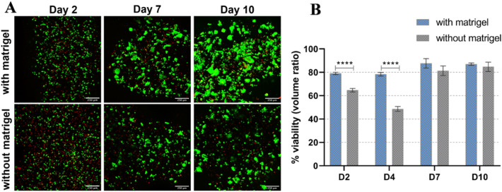
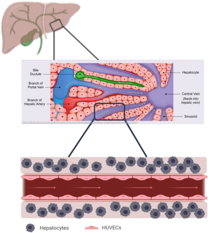
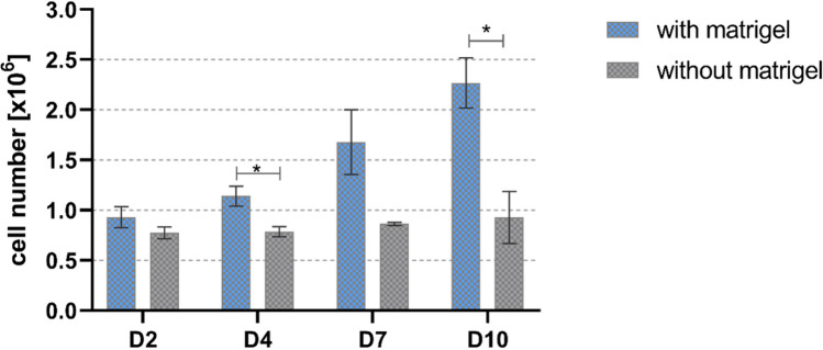
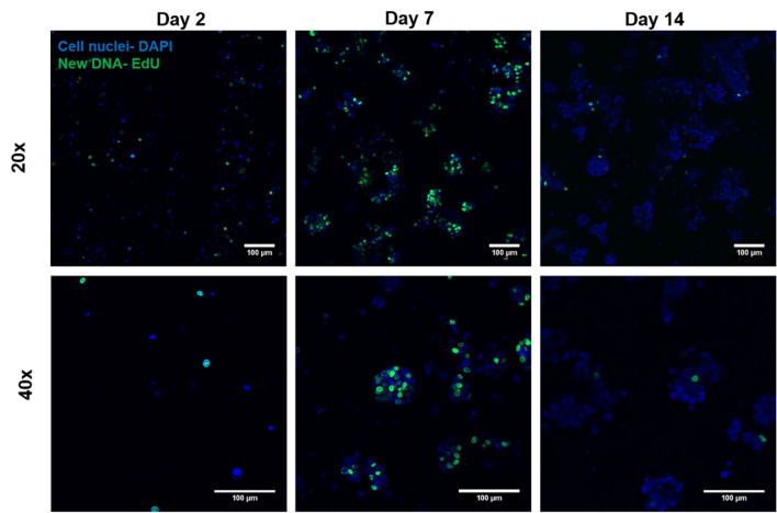
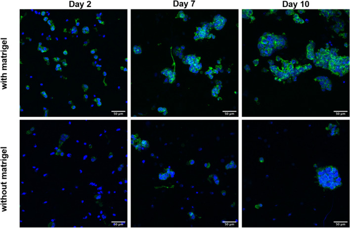
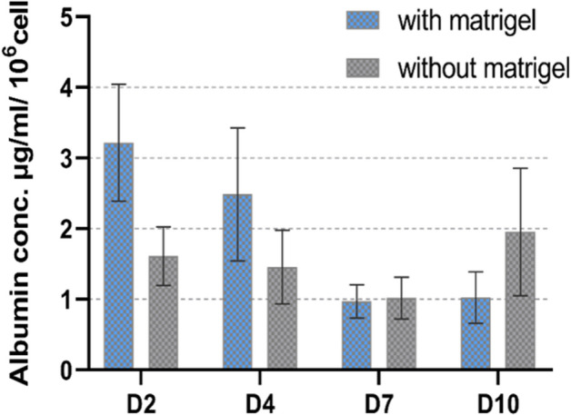
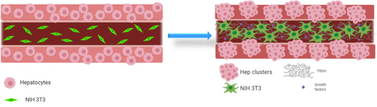
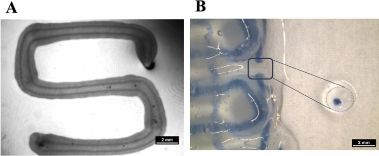
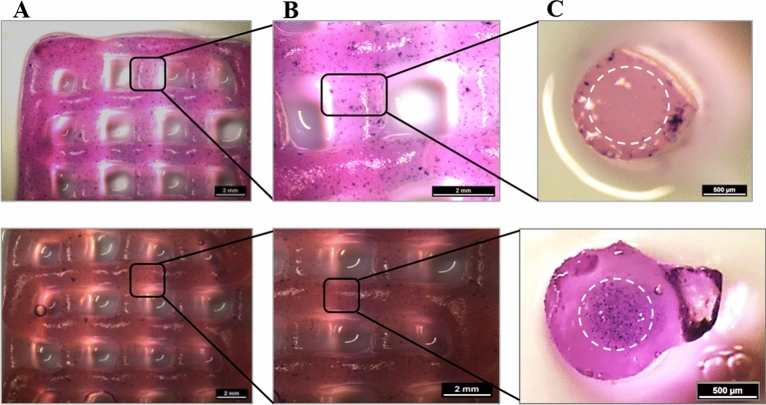
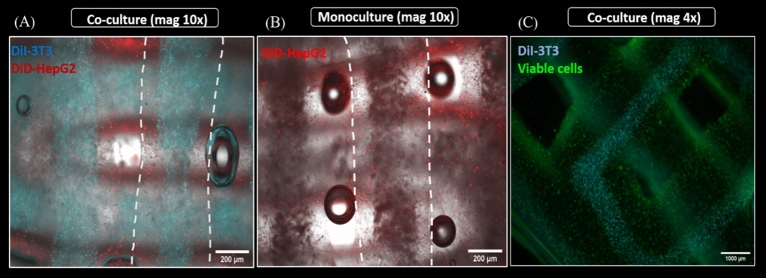
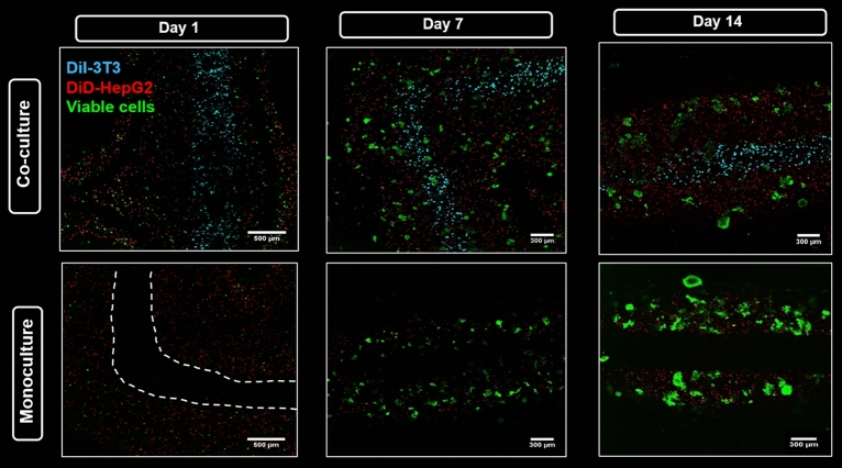
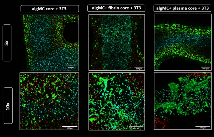
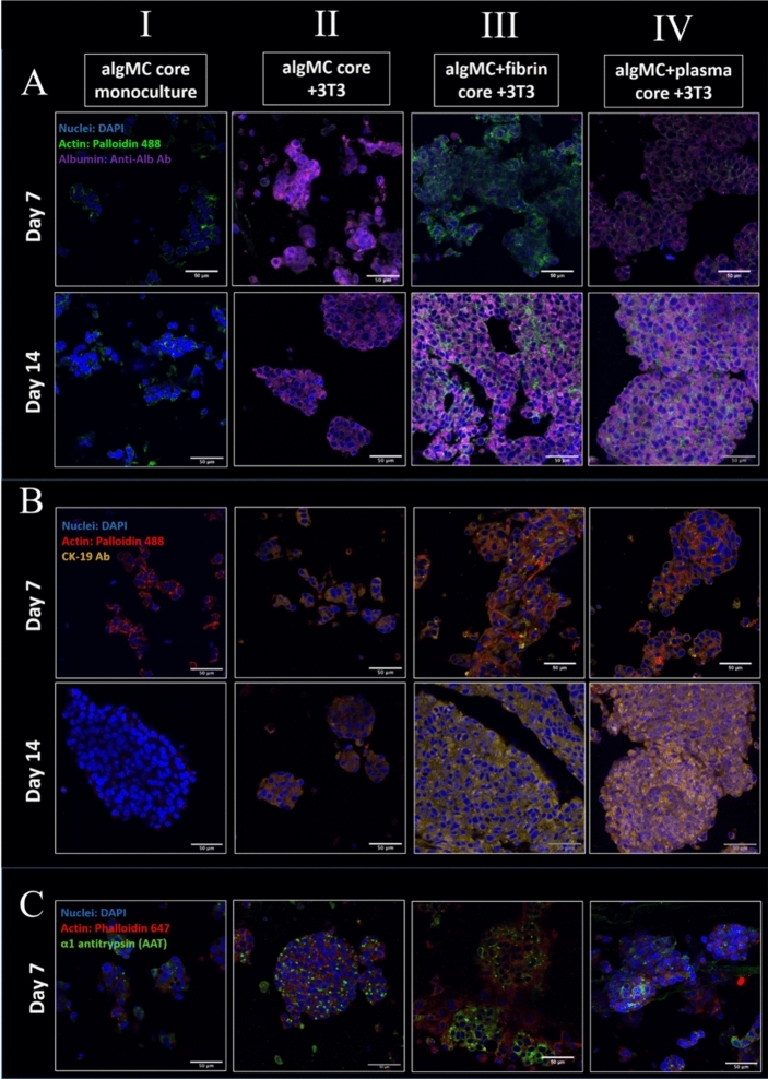
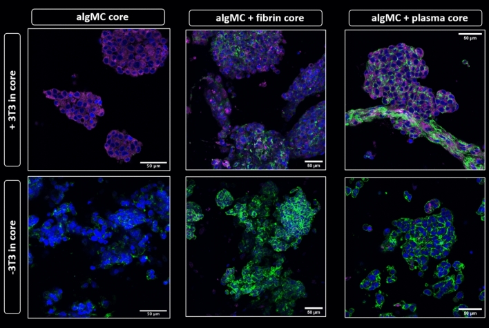
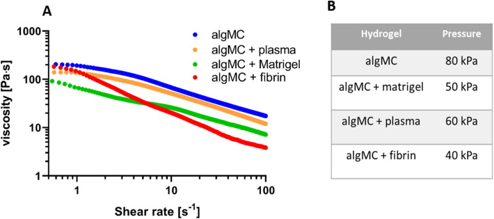
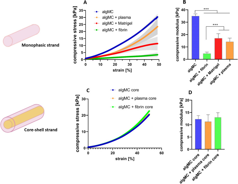
With the aim of understanding and recapitulating cellular interactions of hepatocytes in their physiological microenvironment and to generate an artificial 3D in vitro model, a co-culture system using 3D extrusion bioprinting was developed. A bioink based on alginate and methylcellulose (algMC) was first shown to be suitable for bioprinting of hepatocytes; the addition of Matrigel to algMC enhanced proliferation and morphology of them in monophasic scaffolds. Towards a more complex system that allows studying cellular interactions, we applied core-shell bioprinting to establish tailored 3D co-culture models for hepatocytes. The bioinks were specifically functionalized with natural matrix components (based on human plasma, fibrin or Matrigel) and used to co-print fibroblasts and hepatocytes in a spatially defined, coaxial manner. Fibroblasts acted as supportive cells for co-cultured hepatocytes, stimulating the expression of certain biomarkers of hepatocytes like albumin. Furthermore, matrix functionalization positively influenced both cell types in their respective compartments by enhancing their adhesion, viability, proliferation and function. In conclusion, we established a functional co-culture model with independently tunable compartments for different cell types via core-shell bioprinting. This provides the basis for more complex in vitro models allowing co-cultivation of hepatocytes with other liver-specific cell types to closely resemble the liver microenvironment.

摘要

为了理解和再现肝细胞在生理微环境中的细胞相互作用,并生成人工 3D 体外模型,开发了一种使用 3D 挤出式生物打印的共培养系统。首先,基于海藻酸盐和甲基纤维素(algMC)的生物墨水被证明适合肝细胞的生物打印;algMC 中添加 Matrigel 可增强它们在单相支架中的增殖和形态。为了建立更复杂的系统,以研究细胞相互作用,我们应用核壳生物打印技术来建立用于肝细胞的定制 3D 共培养模型。生物墨水专门用天然基质成分(基于人血浆、纤维蛋白或 Matrigel)进行功能化,并用于以空间定义的同轴方式共打印成纤维细胞和肝细胞。成纤维细胞作为共培养的肝细胞的支持细胞,刺激了白蛋白等肝细胞特定生物标志物的表达。此外,基质功能化通过增强细胞在各自隔室中的粘附、活力、增殖和功能,对两种细胞类型都产生了积极的影响。总之,我们通过核壳生物打印技术建立了一种具有独立可调隔室的功能性共培养模型,用于不同类型的细胞。这为更复杂的体外模型提供了基础,允许肝细胞与其他肝脏特异性细胞类型共培养,以更接近肝脏微环境。

https://cdn.ncbi.nlm.nih.gov/pmc/blobs/b9b3/7933206/b2fd7db06c55/41598_2021_84384_Fig16_HTML.jpg
https://cdn.ncbi.nlm.nih.gov/pmc/blobs/b9b3/7933206/9a11b5e6e1a7/41598_2021_84384_Fig1_HTML.jpg
https://cdn.ncbi.nlm.nih.gov/pmc/blobs/b9b3/7933206/018775a68ed5/41598_2021_84384_Fig2_HTML.jpg
https://cdn.ncbi.nlm.nih.gov/pmc/blobs/b9b3/7933206/d2924c50e23b/41598_2021_84384_Fig3_HTML.jpg
https://cdn.ncbi.nlm.nih.gov/pmc/blobs/b9b3/7933206/cbbc1a2ac235/41598_2021_84384_Fig4_HTML.jpg
https://cdn.ncbi.nlm.nih.gov/pmc/blobs/b9b3/7933206/356e45b271b2/41598_2021_84384_Fig5_HTML.jpg
https://cdn.ncbi.nlm.nih.gov/pmc/blobs/b9b3/7933206/73aed4613052/41598_2021_84384_Fig6_HTML.jpg
https://cdn.ncbi.nlm.nih.gov/pmc/blobs/b9b3/7933206/1b6e81268291/41598_2021_84384_Fig7_HTML.jpg
https://cdn.ncbi.nlm.nih.gov/pmc/blobs/b9b3/7933206/da20a011dffb/41598_2021_84384_Fig8_HTML.jpg
https://cdn.ncbi.nlm.nih.gov/pmc/blobs/b9b3/7933206/dc71de6cb6d8/41598_2021_84384_Fig9_HTML.jpg
https://cdn.ncbi.nlm.nih.gov/pmc/blobs/b9b3/7933206/b8a37e29422e/41598_2021_84384_Fig10_HTML.jpg
https://cdn.ncbi.nlm.nih.gov/pmc/blobs/b9b3/7933206/e1c988da6a70/41598_2021_84384_Fig11_HTML.jpg
https://cdn.ncbi.nlm.nih.gov/pmc/blobs/b9b3/7933206/e81fdefa3e86/41598_2021_84384_Fig12_HTML.jpg
https://cdn.ncbi.nlm.nih.gov/pmc/blobs/b9b3/7933206/331540936d0a/41598_2021_84384_Fig13_HTML.jpg
https://cdn.ncbi.nlm.nih.gov/pmc/blobs/b9b3/7933206/c0be9f8eca9b/41598_2021_84384_Fig14_HTML.jpg
https://cdn.ncbi.nlm.nih.gov/pmc/blobs/b9b3/7933206/7c97b55d57bb/41598_2021_84384_Fig15_HTML.jpg
https://cdn.ncbi.nlm.nih.gov/pmc/blobs/b9b3/7933206/b2fd7db06c55/41598_2021_84384_Fig16_HTML.jpg

相似文献

1
3D bioprinting of hepatocytes: core-shell structured co-cultures with fibroblasts for enhanced functionality.三维肝细胞生物打印:具有成纤维细胞的核壳结构共培养以增强功能。
Sci Rep. 2021 Mar 4;11(1):5130. doi: 10.1038/s41598-021-84384-6.
2
Core-shell bioprinting of vascularizedliver sinusoid models.核心壳层生物打印构建血管化肝脏窦模型。
Biofabrication. 2022 Sep 27;14(4). doi: 10.1088/1758-5090/ac9019.
3
Egg white improves the biological properties of an alginate-methylcellulose bioink for 3D bioprinting of volumetric bone constructs.蛋清改善了海藻酸盐-甲基纤维素生物墨水的生物学性能,使其可用于 3D 生物打印体积骨构建体。
Biofabrication. 2023 Feb 15;15(2). doi: 10.1088/1758-5090/acb8dc.
4
Coaxial 3D bioprinting of tri-polymer scaffolds to improve the osteogenic and vasculogenic potential of cells in co-culture models.同轴 3D 生物打印三聚物支架以提高共培养模型中细胞的成骨和成血管潜力。
J Biomed Mater Res A. 2022 May;110(5):1077-1089. doi: 10.1002/jbm.a.37354. Epub 2022 Jan 13.
5
Current Status of Bioinks for Micro-Extrusion-Based 3D Bioprinting.基于微挤压的3D生物打印生物墨水的现状
Molecules. 2016 May 25;21(6):685. doi: 10.3390/molecules21060685.
6
3D bioprinting of bicellular liver lobule-mimetic structures via microextrusion of cellulose nanocrystal-incorporated shear-thinning bioink.通过微挤出纤维素纳米晶掺入的剪切变稀生物墨水来 3D 打印双细胞肝小叶模拟结构。
Sci Rep. 2020 Nov 26;10(1):20648. doi: 10.1038/s41598-020-77146-3.
7
Coaxial extrusion bioprinting of 3D microfibrous constructs with cell-favorable gelatin methacryloyl microenvironments.同轴挤出生物打印具有细胞亲和性明胶甲基丙烯酰微环境的 3D 微纤维构建体。
Biofabrication. 2018 Jan 12;10(2):024102. doi: 10.1088/1758-5090/aa9d44.
8
Generation of a 3D Liver Model Comprising Human Extracellular Matrix in an Alginate/Gelatin-Based Bioink by Extrusion Bioprinting for Infection and Transduction Studies.通过挤出式生物打印在藻酸盐/明胶基生物墨水中生成包含人细胞外基质的 3D 肝脏模型,用于感染和转导研究。
Int J Mol Sci. 2018 Oct 12;19(10):3129. doi: 10.3390/ijms19103129.
9
Bioprinting of 3D Tissue Models Using Decellularized Extracellular Matrix Bioink.使用脱细胞细胞外基质生物墨水进行3D组织模型的生物打印。
Methods Mol Biol. 2017;1612:381-390. doi: 10.1007/978-1-4939-7021-6_27.
10
3D Bioprinting of osteochondral tissue substitutes - in vitro-chondrogenesis in multi-layered mineralized constructs.3D 生物打印骨软骨组织替代物 - 多层矿化构建体中的体外软骨生成。
Sci Rep. 2020 May 19;10(1):8277. doi: 10.1038/s41598-020-65050-9.

引用本文的文献

1
Innovations in 3D bioprinting and biomaterials for liver tissue engineering: Paving the way for tissue-engineered liver.用于肝脏组织工程的3D生物打印和生物材料创新:为组织工程肝脏铺平道路。
ILIVER. 2024 Feb 8;3(1):100080. doi: 10.1016/j.iliver.2024.100080. eCollection 2024 Mar.
2
Bioprinting of hepatic tissue model using photocrosslinkable dECM-containing composite hydrogel.使用含光交联脱细胞外基质的复合水凝胶进行肝组织模型的生物打印。
Mater Today Bio. 2025 May 3;32:101824. doi: 10.1016/j.mtbio.2025.101824. eCollection 2025 Jun.
3
Recent Advances in Three-Dimensional In Vitro Models for Studies of Liver Fibrosis.

本文引用的文献

1
Bioprinting of 3D tissues/organs combined with microfluidics.3D组织/器官的生物打印与微流体技术相结合。
RSC Adv. 2018 Jun 19;8(39):21712-21727. doi: 10.1039/c8ra03022g. eCollection 2018 Jun 13.
2
Liver Tissue Engineering: Challenges and Opportunities.肝脏组织工程:挑战与机遇
ACS Biomater Sci Eng. 2019 Sep 9;5(9):4167-4182. doi: 10.1021/acsbiomaterials.9b00745. Epub 2019 Aug 19.
3
Crosslinking Strategies for 3D Bioprinting of Polymeric Hydrogels.用于聚合物水凝胶3D生物打印的交联策略
用于肝纤维化研究的三维体外模型的最新进展
Tissue Eng Regen Med. 2025 May 13. doi: 10.1007/s13770-025-00719-8.
4
Hydrogel-Based Bioinks for Coaxial and Triaxial Bioprinting: A Review of Material Properties, Printing Techniques, and Applications.用于同轴和三轴生物打印的水凝胶基生物墨水:材料特性、打印技术及应用综述
Polymers (Basel). 2025 Mar 28;17(7):917. doi: 10.3390/polym17070917.
5
Scaffold-mediated liver regeneration: A comprehensive exploration of current advances.支架介导的肝脏再生:当前进展的全面探索
J Tissue Eng. 2024 Oct 13;15:20417314241286092. doi: 10.1177/20417314241286092. eCollection 2024 Jan-Dec.
6
The cutting-edge progress in bioprinting for biomedicine: principles, applications, and future perspectives.生物打印在生物医学领域的前沿进展:原理、应用及未来展望。
MedComm (2020). 2024 Sep 23;5(10):e753. doi: 10.1002/mco2.753. eCollection 2024 Oct.
7
Growing Role of 3D In Vitro Cell Cultures in the Study of Cellular and Molecular Mechanisms: Short Focus on Breast Cancer, Endometriosis, Liver and Infectious Diseases.三维体外细胞培养在细胞和分子机制研究中的作用日益增大:以乳腺癌、子宫内膜异位症、肝脏和传染病为例的简要焦点观察。
Cells. 2024 Jun 18;13(12):1054. doi: 10.3390/cells13121054.
8
3D bioprinting of liver models: A systematic scoping review of methods, bioinks, and reporting quality.肝脏模型的3D生物打印:方法、生物墨水及报告质量的系统综述
Mater Today Bio. 2024 Feb 15;26:100991. doi: 10.1016/j.mtbio.2024.100991. eCollection 2024 Jun.
9
High-Scale 3D-Bioprinting Platform for the Automated Production of Vascularized Organs-on-a-Chip.用于自动化生产血管化器官芯片的高通量 3D 生物打印平台。
Adv Healthc Mater. 2024 Jul;13(17):e2304028. doi: 10.1002/adhm.202304028. Epub 2024 Apr 3.
10
Advanced PEG-tyramine biomaterial ink for precision engineering of perfusable and flexible small-diameter vascular constructs via coaxial printing.用于通过同轴打印对可灌注且灵活的小直径血管构建体进行精密工程的先进聚乙二醇化酪胺生物材料墨水。
Bioact Mater. 2024 Mar 5;36:168-184. doi: 10.1016/j.bioactmat.2024.02.019. eCollection 2024 Jun.
Small. 2020 Sep;16(35):e2002931. doi: 10.1002/smll.202002931. Epub 2020 Jul 30.
4
3D Bioprinting of osteochondral tissue substitutes - in vitro-chondrogenesis in multi-layered mineralized constructs.3D 生物打印骨软骨组织替代物 - 多层矿化构建体中的体外软骨生成。
Sci Rep. 2020 May 19;10(1):8277. doi: 10.1038/s41598-020-65050-9.
5
A simultaneous 3D printing process for the fabrication of bioceramic and cell-laden hydrogel core/shell scaffolds with potential application in bone tissue regeneration.一种用于制造生物陶瓷和载细胞水凝胶核/壳支架的同步3D打印工艺,在骨组织再生中具有潜在应用。
J Mater Chem B. 2016 Jul 21;4(27):4707-4716. doi: 10.1039/c6tb00849f. Epub 2016 Jun 23.
6
A Novel Plasma-Based Bioink Stimulates Cell Proliferation and Differentiation in Bioprinted, Mineralized Constructs.一种新型基于等离子体的生物墨水可刺激生物打印、矿化构建体中的细胞增殖和分化。
ACS Appl Mater Interfaces. 2020 Mar 18;12(11):12557-12572. doi: 10.1021/acsami.0c00710. Epub 2020 Mar 5.
7
Engineering considerations on extrusion-based bioprinting: interactions of material behavior, mechanical forces and cells in the printing needle.基于挤出的生物打印工程学考量:打印针中材料行为、机械力和细胞的相互作用。
Biofabrication. 2020 Mar 11;12(2):025022. doi: 10.1088/1758-5090/ab7553.
8
Hydrogel Bioink Reinforcement for Additive Manufacturing: A Focused Review of Emerging Strategies.水凝胶生物墨水增强的增材制造:新兴策略的重点综述。
Adv Mater. 2020 Jan;32(1):e1902026. doi: 10.1002/adma.201902026. Epub 2019 Oct 10.
9
Development of in vitro 3D cell model from hepatocellular carcinoma (HepG2) cell line and its application for genotoxicity testing.从肝癌 (HepG2) 细胞系开发体外 3D 细胞模型及其在遗传毒性检测中的应用。
Arch Toxicol. 2019 Nov;93(11):3321-3333. doi: 10.1007/s00204-019-02576-6. Epub 2019 Sep 21.
10
Review of alginate-based hydrogel bioprinting for application in tissue engineering.海藻酸盐基水凝胶生物打印在组织工程中的应用综述。
Biofabrication. 2019 Sep 10;11(4):042001. doi: 10.1088/1758-5090/ab331e.