Centre for Translational Bone Joint and Soft Tissue Research, TU Dresden, Medical Faculty and University Hospital "Carl Gustav Carus", 01307 Dresden, Germany.
Department for Functional Materials in Medicine and Dentistry, University of Würzburg, 97070 Würzburg, Germany.
Int J Mol Sci. 2021 Feb 28;22(5):2451. doi: 10.3390/ijms22052451.
Copper-containing biomaterials are increasingly applied for bone regeneration due to their pro-angiogenetic, pro-osteogenetic and antimicrobial properties. Therefore, the effect of Cu on osteoclasts, which play a major role in bone remodeling was studied in detail.
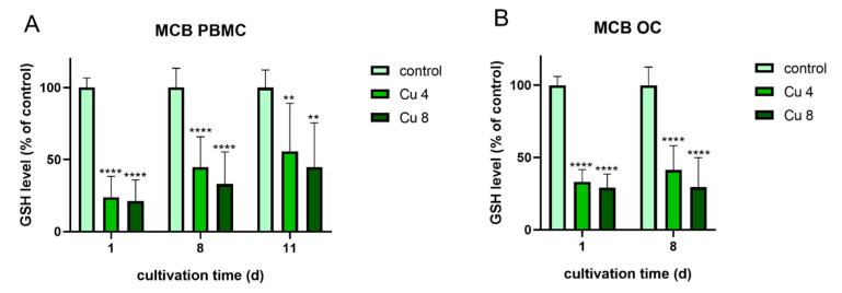
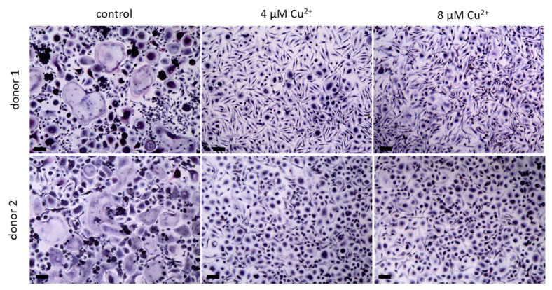
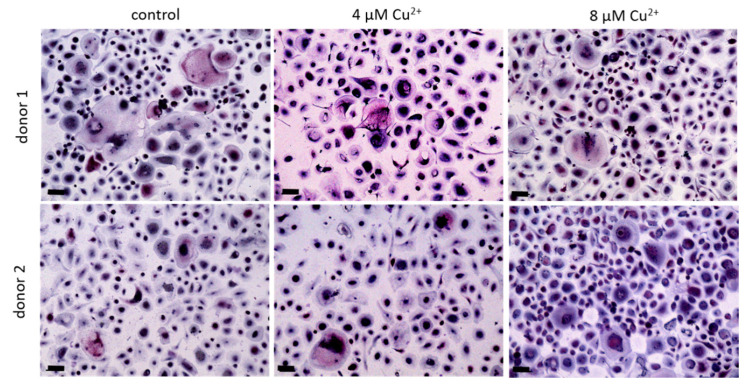
Human primary osteoclasts, differentiated from human monocytes were differentiated or cultivated in the presence of Cu. Osteoclast formation and activity were analyzed by measurement of osteoclast-specific enzyme activities, gene expression analysis and resorption assays. Furthermore, the glutathione levels of the cells were checked to evaluate oxidative stress induced by Cu.
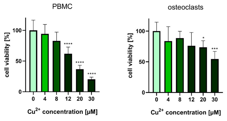
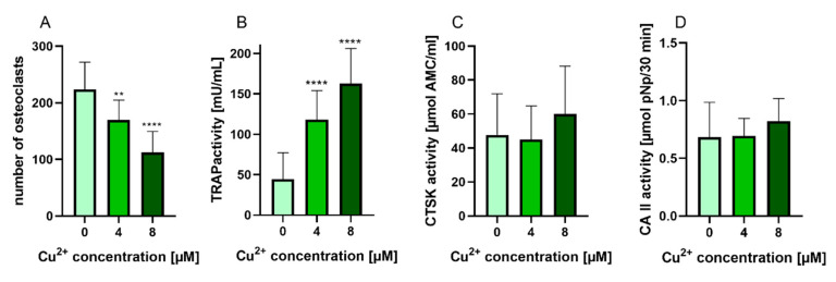
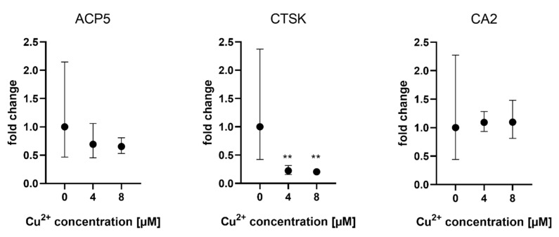
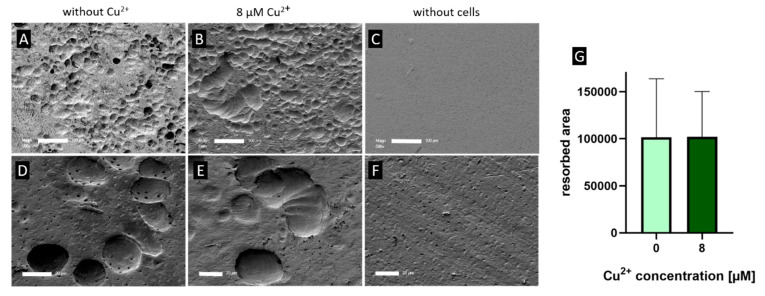
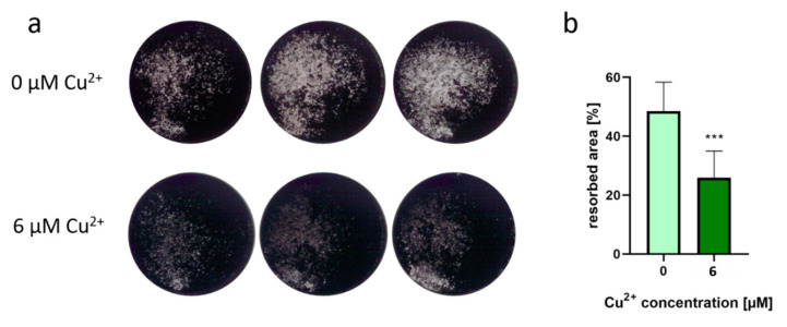
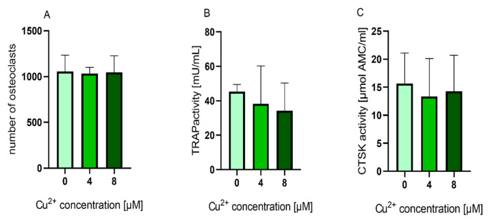
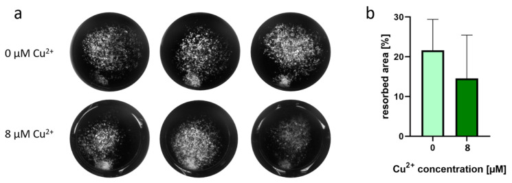
Up to 8 µM Cu did not induce cytotoxic effects. Activity of tartrate-resistant acid phosphatase (TRAP) was significantly increased, while other osteoclast specific enzyme activities were not affected. However, gene expression of TRAP was not upregulated. Resorptive activity of osteoclasts towards dentin was not changed in the presence of 8 µM Cu but decreased in the presence of extracellular bone matrix. When Cu was added to mature osteoclasts TRAP activity was not increased and resorption decreased only moderately. The glutathione level of both differentiating and mature osteoclasts was significantly decreased in the presence of Cu.
Differentiating and mature osteoclasts react differently to Cu. High TRAP activities are not necessarily related to high resorption.
由于铜具有促血管生成、促成骨和抗菌的特性,含铜生物材料在骨再生中的应用越来越广泛。因此,本研究详细研究了铜对破骨细胞的作用,破骨细胞在骨重塑中起着重要作用。
用人单核细胞分化得到的原代人破骨细胞,在铜存在的情况下进行分化或培养。通过测定破骨细胞特异性酶活性、基因表达分析和吸收试验来分析破骨细胞的形成和活性。此外,还检查了细胞的谷胱甘肽水平,以评估铜诱导的氧化应激。
高达 8µM 的铜不会引起细胞毒性作用。抗酒石酸酸性磷酸酶(TRAP)的活性显著增加,而其他破骨细胞特异性酶活性不受影响。然而,TRAP 的基因表达没有上调。在 8µM 的铜存在下,破骨细胞对牙本质的吸收活性没有改变,但在细胞外骨基质存在下则减少。当铜被添加到成熟的破骨细胞中时,TRAP 活性没有增加,吸收只是适度减少。在铜存在的情况下,分化和成熟的破骨细胞的谷胱甘肽水平都显著降低。
分化和成熟的破骨细胞对铜的反应不同。高 TRAP 活性不一定与高吸收有关。