Loeffler Henrike, Jonitz-Heincke Anika, Peters Kirsten, Mueller-Hilke Brigitte, Fiedler Tomas, Bader Rainer, Klinder Annett
Biomechanics and Implant Technology Research Laboratory, Department of Orthopedics, Rostock University Medical Centre, Doberaner Strasse 142, 18057 Rostock, Germany.
Department of Cell Biology, Rostock University Medical Center, Schillingallee 69, 18057 Rostock, Germany.
Materials (Basel). 2020 Mar 5;13(5):1150. doi: 10.3390/ma13051150.
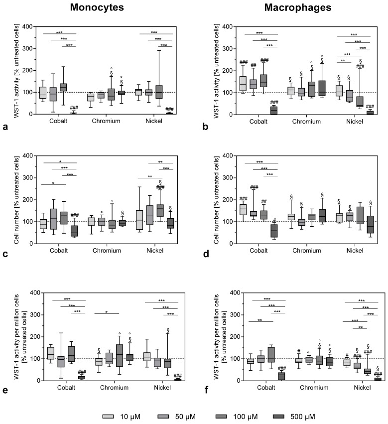
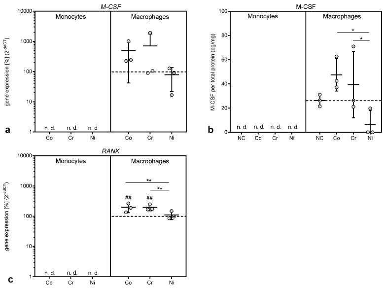
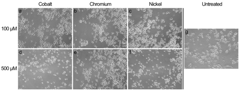
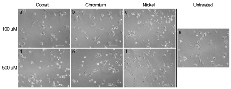
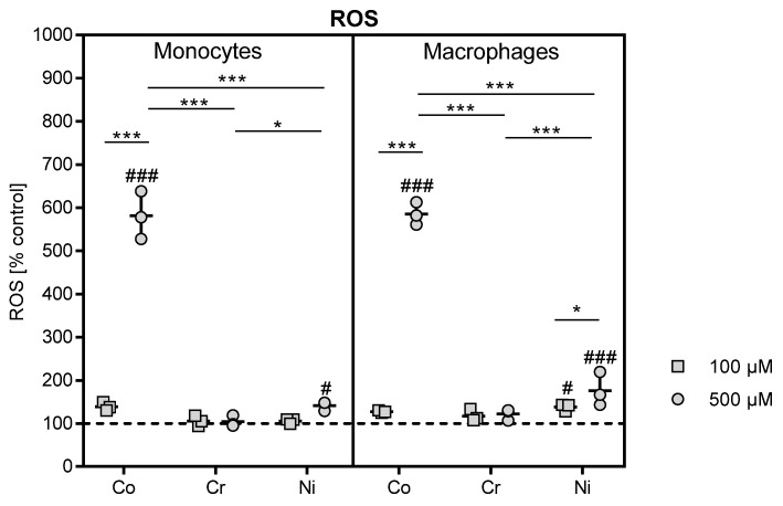
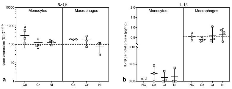
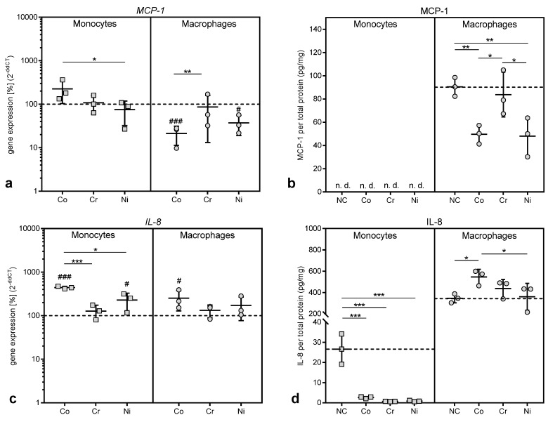
Monocytes and macrophages are the first barrier of the innate immune system, which interact with abrasion and corrosion products, leading to the release of proinflammatory mediators and free reactive molecules. The aim of this study was to understand inflammation-relevant changes in monocytes and macrophages after exposure to corrosion products. To do this, the THP-1 cell line was used to analyze the effects of metal ions simultaneously in monocytes and differentiated macrophages. Cells were stimulated with several concentrations of metal salts (CoCl, NiCl, CrCl × 6HO) to analyze viability, gene expression, protein release and ROS production. Untreated cells served as negative controls. While exposure to Cr(3+) did not influence cell viability in both cell types, the highest concentration (500 µM) of Co(2+) and Ni(2+) showed cytotoxic effects mirrored by significantly reduced metabolism, cell number and a concomitant increase of ROS. The release of IL-1β, IL-8, MCP-1 and M-CSF proteins was mainly affected in macrophages after metal ion exposure (100 µM), indicating a higher impact on pro-inflammatory activity. Our results prove that monocytes and macrophages react very sensitively to corrosion products. High concentrations of bivalent ions lead to cell death, while lower concentrations trigger the release of inflammatory mediators, mainly in macrophages.
单核细胞和巨噬细胞是先天性免疫系统的第一道防线,它们与磨损和腐蚀产物相互作用,导致促炎介质和游离反应性分子的释放。本研究的目的是了解暴露于腐蚀产物后单核细胞和巨噬细胞中与炎症相关的变化。为此,使用THP-1细胞系同时分析单核细胞和分化巨噬细胞中金属离子的作用。用几种浓度的金属盐(CoCl、NiCl、CrCl×6H₂O)刺激细胞,以分析细胞活力、基因表达、蛋白质释放和活性氧生成。未处理的细胞用作阴性对照。虽然暴露于Cr(3+)对两种细胞类型的细胞活力均无影响,但最高浓度(500µM)的Co(2+)和Ni(2+)显示出细胞毒性作用,表现为代谢显著降低、细胞数量减少以及活性氧随之增加。金属离子暴露(100µM)后,IL-1β、IL-8、MCP-1和M-CSF蛋白的释放主要在巨噬细胞中受到影响,表明对促炎活性的影响更大。我们的结果证明,单核细胞和巨噬细胞对腐蚀产物反应非常敏感。高浓度的二价离子导致细胞死亡,而较低浓度则主要在巨噬细胞中触发炎症介质的释放。