• 文献检索
  • 文档翻译
  • 深度研究
  • 学术资讯
  • Suppr Zotero 插件Zotero 插件
  • 邀请有礼
  • 套餐&价格
  • 历史记录
应用&插件
Suppr Zotero 插件Zotero 插件浏览器插件Mac 客户端Windows 客户端微信小程序
定价
高级版会员购买积分包购买API积分包
服务
文献检索文档翻译深度研究API 文档MCP 服务
关于我们
关于 Suppr公司介绍联系我们用户协议隐私条款
关注我们

Suppr 超能文献

核心技术专利:CN118964589B侵权必究
粤ICP备2023148730 号-1Suppr @ 2026

文献检索

告别复杂PubMed语法,用中文像聊天一样搜索,搜遍4000万医学文献。AI智能推荐,让科研检索更轻松。

立即免费搜索

文件翻译

保留排版,准确专业,支持PDF/Word/PPT等文件格式,支持 12+语言互译。

免费翻译文档

深度研究

AI帮你快速写综述,25分钟生成高质量综述,智能提取关键信息,辅助科研写作。

立即免费体验

使用改进的主动形状模型方法对贴近胸膜肿瘤的肺部进行三维分割。

3D segmentation of lungs with juxta-pleural tumor using the improved active shape model approach.

机构信息

College of Information and Engineering, Shenyang University, Shenyang, Liaoning, China.

College of Mechanical Engineering, Shenyang University of Technology, Shenyang, Liaoning, China.

出版信息

Technol Health Care. 2021;29(S1):385-398. doi: 10.3233/THC-218037.

DOI:10.3233/THC-218037
PMID:33682776
原文链接:https://pmc.ncbi.nlm.nih.gov/articles/PMC8150541/
Abstract

BACKGROUND AND OBJECTIVE

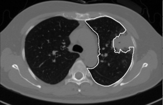
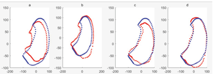
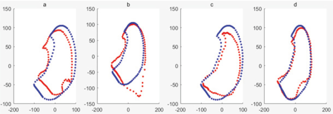
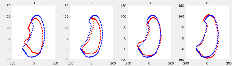
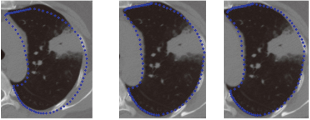
At present, there are many methods for pathological lung segmentation. However, there are still two unresolved problems. (1) The search steps in traditional ASM is a least square optimization method, which is sensitive to outlier marker points, and it makes the profile update to the transition area in the middle of normal lung tissue and tumor rather than a true lung contour. (2) If the noise images exist in the training dataset, the corrected shape model cannot be constructed.

METHODS

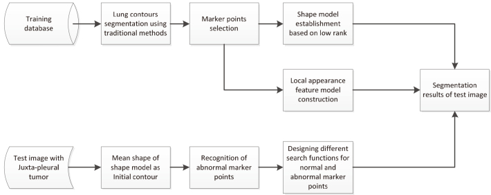
To solve the first problem, we proposed a new ASM algorithm. Firstly, we detected these outlier marker points by a distance method, and then the different searching functions to the abnormal and normal marker points are applied. To solve the second problem, robust principal component analysis (RPCA) of low rank theory can remove noise, so the proposed method combines RPCA instead of PCA with ASM to solve this problem. Low rank decompose for marker points matrix of training dataset and covariance matrix of PCA will be done before segmentation using ASM.

RESULTS

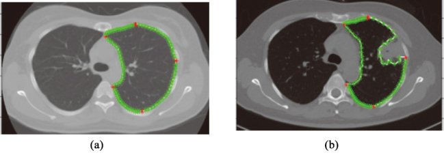
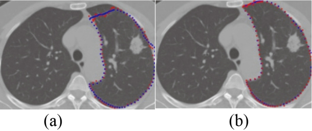
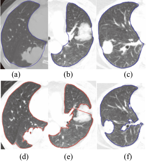
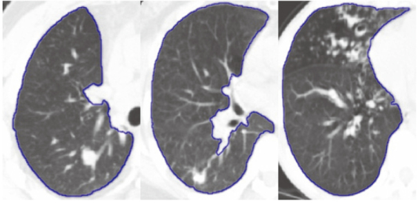
Using the proposed method to segment 122 lung images with juxta-pleural tumors of EMPIRE10 database, got the overlap rate with the gold standard as 94.5%. While the accuracy of ASM based on PCA is only 69.5%.

CONCLUSIONS

The results showed that when the noise sample is contained in the training sample set, a good segmentation result for the lungs with juxta-pleural tumors can be obtained by the ASM based on RPCA.

摘要

背景与目的

目前,有许多用于肺部病理分割的方法。但是,仍然存在两个未解决的问题。(1)传统的 ASM 中的搜索步骤是最小二乘优化方法,该方法对异常标记点敏感,并且使轮廓更新到正常肺组织和肿瘤的中间过渡区域,而不是真实的肺轮廓。(2)如果训练数据集存在噪声图像,则无法构建校正的形状模型。

方法

为了解决第一个问题,我们提出了一种新的 ASM 算法。首先,我们通过距离方法检测这些异常标记点,然后对异常和正常标记点应用不同的搜索功能。为了解决第二个问题,低秩理论的稳健主成分分析(RPCA)可以去除噪声,因此该方法将 RPCA 而不是 PCA 与 ASM 结合使用来解决此问题。在使用 ASM 进行分割之前,将对训练数据集的标记点矩阵和 PCA 的协方差矩阵进行低秩分解。

结果

使用所提出的方法对 EMPIRE10 数据库中 122 个胸膜旁肿瘤的肺部图像进行分割,与金标准的重叠率为 94.5%。而基于 PCA 的 ASM 的准确率仅为 69.5%。

结论

结果表明,当噪声样本包含在训练样本集中时,基于 RPCA 的 ASM 可以获得胸膜旁肿瘤肺部的良好分割结果。

https://cdn.ncbi.nlm.nih.gov/pmc/blobs/ad45/8150541/9fa156698831/thc-29-thc218037-g015.jpg
https://cdn.ncbi.nlm.nih.gov/pmc/blobs/ad45/8150541/8d979e55efdc/thc-29-thc218037-g001.jpg
https://cdn.ncbi.nlm.nih.gov/pmc/blobs/ad45/8150541/d249e64f9baa/thc-29-thc218037-g002.jpg
https://cdn.ncbi.nlm.nih.gov/pmc/blobs/ad45/8150541/895b970bd619/thc-29-thc218037-g003.jpg
https://cdn.ncbi.nlm.nih.gov/pmc/blobs/ad45/8150541/1872f54edcc7/thc-29-thc218037-g004.jpg
https://cdn.ncbi.nlm.nih.gov/pmc/blobs/ad45/8150541/025112ae7a3e/thc-29-thc218037-g005.jpg
https://cdn.ncbi.nlm.nih.gov/pmc/blobs/ad45/8150541/909080281930/thc-29-thc218037-g006.jpg
https://cdn.ncbi.nlm.nih.gov/pmc/blobs/ad45/8150541/9d8249d2d4bc/thc-29-thc218037-g007.jpg
https://cdn.ncbi.nlm.nih.gov/pmc/blobs/ad45/8150541/1ff8137176d4/thc-29-thc218037-g008.jpg
https://cdn.ncbi.nlm.nih.gov/pmc/blobs/ad45/8150541/ea1268c6d979/thc-29-thc218037-g009.jpg
https://cdn.ncbi.nlm.nih.gov/pmc/blobs/ad45/8150541/862b2e38c422/thc-29-thc218037-g010.jpg
https://cdn.ncbi.nlm.nih.gov/pmc/blobs/ad45/8150541/4245ed020957/thc-29-thc218037-g011.jpg
https://cdn.ncbi.nlm.nih.gov/pmc/blobs/ad45/8150541/04776382837b/thc-29-thc218037-g012.jpg
https://cdn.ncbi.nlm.nih.gov/pmc/blobs/ad45/8150541/0d9899e02d7a/thc-29-thc218037-g013.jpg
https://cdn.ncbi.nlm.nih.gov/pmc/blobs/ad45/8150541/99d1b7097673/thc-29-thc218037-g014.jpg
https://cdn.ncbi.nlm.nih.gov/pmc/blobs/ad45/8150541/9fa156698831/thc-29-thc218037-g015.jpg

相似文献

1
3D segmentation of lungs with juxta-pleural tumor using the improved active shape model approach.使用改进的主动形状模型方法对贴近胸膜肿瘤的肺部进行三维分割。
Technol Health Care. 2021;29(S1):385-398. doi: 10.3233/THC-218037.
2
[Research on Segmentation Method of Lung with Juxta-pleural Tumor Based on the Improved Active Shape Model].基于改进主动形状模型的近胸膜肿瘤肺部分割方法研究
Sheng Wu Yi Xue Gong Cheng Xue Za Zhi. 2016 Oct;33(5):879-84.
3
Automatic Lung Segmentation With Juxta-Pleural Nodule Identification Using Active Contour Model and Bayesian Approach.基于主动轮廓模型和贝叶斯方法的具有胸膜旁结节识别功能的自动肺分割
IEEE J Transl Eng Health Med. 2018 May 18;6:1800513. doi: 10.1109/JTEHM.2018.2837901. eCollection 2018.
4
A Minimal Path Searching Approach for Active Shape Model (ASM)-based Segmentation of the Lung.一种基于主动形状模型(ASM)的肺部分割的最小路径搜索方法。
Proc SPIE Int Soc Opt Eng. 2009 Mar 27;7259. doi: 10.1117/12.812575.
5
Global optimal hybrid geometric active contour for automated lung segmentation on CT images.基于全局最优混合几何活动轮廓模型的 CT 图像肺自动分割
Comput Biol Med. 2017 Dec 1;91:168-180. doi: 10.1016/j.compbiomed.2017.10.005. Epub 2017 Oct 12.
6
Automatic segmentation of juxta-pleural tumors from CT images based on morphological feature analysis.基于形态特征分析的CT图像中近胸膜肿瘤自动分割
Biomed Mater Eng. 2014;24(6):3137-44. doi: 10.3233/BME-141136.
7
A generalized active shape model for segmentation of liver in low-contrast CT volumes.一种用于低对比度CT容积中肝脏分割的广义主动形状模型。
Comput Biol Med. 2017 Mar 1;82:59-70. doi: 10.1016/j.compbiomed.2017.01.009. Epub 2017 Jan 24.
8
2D Statistical Lung Shape Analysis Using Chest Radiographs: Modelling and Segmentation.基于胸部 X 光片的 2D 肺部形状统计分析:建模与分割。
J Digit Imaging. 2021 Jun;34(3):523-540. doi: 10.1007/s10278-021-00440-7. Epub 2021 Mar 22.
9
Evaluation of image features and search strategies for segmentation of bone structures in radiographs using Active Shape Models.使用主动形状模型评估X光片中骨结构分割的图像特征和搜索策略。
Med Image Anal. 2002 Mar;6(1):47-62. doi: 10.1016/s1361-8415(01)00051-2.
10
Low-rank and sparse decomposition based shape model and probabilistic atlas for automatic pathological organ segmentation.基于低秩稀疏分解的形状模型和概率图谱的自动病理性器官分割。
Med Image Anal. 2017 May;38:30-49. doi: 10.1016/j.media.2017.02.008. Epub 2017 Feb 22.

引用本文的文献

1
How AI Can Help in the Diagnostic Dilemma of Pulmonary Nodules.人工智能如何帮助解决肺结节的诊断难题。
Cancers (Basel). 2022 Apr 6;14(7):1840. doi: 10.3390/cancers14071840.
2
Data Enhancement via Low-Rank Matrix Reconstruction in Pulsed Thermography for Carbon-Fibre-Reinforced Polymers.基于低秩矩阵重构的脉冲热成像技术在碳纤维增强聚合物中的数据增强
Sensors (Basel). 2021 Oct 29;21(21):7185. doi: 10.3390/s21217185.

本文引用的文献

1
Automatic lung nodule detection using a 3D deep convolutional neural network combined with a multi-scale prediction strategy in chest CTs.基于三维深度学习卷积神经网络与多尺度预测策略的胸部 CT 自动肺结节检测。
Comput Biol Med. 2018 Dec 1;103:220-231. doi: 10.1016/j.compbiomed.2018.10.011. Epub 2018 Oct 12.
2
Semantic Segmentation of Pathological Lung Tissue With Dilated Fully Convolutional Networks.基于扩张全卷积网络的病理性肺组织语义分割。
IEEE J Biomed Health Inform. 2019 Mar;23(2):714-722. doi: 10.1109/JBHI.2018.2818620. Epub 2018 Mar 26.
3
Self-supervised sparse coding scheme for image classification based on low rank representation.
基于低秩表示的图像分类自监督稀疏编码方案。
PLoS One. 2018 Jun 20;13(6):e0199141. doi: 10.1371/journal.pone.0199141. eCollection 2018.
4
Low-rank and sparse decomposition based shape model and probabilistic atlas for automatic pathological organ segmentation.基于低秩稀疏分解的形状模型和概率图谱的自动病理性器官分割。
Med Image Anal. 2017 May;38:30-49. doi: 10.1016/j.media.2017.02.008. Epub 2017 Feb 22.
5
Hierarchical lung field segmentation with joint shape and appearance sparse learning.基于联合形状和外观稀疏学习的肺部区域分割。
IEEE Trans Med Imaging. 2014 Sep;33(9):1761-80. doi: 10.1109/TMI.2014.2305691.
6
Automated 3-D segmentation of lungs with lung cancer in CT data using a novel robust active shape model approach.利用一种新颖的稳健主动形状模型方法对 CT 数据中的肺癌进行自动三维肺分割。
IEEE Trans Med Imaging. 2012 Feb;31(2):449-60. doi: 10.1109/TMI.2011.2171357. Epub 2011 Oct 13.
7
Towards robust and effective shape modeling: sparse shape composition.走向健壮而有效的形状建模:稀疏形状组合。
Med Image Anal. 2012 Jan;16(1):265-77. doi: 10.1016/j.media.2011.08.004. Epub 2011 Sep 5.
8
Evaluation of registration methods on thoracic CT: the EMPIRE10 challenge.胸部 CT 配准方法评估:EMPIRE10 挑战赛。
IEEE Trans Med Imaging. 2011 Nov;30(11):1901-20. doi: 10.1109/TMI.2011.2158349. Epub 2011 May 31.
9
Active shape model segmentation with optimal features.具有最优特征的主动形状模型分割
IEEE Trans Med Imaging. 2002 Aug;21(8):924-33. doi: 10.1109/TMI.2002.803121.
10
Automatic lung segmentation for accurate quantitation of volumetric X-ray CT images.用于准确量化容积式X射线CT图像的自动肺部分割
IEEE Trans Med Imaging. 2001 Jun;20(6):490-8. doi: 10.1109/42.929615.