• 文献检索
  • 文档翻译
  • 深度研究
  • 学术资讯
  • Suppr Zotero 插件Zotero 插件
  • 邀请有礼
  • 套餐&价格
  • 历史记录
应用&插件
Suppr Zotero 插件Zotero 插件浏览器插件Mac 客户端Windows 客户端微信小程序
定价
高级版会员购买积分包购买API积分包
服务
文献检索文档翻译深度研究API 文档MCP 服务
关于我们
关于 Suppr公司介绍联系我们用户协议隐私条款
关注我们

Suppr 超能文献

核心技术专利:CN118964589B侵权必究
粤ICP备2023148730 号-1Suppr @ 2026

文献检索

告别复杂PubMed语法,用中文像聊天一样搜索,搜遍4000万医学文献。AI智能推荐,让科研检索更轻松。

立即免费搜索

文件翻译

保留排版,准确专业,支持PDF/Word/PPT等文件格式,支持 12+语言互译。

免费翻译文档

深度研究

AI帮你快速写综述,25分钟生成高质量综述,智能提取关键信息,辅助科研写作。

立即免费体验

基于主动轮廓模型和贝叶斯方法的具有胸膜旁结节识别功能的自动肺分割

Automatic Lung Segmentation With Juxta-Pleural Nodule Identification Using Active Contour Model and Bayesian Approach.

作者信息

Chung Heewon, Ko Hoon, Jeon Se Jeong, Yoon Kwon-Ha, Lee Jinseok

机构信息

Department of Biomedical EngineeringWonkwang University College of MedicineIksan54538South Korea.

Department of RadiologyWonkwang University College of MedicineIksan54538South Korea.

出版信息

IEEE J Transl Eng Health Med. 2018 May 18;6:1800513. doi: 10.1109/JTEHM.2018.2837901. eCollection 2018.

DOI:10.1109/JTEHM.2018.2837901
PMID:29910995
原文链接:https://pmc.ncbi.nlm.nih.gov/articles/PMC6001848/
Abstract

OBJECTIVE

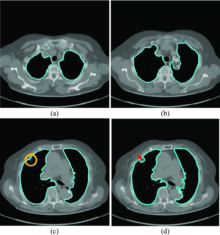
chest computed tomography (CT) images and their quantitative analyses have become increasingly important for a variety of purposes, including lung parenchyma density analysis, airway analysis, diaphragm mechanics analysis, and nodule detection for cancer screening. Lung segmentation is an important prerequisite step for automatic image analysis. We propose a novel lung segmentation method to minimize the juxta-pleural nodule issue, a notorious challenge in the applications.

METHOD

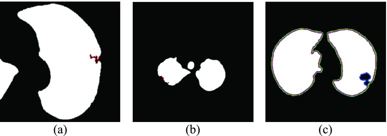
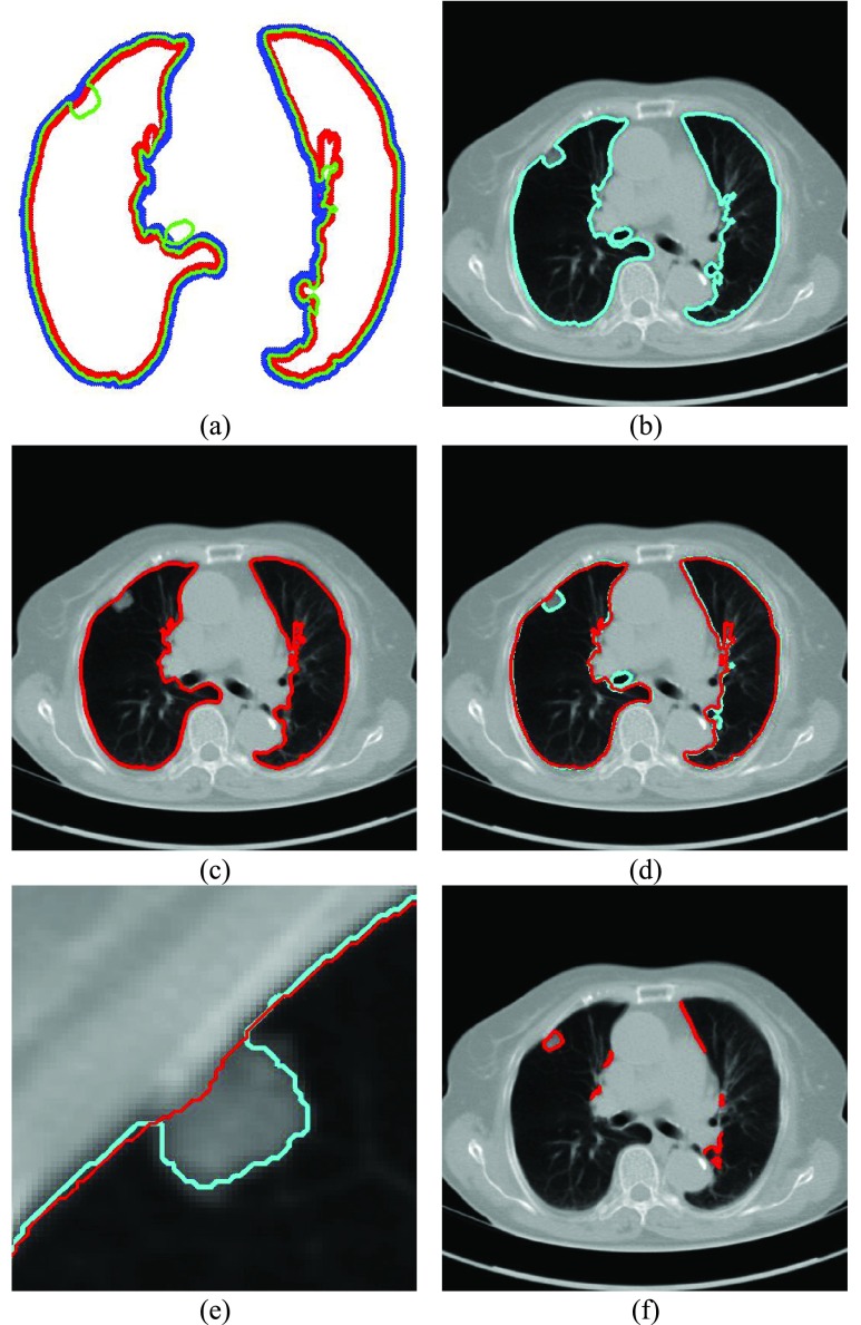
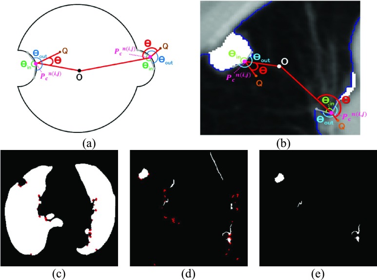
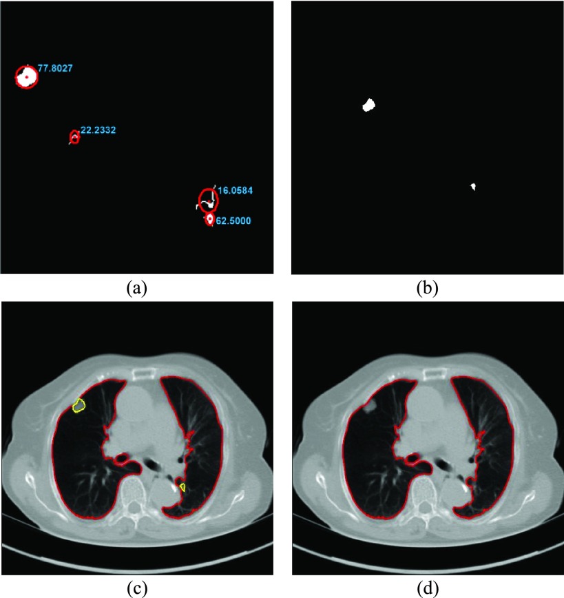
we initially used the Chan-Vese (CV) model for active lung contours and adopted a Bayesian approach based on the CV model results, which predicts the lung image based on the segmented lung contour in the previous frame image or neighboring upper frame image. Among the resultant juxta-pleural nodule candidates, false positives were eliminated through concave points detection and circle/ellipse Hough transform. Finally, the lung contour was modified by adding the final nodule candidates to the area of the CV model results.

RESULTS

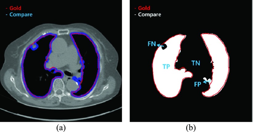
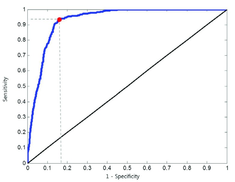
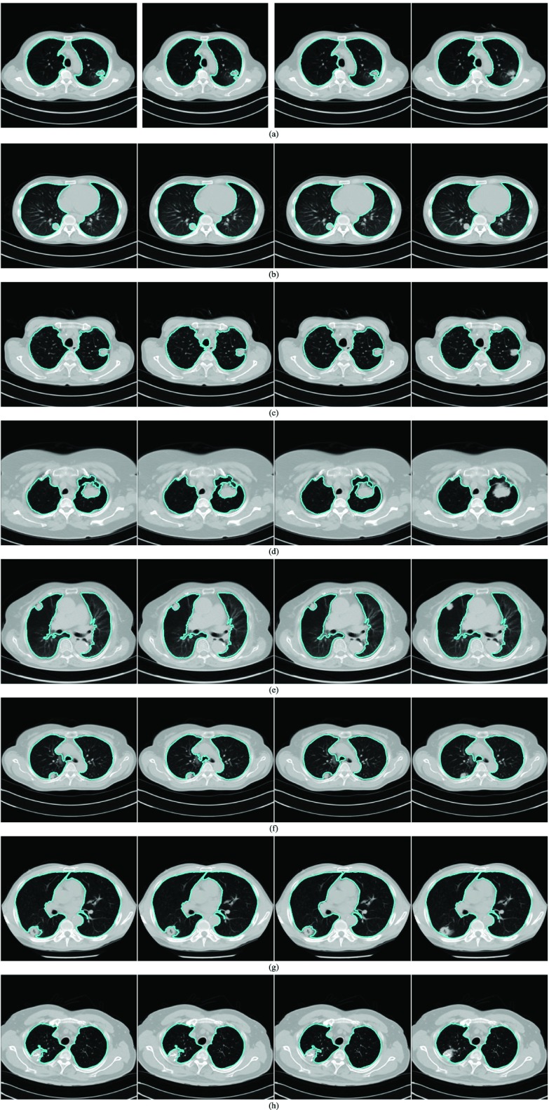
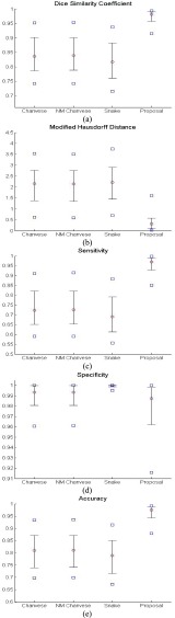
to evaluate the proposed method, we collected chest CT digital imaging and communications in medicine images of 84 anonymous subjects, including 42 subjects with juxta-pleural nodules. There were 16 873 images in total. Among the images, 314 included juxta-pleural nodules. Our method exhibited a disc similarity coefficient of 0.9809, modified hausdorff distance of 0.4806, sensitivity of 0.9785, specificity of 0.9981, accuracy of 0.9964, and juxta-pleural nodule detection rate of 96%. It outperformed existing methods, such as the CV model used alone, the normalized CV model, and the snake algorithm. Clinical impact: the high accuracy with the juxta-pleural nodule detection in the lung segmentation can be beneficial for any computer aided diagnosis system that uses lung segmentation as an initial step.

摘要

目的

胸部计算机断层扫描(CT)图像及其定量分析在多种用途中变得越来越重要,包括肺实质密度分析、气道分析、膈肌力学分析以及癌症筛查中的结节检测。肺分割是自动图像分析的重要前提步骤。我们提出了一种新颖的肺分割方法,以尽量减少胸膜旁结节问题,这是应用中一个臭名昭著的挑战。

方法

我们最初使用Chan-Vese(CV)模型进行主动肺轮廓提取,并基于CV模型结果采用贝叶斯方法,该方法根据前一帧图像或相邻上一帧图像中的分割肺轮廓来预测肺图像。在所得的胸膜旁结节候选区域中,通过凹点检测和圆形/椭圆形霍夫变换消除假阳性。最后,通过将最终的结节候选区域添加到CV模型结果区域来修改肺轮廓。

结果

为了评估所提出的方法,我们收集了84名匿名受试者的胸部CT医学数字成像和通信图像,其中包括42名有胸膜旁结节的受试者。总共有16873张图像。在这些图像中,314张包含胸膜旁结节。我们的方法表现出圆盘相似系数为0.9809,修正豪斯多夫距离为0.4806,灵敏度为0.9785,特异性为0.9981,准确率为0.9964,胸膜旁结节检测率为96%。它优于现有方法,如单独使用的CV模型、归一化CV模型和蛇形算法。临床影响:在肺分割中对胸膜旁结节检测具有高准确性,这对任何将肺分割作为初始步骤的计算机辅助诊断系统都可能有益。

https://cdn.ncbi.nlm.nih.gov/pmc/blobs/d896/6001848/f67389153d94/lee10ab-2837901.jpg
https://cdn.ncbi.nlm.nih.gov/pmc/blobs/d896/6001848/650f6467126f/lee1abcd-2837901.jpg
https://cdn.ncbi.nlm.nih.gov/pmc/blobs/d896/6001848/e24f6e01f7b6/lee2abcdef-2837901.jpg
https://cdn.ncbi.nlm.nih.gov/pmc/blobs/d896/6001848/fe0f5936c637/lee3abcdef-2837901.jpg
https://cdn.ncbi.nlm.nih.gov/pmc/blobs/d896/6001848/be4198d29f14/lee4abcd-2837901.jpg
https://cdn.ncbi.nlm.nih.gov/pmc/blobs/d896/6001848/ab57142db33a/lee5ab-2837901.jpg
https://cdn.ncbi.nlm.nih.gov/pmc/blobs/d896/6001848/70e1ec059e25/lee6-2837901.jpg
https://cdn.ncbi.nlm.nih.gov/pmc/blobs/d896/6001848/1ef542993d76/lee7abcdefgh-2837901.jpg
https://cdn.ncbi.nlm.nih.gov/pmc/blobs/d896/6001848/eee12e1569f9/lee8-2837901.jpg
https://cdn.ncbi.nlm.nih.gov/pmc/blobs/d896/6001848/0ae785b65344/lee9ab-2837901.jpg
https://cdn.ncbi.nlm.nih.gov/pmc/blobs/d896/6001848/f67389153d94/lee10ab-2837901.jpg

相似文献

1
Automatic Lung Segmentation With Juxta-Pleural Nodule Identification Using Active Contour Model and Bayesian Approach.基于主动轮廓模型和贝叶斯方法的具有胸膜旁结节识别功能的自动肺分割
IEEE J Transl Eng Health Med. 2018 May 18;6:1800513. doi: 10.1109/JTEHM.2018.2837901. eCollection 2018.
2
Juxta-Vascular Pulmonary Nodule Segmentation in PET-CT Imaging Based on an LBF Active Contour Model with Information Entropy and Joint Vector.基于具有信息熵和联合向量的LBF主动轮廓模型的PET-CT成像中的血管旁肺结节分割
Comput Math Methods Med. 2018 Jan 8;2018:2183847. doi: 10.1155/2018/2183847. eCollection 2018.
3
Fully automatic deep learning-based lung parenchyma segmentation and boundary correction in thoracic CT scans.基于深度学习的全自动化肺部组织分割和胸 CT 扫描边界修正。
Int J Comput Assist Radiol Surg. 2024 Feb;19(2):261-272. doi: 10.1007/s11548-023-03010-0. Epub 2023 Aug 18.
4
Automatic detection of lung nodules in CT datasets based on stable 3D mass-spring models.基于稳定的三维质量-弹簧模型的 CT 数据集肺结节自动检测。
Comput Biol Med. 2012 Nov;42(11):1098-109. doi: 10.1016/j.compbiomed.2012.09.002. Epub 2012 Sep 26.
5
A Segmentation Framework of Pulmonary Nodules in Lung CT Images.肺部CT图像中肺结节的分割框架
J Digit Imaging. 2016 Feb;29(1):86-103. doi: 10.1007/s10278-015-9801-9.
6
Pulmonary nodule detection in CT images based on shape constraint CV model.基于形状约束CV模型的CT图像肺结节检测
Med Phys. 2015 Mar;42(3):1241-54. doi: 10.1118/1.4907961.
7
Segmentation of pulmonary nodules using adaptive local region energy with probability density function-based similarity distance and multi-features clustering.基于概率密度函数相似性距离和多特征聚类的自适应局部区域能量法用于肺结节分割
Biomed Eng Online. 2016 May 5;15(1):49. doi: 10.1186/s12938-016-0164-3.
8
Three-dimensional lung nodule segmentation and shape variance analysis to detect lung cancer with reduced false positives.用于检测肺癌并减少假阳性的三维肺结节分割与形状变异分析
Proc Inst Mech Eng H. 2016 Jan;230(1):58-70. doi: 10.1177/0954411915619951.
9
Automatic Approach for Lung Segmentation with Juxta-Pleural Nodules from Thoracic CT Based on Contour Tracing and Correction.基于轮廓追踪与校正的胸部CT中含胸膜下结节的肺分割自动方法
Comput Math Methods Med. 2016;2016:2962047. doi: 10.1155/2016/2962047. Epub 2016 Nov 16.
10
3D skeletonization feature based computer-aided detection system for pulmonary nodules in CT datasets.基于 3D 骨架特征的 CT 数据集肺结节计算机辅助检测系统。
Comput Biol Med. 2018 Jan 1;92:64-72. doi: 10.1016/j.compbiomed.2017.11.008. Epub 2017 Nov 11.

引用本文的文献

1
Factors influencing the outcome of volumetry tools for pulmonary nodule analysis: a systematic review and attempted meta-analysis.影响肺结节分析容积测量工具结果的因素:系统评价与尝试性荟萃分析
Insights Imaging. 2023 Sep 23;14(1):152. doi: 10.1186/s13244-023-01480-z.
2
[Corona virus disease 2019 lesion segmentation network based on an adaptive joint loss function].基于自适应联合损失函数的2019冠状病毒病病变分割网络
Sheng Wu Yi Xue Gong Cheng Xue Za Zhi. 2023 Aug 25;40(4):743-752. doi: 10.7507/1001-5515.202206051.
3
SIFT-GVF-based lung edge correction method for correcting the lung region in CT images.

本文引用的文献

1
Guidelines for Management of Incidental Pulmonary Nodules Detected on CT Images: From the Fleischner Society 2017.CT 图像上偶然发现的肺结节管理指南:来自 2017 年 Fleischner 学会。
Radiology. 2017 Jul;284(1):228-243. doi: 10.1148/radiol.2017161659. Epub 2017 Feb 23.
2
Automatic Approach for Lung Segmentation with Juxta-Pleural Nodules from Thoracic CT Based on Contour Tracing and Correction.基于轮廓追踪与校正的胸部CT中含胸膜下结节的肺分割自动方法
Comput Math Methods Med. 2016;2016:2962047. doi: 10.1155/2016/2962047. Epub 2016 Nov 16.
3
Computer-aided diagnosis (CAD) of subsolid nodules: Evaluation of a commercial CAD system.
基于 SIFT-GVF 的肺边缘校正方法,用于校正 CT 图像中的肺区域。
PLoS One. 2023 Feb 28;18(2):e0282107. doi: 10.1371/journal.pone.0282107. eCollection 2023.
4
A Systematic Review of Automated Segmentation Methods and Public Datasets for the Lung and its Lobes and Findings on Computed Tomography Images.基于 CT 图像的肺部及其叶部自动分割方法和公共数据集的系统评价及相关发现。
Yearb Med Inform. 2022 Aug;31(1):277-295. doi: 10.1055/s-0042-1742517. Epub 2022 Dec 4.
5
Present and future of extraoral maxillofacial prosthodontics: Cancer rehabilitation.口腔外颌面修复学的现状与未来:癌症康复
Front Oral Health. 2022 Oct 19;3:1003430. doi: 10.3389/froh.2022.1003430. eCollection 2022.
6
CardioNet: Automatic Semantic Segmentation to Calculate the Cardiothoracic Ratio for Cardiomegaly and Other Chest Diseases.心脏网络:用于计算心脏肥大及其他胸部疾病心胸比率的自动语义分割
J Pers Med. 2022 Jun 17;12(6):988. doi: 10.3390/jpm12060988.
7
How AI Can Help in the Diagnostic Dilemma of Pulmonary Nodules.人工智能如何帮助解决肺结节的诊断难题。
Cancers (Basel). 2022 Apr 6;14(7):1840. doi: 10.3390/cancers14071840.
8
Artificial Intelligence Systems for Diagnosis and Clinical Classification of COVID-19.用于COVID-19诊断和临床分类的人工智能系统
Front Microbiol. 2021 Sep 27;12:729455. doi: 10.3389/fmicb.2021.729455. eCollection 2021.
9
[Research progress in lung parenchyma segmentation based on computed tomography].基于计算机断层扫描的肺实质分割研究进展
Sheng Wu Yi Xue Gong Cheng Xue Za Zhi. 2021 Apr 25;38(2):379-386. doi: 10.7507/1001-5515.202008032.
10
COVID-19 Pneumonia Diagnosis Using a Simple 2D Deep Learning Framework With a Single Chest CT Image: Model Development and Validation.使用具有单张胸部CT图像的简单二维深度学习框架诊断COVID-19肺炎:模型开发与验证
J Med Internet Res. 2020 Jun 29;22(6):e19569. doi: 10.2196/19569.
亚实性结节的计算机辅助诊断(CAD):对一种商用CAD系统的评估
Eur J Radiol. 2016 Oct;85(10):1728-1734. doi: 10.1016/j.ejrad.2016.07.011. Epub 2016 Jul 18.
4
Automatic detection of large pulmonary solid nodules in thoracic CT images.胸部CT图像中大型肺实性结节的自动检测
Med Phys. 2015 Oct;42(10):5642-53. doi: 10.1118/1.4929562.
5
Segmentation and Image Analysis of Abnormal Lungs at CT: Current Approaches, Challenges, and Future Trends.CT 图像中异常肺部的分割与图像分析:当前方法、挑战及未来趋势
Radiographics. 2015 Jul-Aug;35(4):1056-76. doi: 10.1148/rg.2015140232.
6
Automated pulmonary nodule detection based on three-dimensional shape-based feature descriptor.基于三维形状特征描述符的自动肺结节检测。
Comput Methods Programs Biomed. 2014;113(1):37-54. doi: 10.1016/j.cmpb.2013.08.015. Epub 2013 Sep 7.
7
Establishing a normative atlas of the human lung: computing the average transformation and atlas construction.建立人类肺部的规范图谱:计算平均变换和图谱构建。
Acad Radiol. 2012 Nov;19(11):1368-81. doi: 10.1016/j.acra.2012.04.025. Epub 2012 Aug 28.
8
Medical image segmentation by combining graph cuts and oriented active appearance models.基于图割和有向主动表观模型的医学图像分割。
IEEE Trans Image Process. 2012 Apr;21(4):2035-46. doi: 10.1109/TIP.2012.2186306. Epub 2012 Jan 31.
9
Active contours without edges.无边缘活动轮廓。
IEEE Trans Image Process. 2001;10(2):266-77. doi: 10.1109/83.902291.
10
Pulmonary nodules detected at lung cancer screening: interobserver variability of semiautomated volume measurements.肺癌筛查中检测到的肺结节:半自动体积测量的观察者间变异性
Radiology. 2006 Oct;241(1):251-7. doi: 10.1148/radiol.2411050860. Epub 2006 Aug 14.