Division of Surgical Research, University Hospital of Zurich, Zurich, Switzerland.
Plastic Surgery and Hand Surgery, University Hospital of Zurich, Sternwartstrasse 14, 8091, Zurich, Switzerland.
Sci Rep. 2021 Mar 8;11(1):5418. doi: 10.1038/s41598-021-84123-x.
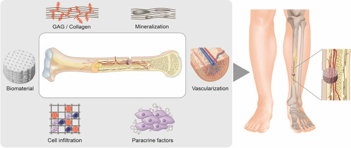
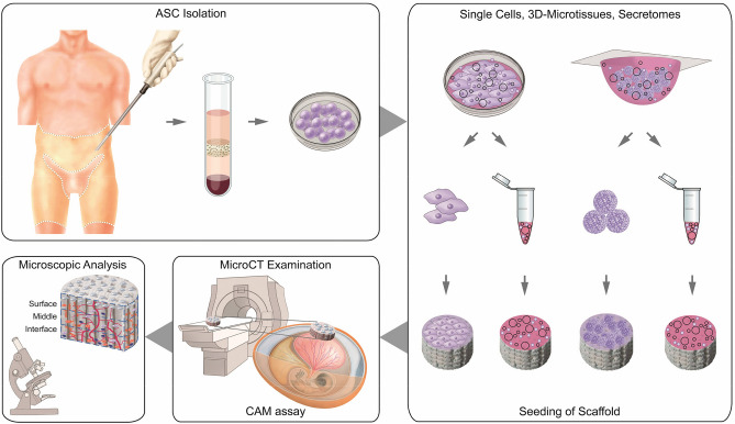
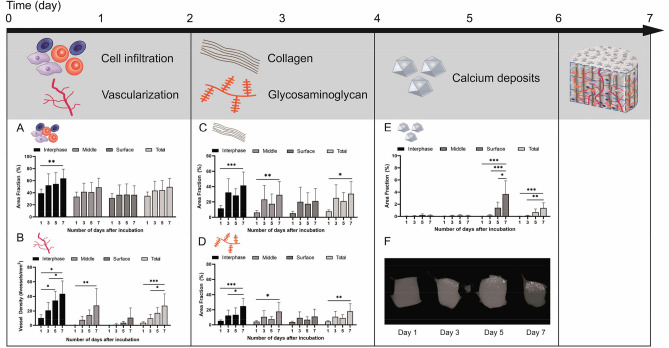
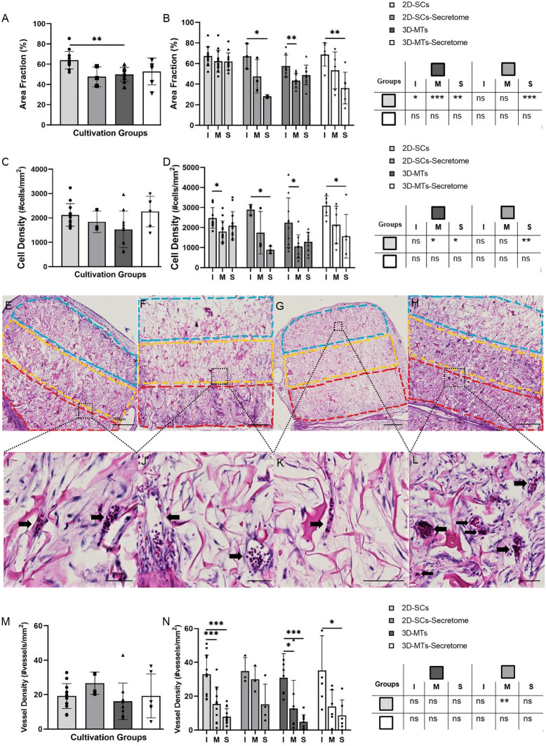
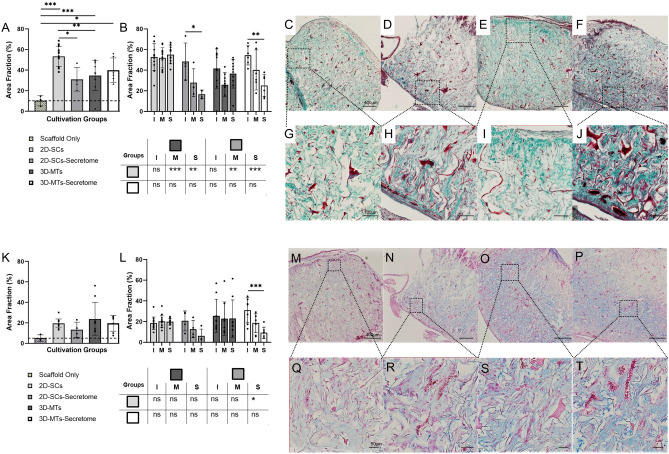
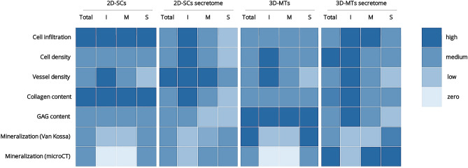
Bone regeneration is a complex process and the clinical translation of tissue engineered constructs (TECs) remains a challenge. The combination of biomaterials and mesenchymal stem cells (MSCs) may enhance the healing process through paracrine effects. Here, we investigated the influence of cell format in combination with a collagen scaffold on key factors in bone healing process, such as mineralization, cell infiltration, vascularization, and ECM production. MSCs as single cells (2D-SCs), assembled into microtissues (3D-MTs) or their corresponding secretomes were combined with a collagen scaffold and incubated on the chicken embryo chorioallantoic membrane (CAM) for 7 days. A comprehensive quantitative analysis was performed on a cellular level by histology and by microcomputed tomography (microCT). In all experimental groups, accumulation of collagen and glycosaminoglycan within the scaffold was observed over time. A pronounced cell infiltration and vascularization from the interface to the surface region of the CAM was detected. The 3D-MT secretome showed a significant mineralization of the biomaterial using microCT compared to all other conditions. Furthermore, it revealed a homogeneous distribution pattern of mineralization deposits in contrast to the cell-based scaffolds, where mineralization was only at the surface. Therefore, the secretome of MSCs assembled into 3D-MTs may represent an interesting therapeutic strategy for a next-generation bone healing concept.
骨再生是一个复杂的过程,组织工程构建体(TECs)的临床转化仍然是一个挑战。生物材料和间充质干细胞(MSCs)的结合可能通过旁分泌作用增强愈合过程。在这里,我们研究了细胞形式与胶原支架相结合对骨愈合过程中的关键因素的影响,如矿化、细胞浸润、血管生成和细胞外基质(ECM)的产生。MSCs 作为单细胞(2D-SCs)、组装成微组织(3D-MTs)或其相应的分泌组,与胶原支架结合并在鸡胚绒毛尿囊膜(CAM)上孵育 7 天。通过组织学和微计算机断层扫描(microCT)在细胞水平上进行了全面的定量分析。在所有实验组中,随着时间的推移,支架内的胶原和糖胺聚糖不断积累。从界面到 CAM 表面区域,观察到明显的细胞浸润和血管生成。与所有其他条件相比,3D-MT 分泌组使用 microCT 显示出生物材料的显著矿化。此外,与基于细胞的支架相比,矿化沉积物呈现出均匀的分布模式,而在基于细胞的支架中,矿化仅在表面。因此,组装成 3D-MT 的 MSCs 的分泌组可能代表下一代骨愈合概念的一种有前途的治疗策略。